 E-submission
 TOTA
TOTA TOTS
TOTS
Articles
- Page Path
- HOME > J Musculoskelet Trauma > Volume 33(2); 2020 > Article
- Case Report Femoral Head Fracture with Hip Dislocation Treated by Autologous Osteochondral Transfer (Mosaicplasty) - A Case Report -
- Eui-Sung Choi, Hyun-Chul Shon, Ho-Seung Jeong, Jae-Young Yang, Seok-Hyun Hong, Byung-Hyun Ahn
-
Journal of Musculoskeletal Trauma 2020;33(2):96-100.
DOI: https://doi.org/10.12671/jkfs.2020.33.2.96
Published online: April 30, 2020
Department of Orthopedic Surgery, Chungbuk National University College of Medicine, Cheongju, Korea �����JFIF���d�d�����Ducky�����d����qhttp://ns.adobe.com/xap/1.0/� ���Adobe�d���������������������������
�����������
����! 1AQa"q�
2#w8���B3��6v�7�X�Rr�$�9��b�C��t%u&��W��s'(xy���4T�5f���H����
�!1AQaq"���2B ��Rbr#��u67�Ѳ3sTt5v8���Sc$4Ă��C�Ô�%U���Ӆ�FV����
��?����������������������������������������������������������������������������������������������������������������������������������������������������������������������������������������������������������������������������������������������������������������������������������������������������������������������������������������������������������������������������������������������������������������������������������������������������������������������������������������������������������������������������������������������������������������������������������������������������������������������������������������������������������������������������������������������������������������������������������������������������������������������������������������������������������������������������������������������������������������������������������������������������������������������������������������������������������������������������������������������������������������������������������������������������������������������������������������������������������������������������������������������������������������������������������������������������������������������������������������������������������������������������������������������������������������������������������������������������������������������������������������������������������������������������������������������������������������������������������������������������������������������������������������������������������������������������������������������������������������������������������������������������������������������������������������������������������������������������������������������������������������������������������������������������������������������������������������������������������������������������������������������������������������������������������������������������������������������������������������������������������������������������������������������������������������������������������������������������������������������������������������������������������������������������������������������������������������������������������������������������������������������������������������������������������������������������������������������������������������������������������������������������������������������������������������������������������������������������������������������������������������������������������������������������������������������������������������������������������������������������������������������������������������������������������������������������������������������������������������������������������������������������������������������������������������������������������������������������������������������������������������������������������������������������������������������������������������������������������������������������������������������������������������������������������������_A���נ�j-
�H���>�>�,�m*>
��
���fzp"�����T������r�Kkr����^����r.�����������������������������������������������������������������������������������������������������������|_&��]�|*��v�P��uܶv�o���Q�1������m
��w�VJU�hu-�I�"=L��n���i��Aƕ8���"�۲����
�k�*ҿ[y�u:�.��v�U���Q +�)��%F� D�Hy�V���B� ��k�>�H�y8��j�ݹ
q~�9D��4�KR��m�z���Q����)�^ʔ�.J%k_tVi5NT��jg!'�k��y|5as����O��Ȼ)R��۸ߩFMԿ3L�4�j�6d��ڜ#�NIw UF��]J��q�B���/(�F�a��fJ�Rzq3�\G՛�
?
�~\�
6�)6���W��4�m[O�
�^L0�E�&r�RM�ض*���C
.��]Un�l��-1�
1�r#Rj/����&QɈ˩��s6�Rj=5Tg�.��y�.·Pӡ:�JJ�S���
:��C8��-���2u]�d�&����v���U�z;��7p�
9���
5V��nL֢����"y)��">�iי�(�IDDd|���
Yj0��;
L���R�f����S��:k����tY�K%*�N2^�m�|&dğ
th�":���e��y�)u�P���Q��Z��W)gcC3Pv&�M���M���W���d&�Ŵ�۲��mv �T��Ro�ժM0�3�*�F
3Y��d������ 6����\8�,�\h�ݻ
kߔ���i��<
k� ��NT��wS�Ԫ���mlj���j[>->�p�������������������������������������������������������������������������������������������������������tU���%�'�
L�R>&E�B��H$�
����
�MQAU�x[�$Z6���v��i&�_�a.K�IQ{�h�yƒ�
�j"J�OC���9����e�F��ҝ����f�j���;�˚Ω<[3�_m%����
��l��Q@��4���g���=5$��(J]Y���c-OMq�<����Ǎ��
wS�����z����
ڗ)k�$
��7
�V�I���P�붾ͯnV+卵*t]i�ЎD�31��~S�A��1�
é�C�2u)��ʼn���Qn-U�o�i�3�:��g��rI8���ؓWm*G
z�ܕ)Z�קJ}��
Y� Y�lGeJ6�cB2I �NS3�Q�>k���=KT�� �B�����T]����W�
�����6����+�S�������O��X�QgG�R�?
te�l�ˊ%��-R�e\hѯ2TF�"��C/�O��JΩ��6���r�[N�.0��{����S��pljjX��1�jOs�ӥ;ҭhe}�x�u`�Ք �&�.���)y��O̒
�F�ߑ.$���Qw�;�9�����I�w2o+R��V�JMS�Oj[SoҌ�Z%�;��`d$�b���l�Q{Ro{�Imڌ>3�e�gf��\O�֝U�z��x��"䢸g+mv%G��������ʆ:|V�[N��'&ס-ޝ�'��k��fE�|K���
,G&˳98�J�u�i�n���/����\\Qݿ�̋v�~�����Ǩ!r��t���WU� ��d����|�E��߫R�� 4d�}.��q���Pw*Ӭv5YE���c�n��������~��f5�c�%M�T�M�k��b-F�>�5��JT���,�}�)�
Q��H�g�%�{("Ӕ�ȸ�W�MsY�y�W��NR��rkk��J�r���0�X�ドnͫ�T������ }�r�-j�j,�Ŕʍ\�Q�2Ri>v$5�!�]"�J��B���2����WɅ�)]V�Ԝ�U��c8��i�|��.�j�e�<�X��7��T�=�r[{w�Ol���TUv*�jp��m��8�KŎ}�Xu*A��H�:&�oZ�ljQj�m�����u�C;N���w
ko�ػ@��}_@�*�>�R�O�6�^�V.�����������������������������������������������������������������������������������������������������¸���
��Q�&��#|�ܶ-*�u��O���G%�JA�t��RZR��r]F�FG��\۩���w+?'zչS����ѧ��t
jz�>
�KW&���ot�{7�P�&�2D�;&\�\�����>�Q����2J�zܗ�A�KS����fe�Nn��[��jRr��ԕ�f6,q,F1�t�R�fԗ�>vֶև���j�-�&���R
'�Z��i2=xv���~E�l��bs�vm8������=ӛ"ūB����ȩl�Wa�u[�]ٷ��B��ߨF~�J�!|���I��p���r3�R̴#Y��p)={7�:G{+:\W�}�n�|Q�#%��)7^-�h��
"Ƒ��q���:M*%J�&�$�T軨�I333�g��_�-
��u���c B
�ww������jp�[�6����i�2��5�����$̏�b��U�ٱR�v?G���\~#Iͪ��b��7�<<}��E���zt"��
����q�_��In���w��,�7-���d����������,���G�÷%T�*�
W�g��1��"��䥱�k��q/�A��.�,_���K�h����q�Œ�
x�wvo��� �u�2ۥ�����ۧ.bQ}� Xκ���A��$֣
�+K��״�ZU���NmڸI�I{.��v�{5
�z5�����Ѯ��R�m����e[m��oyƾ��������d~����c��R��ݾ���K'j.\i�&�������/�S�6�f|b�=5��:�� �p!6�i_�
4�j6�=�.s��i˧eƾ�tS�^� ��c�.�Y�^�R�����������������������������������������������������������������������������������������������������J�V��S-Vi3,esi��08�?�H$G�v���Z�gg?���gi䤟2a����dw����릿�:��"۪���l��k�S��N�>q�-4�kI��܋�ێ�e�̊�qۅ�g��DoѨ���9����;�����
#�T��. �Q���
;��7�#�~� �_U�f����s����t���b����_���'w����������~X��w1���X���k,v �cOt��._}��v}�8��"���(�4��Z��� �\��ۘg
k�?��J?��bm�����_��c�����!�g���{��HZV]��F���kk�%~��g�������E�t�)���b秴��
�v�ΰ���B��������|꽸}�m�
�p~E�6ݹ��v���;�7�P٤���v�+��r��i��*3Ԣ��|�'O14�_����~��7����n�P���{7Z�U��\Vű�[�
����+���7o#:�ǥ����Ŭ\�|3�r���%T�
JX]V7���e�z�¨Y�]��lc|O3��V!��
R��
z�b�J'PnGq��V�J"19��WV����eOF�埜E�aEJω qCN5��Z���
��g��-�9[S��<���$����s��U�K�5b|7s�n\7x
����qmv�����������##�FF\
���w�[�=-4���3$^��o�o��VSi�Xօ�v�7iB�۴y�g�>]Vf�"r�$�J3""32!�Z�h���[K������%7�G��vNL��s��+�4��n����B���/B�{vls�obJ�a�Һ���JR�:����0�g�%&�z��R\
S3���T���[�&ִ�or����*ⷳ�����c�3ʊ�O�[ioz��W�٨%�$���gn:�ܶ���WwFB����Թj�H�P���&z���
u&��F�2�\f;ip�W73��
����
[��;���
'_�̽b��;v�i��b�!oe���c�
�d��C����-tS���<���yC7K��Ƶ��۹b�W/ܻu�z���;�q�\4��R3�Mw�����~��Ga
�աn��]�U�Sv}�q�WMj<���o̺�Z�F;�Pj���(y,��� KZ�<��Ө
��ir�%�q���±��V1n�
�
�ܯY�4ݩ��T�iF�d��'��)=�oD,��S�9j:�v���]yy��bٔq��*Ź%~�C�psrn.N1�^7`�����7I���ߘ�c-5���O,}����B�O�L*u
ȧUc�ǔ�K��n$�#�B1�= �/X��Լ
Vռ}F�ܘ|�
�nܨ����j�]��2On�T�l��ye��B��r��]��4'b���
R�E�����{6�pY�nR�%��+Ce!��J������������������i�-�����/���_��H���U�g?*STKF�H�Φ�`B���C�Pj4��'�}�8����-7�������P���y��ͻn�;���0�9� [�
����N)Iʮ�K�ϗN�r?I�e�`r�&4u
�:�ܬ��.�]붡r��ާ
����B���b�U��E���Ʊ�p��pVn�Bٴ�����ܙ5'�W��ǤN�¨�[���VEU5jSl�"��#��iY����[��r���{/L�Dž����e��Jڜ�|�R�j<�m��K:Aȼ��h�M��Llb��'�����'fWa~v�]�Nӄ���5v*�
)�������������������������������������������������������������M4��m-�
{;����L�DG�C">_��_$�X�s��]�l9���&z�$�2/N��>%'�
�����[���}b�{h�/{`�{�J�i����
�YJ��B���/X%���}.|+{(�����S����:q�z���]4���_Kѵo��`���^��t�Y�_�4�����S�#*�
^��z�vݾM�r��+�T���rkQ�
g.8Ͽ�^�<��O�2���'����\\�E���w���^4��蟡z} �-dz~Q�����mkM� ����\�7�w��
���M�w�/3�/�Ǥ��^�|�W�6��lĚ�����
=��WLt��ބ� ���~�}Ve��z��֏��{���'�Q[����'�f+?��c�����b/�������I�==���d�|E�G%�����_��l�����dk�r��r?�na�u����(��O�&g�Y~�яG������,[���!iYvz�� ɭ������0M��~�b����9���3�W������Lw���:���
��������#'��;��f7m]�ɓ?]��NT���*ի-�(Ӫ��/����%�H�D���.����
̋pS����ʎ��W�u��mv������G�8~ky�����p���r�g�����I��^ń"�
�l�Hj��.o_�+��^�p����ܘ�˟Q�F�E\�0�֙�6���*�<�nT�ˮ�v�<ֿ8�G�^˙Zn=�*֓v�ƫEZO�V���y^�+~eu���V*�p���[�J���+.�t�(:UҴ1g��HN;�vE��̳Rȕs��b%��T
�[e� ݼ�R�P��\�\�yt��i>�ӈ��Ǚ��v����i��x�>�
���$o(�
�
�^��qt�*���&���t1oJ��V����u��-��q�l�5��U6��j�CЉmĻ*�"?JT���=K'��O�/�|=Vo���}l�0b}}f�?���X��[�����?
/\�J�S��Be�,�k�P��8E���T�J=��=�?.
p�5ފ��g��b�U9}���Ƕ���dNKk�����_$8�̸͓ۍ8D��i�\B���Կ-1v{FF]�|��.^��ۅ��{v��l1��2�z7-�R������7w�E?�\n�����h\�jN/Kձr_�oBw"�N
Q��M�B�Zq�
e����-m:ӨSn6�j4���%!hQ�����;sv�'�m4�kcM=���
�!8\��m�[�M4�{SMli��ۇ�%�eֽR�&N:��{2A8�)��T�HL�K3Z�j��[�jP�Bx#Bگ�Mf:�G�1\��`edcʮ�?|w�
������(�-�����̮v���X�t�,��b�������W����2�;.ι��NHRR�#��Y�wT��M�"
��<;�mk\���.f�����oI�D�j�m�lJ;v�xy7���o7i\���,�KQ�Ŋ��9d^��M�mg�c�
L�*�.T��
�6t��L�����eIuOH��3S����JQ�3=F��/�ʿ<��9�\�����JM�6��m��N6=�<{x����kP�!�F1�Q�R[�I$�6��ُim��Xu2�A�n2yԒM�U q��
���f�[��I�������B�-'��䤯jY��m�5����2���&�J�G��\���z��д�\~�v��d��g
�Qt�HG��X�w&�1���Lw+�
nD�E�dC�����1�w|YJm��vP)HZ>i��0�BP�βә?��R�:Q�O[�"��]����I_�J�ʏ��
ۍ>�Q��K�yu���^by��cB�q4lXF�~l��
[\*�N>-��J6,�G�q��(Z�r�5h�]CwY��ӤU��~ʶߑ�
����u�*
S��Iv�%���Z�f���J7�)! F����S��*s��_��\�
|I�Ÿ�Z)J
]���ܜ�i4�"��z[�����+��Z��,MOZ�)<�F�32'&����VlUlQǷi(������\�u r�ϕ"l�
�6l���.\�.)��HyKzD�,ֵ��KQ�����W�^ɽ<����E�9JRnR���)JN���m�V��K����c���j�!���J1�RJ1�IF)$�I*c��պ\Xb�h嫞�LK��2о���Ш��xf˴��ǗN7[$�o�݇(҄��D��yC�
N�œ��9�\V2�<�I5N
�r��V�nn�9Q$`�yX�OU4�as/��f��
�V�����Uv��GV�~�&��:��jݙn���.;·���ޑ
��l�$
�-���-��!JRT�i���7�)Q�n4��X��H���uc�V��cR�%o+
��7iU�*.;s^չ�U'��$�/�O��1�u�ͽR���2����²,�IJ3�Z��-�7��K�"�.D���q�6��3��jWk6��G�eK�����-j�-3`�2��Zk�B��,ȖӉQ�k�}
c����]�je���t���v��n�$�IBpjQk����y[Ѵ�`�ʚ.������j����z��pM�ܵr2��T�
���ơߍ�����?��� ��j��T����5��������
����O�������i��c+3��R�Ց��*�v-�wZ�[WX�l�w�Z�G��U�ۥ�rά\�|��JEQ�P���r�������e^{����-i|��fe�cci����W&�r�cf
����
i�Ը�K�W%(�#���_L�7����N���jy��u�?Vm�M��M�cB�7��N2MÇ<�Fۄ�/!�uU���?
�5ժ7�j��R��dXW]^C%f�ͽ.
o]�J�m
?i$��-�9r�v��ۜw��>)}�|��Ʀ���(�RUNII�I���.<��?�fӆ=�?pE�Ry���+i�a�NP�h�%(�����b�vh�9���^���?D�N�����iӦTUy]�UP��V�Ƒ5Q�6ٺ����iN�$����nq��[�
3�u]K��(cdޱKǸ��S�r�J�7D��E���
5玔kZ�:r���j��v�w7.�-�S'�ˍO~<��B��i\����?Է�2���>�˨����r�n}�M)�xx�ӕ�0��
�
eyҵ��7YMAB�]ӣ�U:/ѭ*�6��b��cwP͵
���"���+��q�ēV�j
ŹO��|G����tY�4V�
��j�[mL�V��
M
-m���>�",���B�$�
�G�D1~��j�6O�4|��L�x�nN���mqA�T�N��R�3ε��|DŽ��a����[���f��m��n���-�ڭ+F�i�K��7�P�cm�;�r�5
l�8���r{��#����-<�U��Lgd�y�|����\[F'��
ѩ�@�{�8��nR�ɕ�xi�vo5N9�'8\��$��.�����,r7/Ϭ�*�XZ=�Ў��n��ޚ�o+|(x����b�8����ۄf��d�)�F�7G���3�����G�Q�FR�R)���)��U2�|Wb@�S�D�����Yi m��II�?Q:��l.�k�X\���ó�f�ݸgeB�2n�0�cuF1�RQ�I$�I$zK��B�#�t{��=O��W'R��2�۷t�
�nݹ�bw.\��]'�nrF��h�2ruy��cb;��pW
=�njRqR��J�(��d
�mn�pc���k���N��nʹ+6�]�t�z�~E=ʕ
l ����ZZ�5�jSi3#4��7�.����Lc��fe`���9؏��v囜.��
�F\-�UZ:*�0_���<�Νu��9�Lӵm&<��qX�Ƴ�e\�F
cq[� �N*RJ\)��_�3�\�^ҹ3"���1n1v�����_|�uR�ʞͫr��'��i��ȧN�_��k�H���8���x��Xrj�=������\�М�H)�V���\�ˬ.�X�ʸ
����� oVRC�}��ySU9/��O�BY먌�5
�ٿ�w�ޞ��)��rw8Ӫ���i5*�5Z��Η�c��G�Ʊ
!Z�ۄl�mp�j�J
-�
l� <�R̵�/����J�A�պ�Zuq\������I������dUS
�� �4�8�wXJ����Jtc�g4�cI������~�aqߓ�w��ŷrm��-�v�)G�7��y��S����^���7H�^-�\��m�Ō�Aq|�"�m9�I��B�nF㏉9[N+m�m��y��/!��KK��ۉ%��n �+B�dddfFF6�FQR�N-U5�����;Sv�'�m4�kcM=������M�n���
)�\qιqUd9F���%",���6M�Gd
T%-�
�+�~
f�%+y֛^3�S���r���F>6l���c�(��֪�v�ۊ��N��;���g�.��_0��Sѧ�]
����E���T��W
�رk����Q�Kz�Ge��9��ʨs����KA"yC��
y2\��[�5
��r�ԭ�7Gk��5M���z�w���_4�s��M3��hxЊ����'���oÍ�5jsu�b�
�)�ͪ~�tR�����2�H��]�R�͍>
�̋�m�
6�=�%(˿�(��W�rr-܅�y5(�ܔ��J��Y�unW̹�븹��N��s�qK�
����]���/Q���R#�"�ZM�D�fD|4��3�Qw�|.�_ԡ�S����q���T�Z���B��g��?���?O���
���Ϥ)����/�E�_�U|i}�2
��9Z?¹ �0�:���x��'�3�,�whǣ�?��C
�y�-��A~����=d�a�������J�ј&�M?D1�_P�
��S��+���Oi�&;����a��
�@���������;�Dž�7[������ �z�Z� C"b�v:����j�jMQk�$�M
�RԸ�3u�A\��=wI����.AwC"�^�.�{��?-\����NS�iˏ"�b��}��T�/��}� �q/�
o�.
�1����
M�}R�% :�-���Z�niʒ�L$�S�g�rBW�*��,Mw�'N\�ɇ{s��\��j�]�������V�ryG��'�8f`�}���
'N�<���*/�` U숻�z�
C��wH�q�1�8���J+�vԕK�s�s�4�R�5�3/�&�����XT�t�1b�Z��Ɵ��o\�=�%��nO�)�h��$�r����B�i-�n���K
Ī������^�
���ջڜ�lwkYm��[̑+�/Q�rZo%�TQ�;�T�����Ls(�$2�C:s������.��%��+����eo�
����Ntt�q�۰��k���K�7O0m_�t�_���p������Z��1��SsS�M7"m��e�v��FZ[��w
�-F�J*T*�j�����QRg���
B� S��u|]g�:�ɵz��jq�wm�l���tL.e�3�sRM�چ
�k�Sm���jkmWœިm��++¦�'�t�IL�k*բQ
��D,�P�B��\l�I[�9{<�Y'�L��z�̶p�!�����·j�
��Jݺ�5T�ܔ�oKsG� (<�����C�B4��jyW��?zm�ӍSN����m
[�Ԓ4��=�m� |�۾��%�cZpj|ͩ�Ui�Di��<{l���/���T�=
��Q�^�hO�yH�$�sN� �R�V��:vVnN���+�q���ΰ�77F�v��;[kK*�aUlt�*���[��#���q[�˶��c^���bږ%�Gb� R�6öۍmX��-�Ma���ʉ�A�93��1cs..G<��y
� Iߵ�{'Ÿ�e��g�.|�4���C��u���jQ�տ[9
�}G��-���xwl)I�Q�z
j
��Ó�"�rq�e&��=��]꾧֎���������c)�<��kӳ+��� 0���Jr�R�R3'���T�n��X�i�^x��MF
����Bު�*t��I���L.[h"2"nK�z�Ze�'Z�V/Rr��N�Yz]�8죝���n]���Ķܩ
>��^�Ժ��]�u-7^���\m�Z�j�ܣ9+Rmn
���ߑv��?o����ꋘ?&��ƪ��y^N����4�o=3�
��-�ؔ̿��*��`��}V�݁
���ƒ���P��u���8���%�$���
ݗ]�w�t;\��y���\����>=�'O�jP�Ip/nJ��U8���{϶F����N�M��sf"�ί�Nq�ƹ(�+
ݮ��F2��K��m�
�|jܴ������Z�s%�zf*��eȫ?�]��4)I�۵�nR&F�X
��+
[jD�h��(�#�
�哑9�q9�E�ծj8n�o�Ǖ��Z�f\J�-�l���&��Z������˫}`���ӎh�yrΉ���n\ű�n�]��9p������ʌ����ӣ" ������W���t?N4��_�I��_~�5��4���#/my���1� �Xr�����*척aS#D�T��
�>����q
s��<����F#wM�F�>sΛ�W�;3o���U�aJ��S�����R�M��D��gQnt��:�Ql�,��/ ܷfR���q�iM��
�Ȼ�>����Co�b;A���>ڦ��Wق�M9X���~���/!'�M�W.�}Vrߔꔵ!�5
|���iB����(�0-����z�
F=���}�����okڢ�E$�^w���W�~n�okY�߮�\�6՜
̌
{i�-AF*�9)\t��9�I��V���
������6����۸�5�Z�UF��6�R$ŨQ�Iq砳Y�UZ]e�yv��� �>hI櫥<����w0���N
�)���&��l Ju�
l��wE1��G����D�Ou� F�N2�|
�}馥��uC�1��rޫ�V�+^�g����d�b��&W[4<^e��4�Y�W,d��|h�t�ͮ�sUM)۸�8:{3�d��{A���Ѣ��)��~
��� \#J=N�d�����Ʈ�������ꮓ��90
��|�����1���K $�����v�*?��S�
�]i��$J ���,�C,���SG�?�/�����_՜��pMSƯM��|mG���1V1�$������~�K���>C�S�
v��k����u��j=&�)
-,yLj��uFHK{c駗���.�SO��u��a;��B�r�Sq�j�-ۍZ�#��
�'Jy����s��7��[g��2z
/.u��4�+XV��2V��Q�.ޕ��)$�"�(�%�)#Z������7suZ�%j���
}�B�������Ǭݕe)J��vz�8z�Jf:�hIN�|�svO1�O#I���E�cۍj��ݽ:Sd��Ὦ��v��u^@:o���^�5���c�s>��i/�Vqm�V��m]�ؔ�ܢ���n�6�'v�ޑ��̗J��4�Wn@��O�lKb��X�
��;��n�:h��g��J�9�Ży
Ǒz8�f���܌��q&��Y�
�f��N0��N;[�69�
r�bׅ�
C2��/#�k�E
��l�&2�~�è��MR����.�*%��g�=�Ft�<��N�a��w��R�w��w\���ϊ�k]��5o4^b5�fZ�O9���]�
>.%�؝�e�8<. e�
��=Uv{���~㻏"E�ˑn���vD�ѭ�͜�����Lu��3u0��:�U֝$��[M5<����:o�i��+�V4�V9
���6nXvx��&��_� ��q�
Qq�w�3�W:uϔ����2y���b/�(�ɳ��|5zQ��i��J�#r�|H�w#�.�W�?�4a����D����Ų\ug�W����G�;��Cw��������������������������������������鐢K|x���g)��##�=O���.�d��F��˟�j�MUvW��Ļ�s�r�������.z]�kPc9"]R�)���mkf�Od��*uYf١���R s�B
�A��îh���=k]ʳ���Ur���r�Z�sq����`�d�#�r�$�/<������Yy���F0�nweM�W'��N6�T�q�F��H��y}Qr�y��m
AS�1n �S�`�T�����j�rT >�Ը�3o��^&�l�R�WȍyuW̦��Y�4�Q�DUM�J���6�5��ƒ[�������+��y�g���k
X�K���_±��k#y��:��8(�T��JOSQ�h�Jt2.D��R}�"5 �[�)�
r�)6�V�6�u����5�k:e�XZm�v𭤔!�푊Q[q��Q���}ҹL����E���-
�8�q��IZ�G�|U�M�4�j}Mܕ��[V�wm�{}
N�aqµ"Ԉ�M
��zO�p��KѰ?IA����D�����3I��r�0�'/q1�i�to�B�5{���%�w��kO
��B��n��-ۜd�uqIz�YK6�
��0{+D�ʕ��ܞ�qI����t"��;r����1��m����G����/��\�/�y�m��[�6��J�
ƫ��R�
\L������=S���=OT����@��I�x[T���M�m{>ݾտ֒ݸ�Ӊ�L����Y��Ix�>+"J��V��N�zx����||5r�I?C{�oz�8�۹e\����R-���^\A�2F
��R�+N�9� v�lT]�"ۭ ��d)t֞i ����#�E�2�jB�@=#���/���N�+�!ĕh� x�}I�!�cM��`ą�Z��*ŻɄҒ�߮Y�.�Z}�=�'/oۙ�3��Ip�W�̮�hT�7�c���T�S��uz9�>B}΄�&�h�!��>lӵ�n�~
�j�˅IvU.'�v'�CS�Z�w8��Q���K3G�>�
�,J5��9ٷ���+����HSg��䧎���hJd���zvwv
����-��c�v���x������S�5�[̊n�~ؿ�%��ַ�X?O���0�\�6���n��e�
6��kn��9.ϯ���}����
�*���h��
����8�_���Q��h��L���ݣ
����7q
+=�X����BҲ���5�?�[[�)+�F`�=4���
}B,s��Ng��==����u���*������������Nj��9k_�GJ)�+R�~GS��P�B��ȒZ���:(�����K]h���e��L=vK PӢ��wq�(N��r�G^�ث�ϣ?�#�tC?�.ͼ�[��
���ۅ�o�����؞��y�#%�Ǜ�jVy������LS�w�%T*s92
JTM%�"Y��kQ��О.q� )��gC�Ͳn8cgi�6����j1�M�����Ѿ��[{���9<���Y8�]CK��/�*q�����v�R̃�T��Od��w�����܁9O*e�y��t�0e��
jm]hs�n ��K�BQ��ЏA�y�Y�1s
~�s��76���kz�nJt{ZإJ&ҥ�`rW-��o&�<������7<;��f�UI��V�&����� ����F�T�raJ��h�^�v�Ƙ��ǚ�םidfi.^R�H�m�����g��&�rׇz:}�݃�}x�T����$��ض
k�����'5s-���狶,\��vp��bP�D�،��=�Okf�������.c#c�dz2�F����K�5T���!&�)|��n�t���D�<���+���OŹ�U������
��i��-�G�[�E��E*��FDfe��a��f�
���2�Q�Ƥ���M\�UG_��{
ǹ�m%�\y�r Gy�����:.\�4��wjP�GUJޕ�U�V�7Do\7Vy_13w;��[?�c]�H�\��$IJ,*L��]3b%�L{�y.�J�R�KG2s�q,��B��6��T���}(�#��nW��
|km+��q5]��
�r㪍b���J@y{����by�z ,�b踊3�ϻJ�,'^����xd�،���)JV��w#�.V�ټ<�S}�j�bZ�}�t+�LvAF��"�q\����r㲝
\w!�}jR�����3���ν��d�ҰT��o
�%.�*wo�����7
2m��c
�r���ӓ�
kO��
[�*w��Q⧶���JŘBM�*�o\�TW�'�)b���n�d����}C��zC�����1���)Ŧ<(U��ᰕ(�
4�m:%)$�b��Y����>c��''Ý�պ���Wtb�RؒJ�z��۠8�!�o�9IۄS���95E�9ؔ�-�e9JR{d�m�nッ<�[~�n���$�
<����nZ܈��K~�Y�6{��a$�ҧ
(A$��Qյ�c_�z������8��������s�)I��*�v#�yc��CK���z~���R�q�,ZƱM�NQ�f����&�Y=���>� ���{~�Њ$W���?&���Ր����Y_��? #a.�ߑv��?o����ꋘ?&��ơ|�y^N����4�o=3�t������=��~�7!���/�M���3>n����8�W��홎2�
M`���Qx��+
��z
qy��8�%�]7��_~�5��4���0ۦ�彷��]W��q
C���ѡw�kď���y�������F��5Du���m��_}~�P(��5���.��(�X�,�K9��vᯐ�?�le�B������9;��J����hm����#3�{��CxG��E-��S{��;���@�F�z���� ˙���]=O�'�!ɿ�]'�
r`:��7'��2b����Ж>I��y��,/�e��T�y/�V��<.���H�?�U��Y��Y�{����\�^#�ѣ��r��9������^��7��?x�o�R��Ȇ7��E�o��S�_������������&�?�?��z����������ϾM?��(~Q���-���K&�>"��~a�ߨ
�t7�E����m�s�ϛ��+�?���;f�����Cr���)f�Y�+>��z��$����tIk���j���n_�>�v�nrֳk��i��-˹l=
t;
�'EyC¥�|���/B������LwBJdgjۛ��$s S�1��|ɍV��%JI 6�Kv�əhz���IlBY
ɒ��|�0"�Sy��0F���<����H�b�����ծI}���ы������b|�y5������[��� ��G�
�������c��m��������������
�u¿��q�q�u� ��'�|?�-�:��ꯙ�����w���)`����������������������������������\�g�����K���I{�$�b�ܛ���O-$J�#J�DZ�� z��. �.��T�
[{�ܽ=��W�(ǂ7
�E�lM�k�{-fe̶&
ǗJ��t�m�z�qH�T�*LZ
�:�r���LG#-V�e!
Rz�5�~�ɺ�b��F�"����&�
q��rTj1RM�ټ�
>�>e�o5��W)�O+X˻u�';v�)�2�vV�q۳k�����ۮ������ws?U�ʑBǴYO����漪e
2MIj�PA�ک\�b��1)DD�ؚKm6Z���W�Ψg�ȕ۶yj�ڳ� �2ضN����� ��[C
[|�r�@9J�f�o<_� �e��I7��q.|���c�Ê�V߷:�i��.:$�ȋ�)1�%%�)A�DZ�CEBx�J0MJ���ۥ��y(b���N��s�K���M9��k��43�Iw����N����t.�\���%N簤�I'.��j|ƃ2�$�g���rB�E��ٌ�\}�9:v�*���!n�7��M(��ɽ�]���7c���@�XxƱԨ 37��īf�6�2cT��T��f�FK]9�� wn�����
tQ��
�Hͮ�v�ٱI/f|j��
��=�����7}�\�_V��5U^+:u�l��j�S�ȃ���� Y(X��I��.�ȱ��mo�1�甅j���ڎ�I�Z��2>�#�\����*�:g�Y|4k�\8�Zw�Sq�t�y��A�!�+];бޞ�K���ծ�Ë�¥�����e���)�#�5<�p8I/ڕ�Cc�Z�(�T[B�ʎ
fc$��T�i�
N�r_oap���.�QK�^��8��VdU{*�ѽ�L\�=qm�j�n�
B5>{����� ����Ӟ`��v±5
�����^�k�&O~O�s��hɷ��,�;6n�������O�W>u6�{�R�q�S`�)S%�jp����\ip�dE�BL�fTW���y$G�IYw����~�䲭J���.1�v�SY�5��z.V>^+Ǎvc.�I[���R�{Q�s��NR3ӎ���f�h��d>y?
UJ*}��~�[�e\�i5U^͛�����E�]�G��_F���
S�(�Iɿ]i��8���:4���zj���~շsW,ˆ�s��y�:�%��O}iu�r�]iF�5~3��M:Ӟ#N0�6�)�4 ߧgda�w�I����o�ti��z:�1r����5�YDZL���HBS�i�;NQ�c4��4���la�=��Y� ��kQI�T��*ըl<�S*p[�EZ���R��ϒP�~�W
-T�ԅ�Z�wT4mG�q�
�5�C%ߒNs��i)\s�M��ݷ�� �/,��Ҟ�˜4��r
�~
%K��oۛV�طe]��JTj�6�B�R22R�HQv(��g�n�D���i�5v��+��b�
N������w����ܔT�IxU�b��[h��5��a�pׁ�a���)�dGʒ#�
�gb�c
J�j�����%c��
^+T�J|K�P���U�m��Q��^��)M��2뵓i
<��|�䜃26H�#�\�.��Fw!�����l�+���
�r�3r�d��-�kDtM'��
^h��2�X�l8*q/i��)n��U��˿ӡ�����RQTG��
]�V�ޯ���ҒRt��RE�əcK[M?�������6:g��G�Hxj�����|?�9�_�X�Į&z�N�/�sk��h;�RU�)�uy&�WE
|%\�G�Z����B��UN9�]8~��m�qo�p�~��y~p�\St�j��O�R������^�u��
��^ �Op��zJ�J����Bo[�vu���\x�~U��%���l����~�\\�/ 1'�:ݥNg���Ӧ�j��_/�����b�+���)}���mI�C�j�{]d�*l���� 澩kܓzj5m
O�����b�x�
���.:�H֚�pVm���� ��@��6)�2
�H�A�4�ԕJ�I�>�:tq�2�(�է�9�VO�4뒳��܂�~2��r�q'��nr�V�ZŦ�[��t�7\��oլ���f����b�/�ml��p�
c.I8������콚q^1i��E�~���䰳m�i��[���d�ۧ��w֤�ICfdFe���C��sg:i|���
��6擣�*�
�96��l�u
�s���t�^�{��%��99�UN��RvaM�ܽ�����o��
�a���m���m�i$��e�m4�D�6�DD\�n����A%�$�$�#�}۷/ݕ��r�99JMն�[o����T�����E�"�KT��aP+�HG���kŴj�5��T�M5���x��Ʊ�����OS-k�����`ۛk���ٝWz�;{k�S}�F�;~q�|�~�^��_����|�e�u�wn�E'�pSu��pU�P)V��]v�E��+t�
=Z�R���aVd��G6=� *.ϼ����nj9���:Uɷ�b�ې����mF�_t���ޫ���gH��j���V�
S'
ś���Ǖ�ً��d�k��k��ѻ
�_��]��K��v?�nT>�)��^���e=�A���r�1�
�'�3ԔI�LyD�?�:�-^��i�n):{7.����\���.�:V
}#�����������뺾.3����r̸��*x�b���F�M��
��aȵ��z
�6S�Q��:ײ��j[�
8�n�n
��iFM��w
rR��"5�M���5I旘35��f�^����j�=���'j�:nNW.ʭo�cZ�
����v���Z�K�V��^��ɚJ���.c�M�1�ZI7E'6�rg����탸��5o��Z�=�[�m�
Z`\�h��b�M�UR١����Ȗ�
ĉ)��:Jin!_7��Dй��+����f̷e�KҷvͨB��PR��(V���`��y���6tw����*����MRΝ
���cB����.ڭTn�c;P�
���$8nF��v��m�4�(���D�(�R�#R-�L
-2:��FP�
lx�ZKQc6�I("Km�%�$E��,
7
8�u�X��IF�A$�RQ�I$�J�bInG�]c[ֹ�:Z������M�+�n^�')Jm���JM�JRu{e)7��jQ��Dw��~%��yQ�l}
BZujS�Sf��۩Q�Z+�Dzhd5o����%BI��c'GZ?�}:>Ɵi����vז-%����J5MqG�W��T�Vʦ�����h����������݇�ܟ���~��Օ�_�6�
�n'�{3���~����mϬj�'�������
�J1���1��O�Ȼn߃���r
�Qr����\�3������y�٘�+��� ������W���Ӎ'�W���x�E��s^��O�
3
�o~[�|���7�>]]���H��9�݇����Z�o
�m��T@�]?��������5�B�:��Z ��߂����������'`�����V�_�+/��MS��K���X߆��ޠ��k��3?�o�7�y:4R�/�7��þ]���
�iG�߬�a���������BR
��U��&�?�r�&/��} c����������Q�ߥG�����j��2�?�C�5Y�ś�e�7��h�U����=�?�+����� �x�龳���f��-�܈cz�W�^7���p�%<��Q�/��,�O�9�n�!��@ǯ_���
��
�����r���+|��i���n����.۷�� ���܇CT\��6��<�������^��j��[&V>����-��(\�D4��h��{�U�K&ӡ�n�^m��]F��ݢ��:��`δ��v�j��俜F��+�)�
�y[�{
����{
7
tu��>gv��rěOj��������'��5 ��iRg[��Ͷ�F���j�Ge
�n�~�qT$����c��i�
��ۚ0��oԹc*j�L[s�V���Wq�j�\ݻ�&��6��"W�o��K�:c���nWmr��v
)�o >6��6�(F�>=�W�^�bf�#c�
zz�ʞ��t�پ�y��%��m�ՉPël��
���e���}����J.\��Z�k���4t�tt�>�o�EM���=�q��)h�J���j�I=ͥ(���%]脼���_��8�8ф;���� �͛�g�W����G�;��Cw������������������������������������~��˘�$�4=u���W�����d�Ĝ�Tث�����N�D��kiQ���L9U*��O"4XP`0�2���,Ge�-k5$���h>
ܼ]3�v�r6�!9�RQPIVSnM(��ۓ{�>�;/Qͱ��v�{3&�-[���r�c�)�ܚI$�n�{���Sv�3�[�j00��)��-D�3������z�}�MRzV�Qпj,
T�[u����V�s�0\�����}���Sid;r��(ݝ�����J>æʺ���L�&�c[�j�P�K0�~�d��(FKÝ�W�\�m]����G���Tc���F|�Iׁ��)�I3�~���#�oX�%vҦEݑؼ5Żv���2��qAZT�E�^..�M��{ʐf���ȏ��2#�#.��R�}�*K�ʛZ�z^ӞN*���l����P�����ťL��f\��G6[�W�V�Q��q�uV�]��X�Ai��)5�J�!�,��$��i�����J�6���o$t����P��Z���c;K�
���jx�_�n3�`�qI�e�l�V��~v�L�y{���f���n匋�Ѿ�n�%��;�zV.�n����'���-ұ��dd�2߽1�b�Z�k����s����Pe
�3TI�9�)�$�
ԩ���IN�9V���ơ�\�=2���885N
�\
�p��)�/a柛�w�9g_���l���ױo8ݷ� ��i�ix�JV�&� ғ�R�i�{�N�� ^�_o��AŮE��6�Y�
���7��������I$���N��k���$�|Q)��-�*4�����Z) ���^�¸� �%����4�Qm�
[�I%�.c-OV���+C֧R#%�Ѩ��C�e���3i��;w$G���+_���dy��| Fzj�$�����D�I(��=OA
gj%�v/]�8qԯN��I��S�*֩',Q%�\44��Z��Z%D|Ǧ�ʴ�����6�����&�vֵ�I$�%8�(�Ԭ��ƾS����&�<����S�_�Z�3��>��#�Z��.
��}6z�?b/|J��l�{�ץv&�����m�p����x��4��Z$ڝ4-H%�dGK�fM:sKSRW��eJA�����n]�>�s����6应���-����W�9'H�]'�uȫ�YvgK�^�\cz
p���|M��� y���\鏩w�/ËQ�.�)��]�\Q�iS`8�u�L��뚛̸�=J"�ܻ�i\å�'��-�)�54�Ue��]�:���K�\퓡�v�K���� xw���Bqr���H�\*�֕Tn�z��C.mT�=t-H�]SČ~��Nu╏N���Å�����3�f|��͡�G��~�B��+�X��m[Q�7��U{�9"~��
�jg����K��
Zo�ʰ�7��������"�qJ,ek�S�eN�G��
�g���ϳ]
^�.�6:s}_�,%eR
�g��<���5��z{��ZP�un�
#jRІ����.6�g�
�T�.!]�x��a
�����c�#�j�N$�Zp�l̋H�
������W�Z�����u��8�������W�m� M���R�ýsĮ�?�M�co~�s��x� ��T�U���҆�Q��
�:��K�DG���4���n4��2.<3/'�^?6��/ܠ�ڒ^�����y���r�rÿr2������\D���}}��B���]�^��� E~�^T���
��c�ɛ7϶�Y[���<�֞�[7d��}2%��QP��qOLEQ�R\�C�Isj1?\��}%�t�J�0e~
*�s�k"*)&ۓ��E���i#���{�1����J8��H��rt�|'�ܝR�
�r��8)=Ɣ�N'�RVz:cf�]�F���7�b��ZyZ��UȘ4�x��8�,��#�J��G̒��?.�W���9X��n�O�]�KO��]�%]�ƻ� O�5Γ�/3�q�Ó�j�؍/���r̺�r�Ƶ 5�\��&m�6�h�.����xoeX�����[�=<������3%< l�Z�"2��h\��Z�����[��&��jW�3ejm?�k&[]�ųj+{N{66l����e�u _�+�lj�]��q*�
�
�7g*����knأY��v��=
q� �ە �d��xЬZ|���%�GUr�Q��3jL�ŒqE�T��]�1� %�
q�kX�Yū�Y�����c�� ���[�7Ś]�Q��Y\��
�jk���o�\��<���/Lc�7�+��'hMSU���c6�qX��y��ؙ~
��6��#ѯ�v��.�0��$B�Qi��5�Yy�Ihɍiy��������=�KD�!n3��V�m[V�%��W-B���%sw���
�a������97aj�ۗ�
m�+�9�~����]�f�K�q�|Dda�ˑ0
A���]_v���m�M5*
F�-
B�Y�HJ���5}�q>ʉ.6hy��Dm��p�D�'-_v5;5[��8K�[��v���iJ.3�d���R:o��Y�
�H�H�h9I7����:۽f�i+wm���^�� �[��)���o�dP�ѱ5�2C
���ZU��Ji�c�Sw\�&_s���0uBȍ
�h3�������2�dž��zQ��f��l�c�d^��m|7G���ѹ��E�!�fO�5]]���H��9�݇����Z�o
�m��T@�]?��������5�B�:��Z ��߂����������'`�����V�_�+/��MS��K���X߆��ޠ��k��3?�o�7�y:4R�/�7��þ]���
�iG�߬�a���������BR
��U��&�?�r�&/��} c����������Q�ߥG�����j��2�?�C�5Y�ś�e�7��h�U����=�?�+����� �x�龳���f��-�܈cz�W�^7���p�%<��Q�/��,�O�9�n�!��@ǯ_���
��
�����r���+|��i���n����.۷�� ���܇CT\��6��<�������^��GY
��.�}+tv-ꌻF��{+Ӡ����
C}�跒#��k�o����,�f�52�Y���
����+Wĵԭ۹υ����&ݻ�*��%u�e��&�jMLߣ��
��f_�_5ߍ�}C%�iW&�|�qQ����Idp�x�n���[�n^�k�o��R�H�����
�@�ë�k4�D�S*p D�sa�iIq��m*J���j9�N}�OL�;�=����:J��d��O�;
�j�F���y&��k+H˳;W�݊����E�p�^�E��%J��{w�{r=
e;ܕ�
ݵ�:���C�,&�!��D��<���5,��=[4{M�y��.p�Z�2/�
>5�|Y:S�J�E�\U��-(���a�_�c�ƣ��Uǽ�XX����K�
��
iȞNl����mۊڭڄR�!
*�*���ܤ��M�eȽ$�|���X5��(Ź\�rJ��~�ܮ]�>��'H�B
0�c
p��
X���Fr���_�c�?��f���?��7��<�����������������������������������u<�JM9�RO7a���K��LY5Zlmq*�}=
��p���҃�^��bwa"Dzi;m�*��ȴ�|�p%���?L(����%�[r�˄�kI����H#(�泩�
&�%�W-�C.��M�8�r��d�I%U88J*I��w����
m�՞f�s�&�a�±�t]���#;2�/��U��5(^�ej��$dfkQj�pR�M
�3�Wg���u1�ےO&�����R{k���o��w�-I���mF>�k�Sgov¥i��G��>�>䙗i ��.�+t���+����b�OjI�ܶ ��. ��i^:nm}�s}�(3>�N�Z$��2�Qg��(�[�"��.��>�i�.�ƾ���������)B�̋M8+��"-
���>��eE��6��D�����ݥ�J�nJ��ˣt����
����5��.���˅nJ�GwZD��~�!��i۶�a�,Db�
3�ZQ�3�O#��KO5��/֍o�z��uK����'GbR��i�N��V�_�ҝ��c��v�ם
Z��o�X���}F6�z 7�e��5��_e�:ۓ�j��=���A����B��+��iܔ��ER��a�d��M��B�q*ԯ
DwI/Gy*mĥiRKg�6��s
�kY�/�#�SN��4���e$��-�
�y��X�M�
�Y��L�?^ĸ�N��NӪ{$�r1���JJ��R�SL���O� �]�Aq����m>V/s[��~�i�/j+m>z}eI"Q���vp�]{�Z��Լ:{��vPA�G�2�=T������͡�@����ڐ��#�u"����E*�>�C;o$����~��C#�_d���/�HB�q^��Y��RٽzIKbO�m�\~�ż�����jFFG�diQ(�*/i*#.FF]©�����m=B�m��p�QQ���Q�S�P&Ҫ�!T�&^�>�:
y���)$ˑ�Ð�F�čI
B�ӡ�-t!��b�M�
���W�ҦŶ�'UZ=��}zvn~��oT�/\��ǒ�'nr�8��� A�JIӆ�z�<���^�uߖ4�eFC���1�i+v�!���3q ���N��yߕni�?�4�JZl�m�YF��X�F�ۼ�O��0��B�\���m[�
t�����ʄU��3���s"Sr����(NJ;SK�W72L�4�̏�BV�df^Ҹj\�]��ȱ۪���(ӷm
�?J��-��K�E�mW�ڽ^���4���<8q��u��%9pŹ��W~��877���ܾ��eVг��S(���յe^C�]���yX! �םm4�
���FG��ȋ�\��y'Z����
FX7e�)|��Gj�t�߹�#��������gb��\ŧq_�([R�8[�q��U��$�Z� ��(ʻ
e�zV������2V�!iQ,�i$�
JE�˂��٩�
�a(G�K��'O�{�v��nBvr��yR�d�-RK���4�=��q��xZJM�l���_�C���uuI��z
@�Rt㮽�!|6��8���\-l[џ8���4����-2�P��u"��
�RJ�_^�O��L>G�1�~�X�n�BŬw6�J0�*�Uvlږ�1�N
�G��1�q9���IU�m*�'�o�Wu][��&�Uy�
���YZ�b
BZ���R��ZN�fE�J��f�"+�2��
nF~E�û7n1��xv��.RU��M�$����6
l�Ax��S�Q��J�&�n5ܞ�w
l���Eó�i��"�#>�4Q�.���nEq�7Oko���[1���wg8�ZQ�w�����ZYi�q�tm��&~"�>Bo�?w�͡n�i��2峋<��X�����N��C�E��y
Ҕ���+%��ZJ�
���ʩq*fpˤl,�~��^��Mχ��k1����+�:ݕ
z�&�Y��`K�L�ӪУ�D�r�3�[*Z
:(�S�L����<�����r�8�jQ�Ȼw#r���5(-�j�����K�
e��?`����=o�Q�Dg;#z��!v�Z���q��r
MFI�$kR&Կ�p�����4�:���Ӧ ����|O��5?�<���Q� �]G��U$_n;z�v��i��
J�*�2�Y��J�M�_�:�9ɲ^�J�R(T�
��
5��k��PJ�Q� �Ț7M�f�*�3�s��O���+��ɹ�'n��^�
يN��Î���z��W��������Y�����,[��(E[�۷�.'Yˊ�9ɮ�*��b��^�5��mz��~۔��ԯFTJ�
���%�fJB��
��
8�q��4:˩J�RV�Q~n���z��wHֱ�ei���;w"� /J}�zkjti��?{�9���/]��nT�����I�ڿfn�����g ' Ÿ�.-�C���/O�9f��,I���E�Q!�<(ܒ �T�.�hWo�SF�˝5ĒJ5�ۉ<��ӓ����+�yzb����B9v�:�������;����%ñ�F�O���a��yM�.�ۊ�b���ӕcIݥ��
ӍJ6�Y��ZQ�j}���&�*lTr�Q��1e��hTj<|kB�ڌ�!��-f鸝Cn�Z�Ԡ-|� ���r��^M�2#��:�^��6�8AbZ�h�����:>&ݻ�۬V�q�s��yԭs
x|��i���I߽zZ��r���g��.:mp%���6Ԝ���v�gm�p��IU���t�;�Qb���S.Է)
ǨK�S���V,���*lڌ���|5Jt��3#NP��.�=����+OZ~/��G سI��gbꥹJn
��l_D�UM�\iM!֔�wV�ZuԺ�,yV�.Q�
>f��
v:��݇�W���iaŸN5�Ҕ[��M7S�s����rv���ǣr��MW�=�
\�8��ZW-�js�nڕ.Z���nF2qt� ً[ٻޘ��Y�۷��
Zm�"J�x����r�&�N�AfA-���t����o�9s3�59݆m�Z�+N1��-�q�S�$�D��=��1�7��
x�+%_�
��ve4�ir6�Z��$�F� ����Dڗ��n����F���t������O���r'7��'�{���9C�˨��ꤡa�Yoa
ce�{Ref�n��f�t��
�R�R"4��%ʌ�m:����Sj3)�OdInT�
O��>X�'vxV#jܮ���w9�Fo����g�����;�5.~���Y�5\�~�1�8Y��Q��ܹvj4��+�~���t�7S��
����ﬕs�
%^۵ڴ�DZ�V�6��9��R����^��Y+�rj�$ԇ�o�JK�R5
�wB9C���>��Y:���l+�EǎS���{�ʲ{T��6Wi*�
���^^9k/�y/C���s�\�g*��qڵg��n4T8mE��R�r|Ti+i��P��e;;.��i�\�EBEJ�
丬i�9ɧM�-ԼsG��D��rZ>r#��R>��~X9�y��4��b棇9J�wV۔%m�(b[�T�j���vl}۩�~���nDԺ{�Zo-Y�uK1�vx������.�nWu���O���+��jN�� ��[�ٮ0�%"����C�dT��JK�-RަH�"�$I(�*��v�e�� �&�҉Fz��B,_����Vpqp9��m�8werv�')�E�;�o&��QE��^d9�˦��j\����_�,ڵugZȻ�̧8�k +����j�K{��w���m���r@�3�ӭ��2� �wF���kzFV����qs1�؛�.���v�'�I%$��[��iT��]D�5�D����l���2
n���k7��q�Uxԫ� �L�S��+sا3/Ζ�e�ZYK�<�["%-g��/k�R�s�:�f�����3;*�E�
ت
w�J���%)5�&�+&�������rw*霣�i|�sMҴ�|;R���+���fm�䡩�.���!*��*�d�Ӷ-�6s6����,�]z��A�XM�W�jm�n�z��%SJߴm�2�UXw�7M�Q%<���!t�����K��ys#P�,��W>s;3IYw
x<+i_\\\U��6�
u��7P|���x�b��n_���k&ӓ�V��O�e䦒�
VUr������,-�㘘�"���-
�L�Z�eO�S��Ҡթ��rEv�q�8Kf��%
�5%&K"#%v
���D�/.Z���Y�Y�ŏ+p��$�nZ�
��k��v�އu����W9㓱��Z�
�G
�w�Y�����IF��y�f)?ƎU�m5 �ԉ�/���'�k����8����4���{����K��O:����r���Q���I}��X�R����u�Ԫ|*lu��)��3qZ[�m��S�m�5R�3�".Xcَ5�c®ࢫ��I*�۳��~��wRϿ��QW�ޝ�(�EJrri&ۥ^ʶ齲��Im�|[yb;�mn�m��֩ui�ܘ�q�>
E+�I��kx߄�3r3��3-5��09ϖ�9[T��z~m�r���5N�s��Wl$�o�Pu��sޛ�
^��{Z��;�����);s���ڹf�\3oٹZm���ԉ�/���'�k����8���4���{����N �O:����r���Q���IBø���8�������Bݱ����3n�֤Di�K�4�u&
�
o���fS��Ȓܩ�x<�˘|��N��0�Fչ]q���sp�"�}!� �Q��W�w@�
�t���4ӭ+�cO5%�]'*�{���eM�߲D��RO�1���y*�q�8��w++e!c
�߶��ܪ��l�Z�Wّ���M欼
��CQ��̼빶�l��X��{v�i��b/V�/
ai;x����6��~]�+z]M�W��B>�r�e-��:��lgk}պ��!�#���9?�%�܋�V-c[���z�!W?��c��7YN��m/jRr�[��HO���z�Ի
���e�f�ճ0q1�5Z�p#�r�kQQ��0�tU-�A��m����ڵ�P��/���cȕ?0c�ZY��j;��:�0�Z���M���=D6g��
��?'U�N���+�ձ[K�� ܖ��B2��'���x�q9{�|۫�N0�k�u
7��x�aj;n\� �2[V�z���������������������������������������nM�lW����i�Kb�S�k�))f
.����.)Km)&bGZ��=��>
OR���܍W:j��'rM���'���w�Y�z&����� /鶧{S�ʵb�"��v�ջq[
�I-�����Ռ���
Z���H�.���_x�*�Bag�C'T��(�Q�
�:$ͳQ�cM�C��K�y�?�3g'�ߝ�qnT��);q�s�
#ؤ�Z�}OO���I:c��f�n�c��8W�~qy��.;�^��p���Vl��]Hԓ>^�H�^�@�7-AA܃��n���mL���(�uW��ܻ�S߿����
���T�d95�Bdh4�t6*d��
Dh�!Eh���I��[i���Ũ�\L�.&N��c
ܮf^���;�$�R)�\rip�9I���|ٺ��?#R�.Z��DZ;�/]��nݻqs��\�Q��E9�M��&�Bd ����]N
�mN�*D�>tgbK�>+ˏ.��!�23�]BȔ����R1ɝ���^��j'k2�ƮqBQq�[���$��d���i�]�i����cV�/�e`B����.FIIJ�q���b�i�>Ӥ|�p���;��
�
6�$����
{)��RU���>�_e}^��dz�dfzi� � �������%�e
�k�����
R���V�US���?6'���hׂ)�5�.\+q�U�g�zE2C��˷e���cŏ^���֔ibk �s�he�s�F����W������J�#~>�� Wk��~ݨ�}�ڶ>ơǚ�)��ZƉo���~B�-ڼ�rv��o�E����:Ʃ�3ۣ
�K7+Y�
�`Wi�
�rS��)�:{�>ڛ}�:�w��Ԩ�(J_�";�����6�R��%�
[�u�&ƫ�dZ��_�\'np���|
�RJ�wN�eT�����������W�,�=�r��r����b�nkڄ���[M3ܴz� )3-
�R.?��:o�kۼ�0TU��'�w{�����6&w7�j�1�z�����3��O�N'��fGo�O�?�)S�_��bQ���_¿�R(^�ԴԴ�G��. ��
E���t�M��ڇ��&��RU�i��W
�uQj�����
�U>�
Kiu�1d��<ѥIQ'RQ�1:O�/l�ŗᏩi��ʂv&�J���c���{��D�5
T��t)���1.��n[n�۶�X��}Rj�q�n���O�ʽ�(�~��[N��s{�ސ⛌���uO�,k�go�֢�d�RNQȄ �. �'��6���W��!�P朼t�dZjFG�E�"���]K@�'i�۪�N;����sI�[{SOzk�>��`
��r���R�R�+!σj�8�&TjlvA��̷
Q?Hy��j��y�LH�Nտ���JM��jܶ��T۽������l�G�?��S�n��KN�%<���
�nq[N0�S��q[Ta(&������
�t��(|HG��O�~�
�g����v����kݻ��T�R4��&Z$��#V�iO�Y���1�r�$�6���YF?e4�U�/M�v�x�ų�:z�bU^��g�Q�Q���+NW_'
4�jf��z
^��c�'#`r����v�r�ڡ(IJ/�J��
��ݦ6�� ]�-CW� ����
�����|_��{v*_��q3�^��DZ�}���Ic
6�
��U��ڌ�8p��
�7�{��c�rZq5k�����i���`�)�m�U�6|�
-Z��5�^�i�Ez3P��=:�Cu7�DF�'���k%�}�<�
��C-���ֲ̱#\,(��f�8�8%X�-��N(���c��k��0���V�L���������R~��}
��G�"-8��ӏ/��ϰK�q�?�(���#�nrV��Tm�Z��;�z�ióM4���
m��
|U��T�'C�^�����������������������������������������������_1X�.�gX��M�{�%ʤd��
��4�\ov�N":�"�������y-��,�T)f��LQ�g�
ۢr=�/Cƹ�ǨJV�r�[���a��+!��rT��|%Y\ٱ���zs�
S��>jͱ.o���Oc�6��f�$�q�% �
� �ǒ���Go;��n�[];ߎ�j���r�k�{��~\�Vۓ�NI�G�n�:�i��q���x�o�
�|��~�t� 5���)�Rxע��r�i�{V�i&�N����UOl
_��Ѯ�MfsޕkЄa
�y� .0��P��{7�N((�B�aI�P�$�
��K���"U6�G�l�
����ݙqJ��R�u�������+q��N$���
m���#�*���p����<|{:>-�E�v�=�86�N*�M�M�긭�U*���uѾ�?/^�o7;'�u,h4��xښRM�:5.(��/
� \ի���U.{���F�^��rmF�-�J�ɷ.�>������Q�"[4xT�^�OZ�~m�K}�T0�ݛ^S�A�o9u?�l�X��(�����'��
���q�j��%=X}"^e4w�ˠ|������rܫ
6�I\��Ķ;Ӻ�w!'��ڍ��Wg{ �i��������������������������������
�U�_9�
��Av�h��ۣ��ƾ��+�:v�s��/M��K�[�ɭīe{��`��Z�g��b}r�[i'G
E2���J��7�Ne�z�5�79w�R��q�+�
Un�
�]��J.��c�J4�M:�h���箽��Wx
��x��m^�
�p�c�\wcN���%��'My
�$�$|
:$��F�q����ɏ��¾ ^�қ�P�9J6���Wxv�u}ݵP�>Z'��FFdg�"-;
���[��¢����c�mWk�ÎT�8��n����G���%��ݣ��7*\�խ��C��L�R�Y����Zͤ�i���D�&�J#'��ehbSyX�K|���y�*Ӟ���p�S�̍�R��`�[p�T�r/��E��g�)�K���+
9�2��{_�
�n3zw�z��'�oŸ�ۤ�+s��Oj �J:�����`T�>�C�f*lwd\�fY�O�P"��
R� E�̔��֢L��4��ɥ� :��;
�.b�(B�0�2�r�J蠟9>V'9��M%)Iqn��h��P<���%�,r'P/�����v�NS�wr�#w"ݨa�q��c(|{kd�=^�0jT���MR�2�U�L��N��z
�|�.<�|��^�P��f��Y��22##!�,�K�~E�
B�E�J��ۜ
&�j���RN�sHަ���ޛg\�����r,�v�.�jK�3��)[��E�J2i�i��{KEi��H�P���^&��]�������G�n8�
x�=��K�}Wx�/������K��I9-ϵwQ�%�sp�ܾ[��^�R���}�S3$� q�v���q8M[�
��o�zKxcqmJ/���ӿ��{�_}7���&��ݨ\�f���6Z S��
yQ�z����&���� 7ۉ[�8~U�N��n|n�k�i�T���B+�����4���RI8�'Nc%tn{�!��]Ȋo.�nE�m�ʱn�J��� A�+�wy#���+�i�kǒ�ڂ�;�՛�s�8�5'K�mE:Ђu��""�I�
�ģ5p=Б�b�T���Y����-�ͽ�����ڔ詻n��g�L�
�2Q}$de�#� ��f�s��^�o�{DU���Usf��w���Ӷ�;s1T,Ǥ�t��ޒ�Q�\��J�=�.t�KU,�7čJ5 �N$y3��������k�d��SMQ���U�~�����mO����[��03 �$��<�uJ2|.N*���Am�h����t.�������|�6����5WuI�F�Z��E|]��7V�=[
�NH�'�p>��z��Aڟs�F���5�^���뜞"Կ
��Q�Hm����rR�"ӳ���ηer��+Ҕ�Z�]h����E-�6��J�mt���'�ޒ�=O��[s�Q��j)6�K}?e����4��v�_���K�fZ�heޓ=BV� �[b���Y��}l�ݒTT����Ь��{�ȫv���O�_qp��RAp��Vŗ ��6ju���=*BR�)��g
"�O1yh��b=tqJ�
gt��m�\�b3RY���+J�Q^�Ō���֍���
�\յ��\>
��+�uSi���{=��x �^w;uӘ�#�ĸz�Ln�*$��an���ok߷��C��Bӷ�}��5Yqv�dž<(�
���������������������������������������������"�_�O�Wit����5:EZj�2�
B
��ρ1��̊�fi[n!HQ
�F82q��1牙nqnE�p�T�(��2��RMoM4ϳO�u
':֧�_���X��j�����s��g
�jP�^�(��ڙ�{���2%E�͖��j^}��ZU�[Q�$'U)
�<�܂%!�s"�m
R�'���G5�M�0�<�+�zM�6�q�Ym�����$��ڕ�$����3ǧ�H�]�?o2�N�<8F1�̻r���_m�y[���R�f��59��NjpzBnl7*{��.��Q�P�
3N�&^B�LJP��j�A������HCK2Q����}�$�#~��Y�Mq�8
�k�(��MFMU)8ME�q�Ty�+�Tʞ-�ar5�y�ܕ�OXw!��e;q�-Jq���ܶ䓊��Y��:L�C� �U�E{/�t�>r�"lI��9)3KJjϤA���
6���SEE�$�d߇��3��K�G*�E����n|P�\ԭ��T�n6�I-�ƍ�K�Tj<1����H_z��wG�r19wF� �N��8���ݝ���+� a�9�ɫM6�m��he�P��i�%��m��m��D�! """"""*1�b��R�KrD"��vn���rM�۫m�m����������������������������������������Ľ�m�]�ӡi�G��� ~�e�"˩��
�l��h��R��T�Mk��^�M�X[
"J�
ݱk7���_ޕ*�
�DqĒ�&�flՒ}�����`W�}~Sմ�Z���{��ĕ~�wm*�/�{��{ѹ_���-��0��ط#P��]xlڱ~Tn5�w�i*lڪ� �(Jx�i�o���Ϗ�bq�K�Y����R|!|KN�53�
OS222$�jzw�w%i��}>���N)�E��+����r���ۥ��7���c�$
�O�fl/L�N��ث�\��6�H9:
F�Y���I)fB֔�JI��_ ֣^: ����9��mY��{6���6㒢�7Uj��]�:���.-�os[��R�&�g��������MF3��˸�#�km��jq��^8�W���"�P���ΦU�R��j�ʄWa˧T!����͋ ��lW�48��JB���2�ko+
/N�w
Q�w Q�zQ� �ے���%$�ޓ�7����^��Y��L�|�
��r��7!v��%T�rܥ &������|���M8�~y���b�����rn[
R��V�
g�Sn�{����{*#2#Խ�ᢏ���Ӵ��H� a�k"
�ӌcw��Ҝw&��R��J���0��7ױ>�Ļ
�����=�^
B���Ɇ�)����v��32.�M��1=�#�6�%�̠�tҤ����n�zq��M�w��ԣ~��s�*�%�-����j|���_���������m��*��.�Yx9S��z��=���)qE4� �3p�k+,`�=���kN�R�ڥ=��B��=nŔ��NA��x�)Q�$�ԩ���ȧ��4�z 3t�#Z�2l�ҮYn��$��S�%��y-�
�J�� ����z��G�p�u|L���BV�7�ZW#
;Ww�ipܷ%(6���j�F���G��5#��{$D"�uۭ~�]�֫�Sr��D���܃��f�Ҏ���Ӿ�+��T����u>-Z�T�Q&
N
�|��$���沸�ii>����
�e���R��W�ݳu'[�����O̻�j8���J��ۻEѩ[��]vni=���� ڒ,[��_%��k��C7�I��3N��v$��4Ɏ���Ј�e��ٸoU��u:[����}Do��5�|��zN�����q�=�T�r�e%�ɧ6��&���~DȍF]ƞ��G�5�q�
��m]���/���w�/
\ʲr�8��=�oʔ�e
9U(��W"�|�S]uZ�d�#�?S��e��[�W��"����ֿh�][�-�7Nu�:�T�=)�R�}�.;ml��*5D��l�f� �$fF�(̏T���
�hiI����U�U4Sz�ɕ��
��t(
�����%��_�|���2
������~��6�eM�����;T��ƗK[f�&]L��K^C�E2[��ȏB��O�d;�M�i�|�c�x�,^��6��;s��ی�G�p����Q���\���NuJ����IFT����J�~�đ��Ar�h�*
B"$���H���쉩�eX���PRj?s���l�"ԥ)�su���]�x��������������������������������������������������������������������������������������p�Դ��Y%V�ES��H"Ћ��J�ǰ����
��������K��&���5��^Ukzׄ8kE�g���S2h��&�Se���\
Y�����l�]W��Ҷp-Z�U��vi����7�QS����:4�byq��Oo+[̺�腋[6-���_��Fo.����6����[7$p��&^�
_G�Z�
���Ը�ߍ�k
���c
�������.�qqo�I�[�9m������߸���YxOZ�Ц��1u�o��i�SH�)P9Uʄj�cq=
S����>֙��N�eR�><;+�ڌk�%_q
T���]�.�s
�rNO?�s�[���=�v��H�[�]�RZH�R�Mtᩗ������Vؾ��:
����/~���u�)�ԍdg����%�=��e�d�V�rISb{6vSu=(�ܥ)mT�v/J�}̇�
8���
S3a�d:^h�BS��f؉Oɔ�L��h���I_��1d8�,�L>�<�_��A0y��3r��X�q����"��'(۱;��m�FNII.v5�_(^q�~��X�>y�{3�צ� I�����*V�ܛv��/jW��'
���T'��N�R��'�j�%�ꔩ���:mJ3��S�B}��!-H�-��R��JBТQ�
oe�di9�t�j�E�N��enPp�ke
.%4�]�#{:>����m�kEɱ��dY��Wl\��\���\'nR��M4�&�U>?�Ќˉk÷�!������]�]����5}UqG�~�ݏ�I"��O�~����s���6������(�Ļ)��qO~����h}�uԕ�d}�Q~��G��,o�E��!&�G&�/]_�����H�-����O��=�o��{�k\�̭bk���v�.��Ô܈+;��arZ���x)m�?�M�\�3l<�L��k<��C�q�r�KnV��s�$�\V��B�gD��]w=ʑO�/�>U�$mk�-C������F�X�j�T�v��6�u�'�
��g:V��n_��*��qk:�V�C�
��A%�'�4�J�V%EY)�����#BғO��4���<���e��jQ�Q�]���y����U�r4=w�
���m���[���K�1r�%Iũ-�
�������O�}����|k�C�;/V�c�ݩ�����W
Z�)��EHdž����T��ru��]��8��hgĵ-;=>���������U��_��
I�n�v�Tm�_jBM�+Q���iF�"9�*{�DI/iuo(��=Tz���ϖm��<�ot��?[�i��7�'Q����'�?XY�m�Ү��Kf���ݸ�I24�Ihg���8�a�C�[�ޥ����{䓵mF8�✛k��˱
�RR���
N��A褨��H�z(�1��݅�ӎ
E~���O�XdžN�{u�g�(�o��O��5衴�Q:��mU~�N�F�zk���se��\��ZjfH��7^F
��QIڎ�۪h/��V3j:zT���~�b����� Q��V)7�G�'��]��W
�� 1""��ǶM�/b5~��s���v�/|͝��'K��\�RI�_q)��DJn�f�JI���%�9� *��p�fr����
-z�B���U�W�Ԕ%
�OkM�n-Q�?�CZ�f�
'
����=��
��cj�.�ioi���=9�E��O]>�P�Q��l�_�v4��z�>��T������������������������������������������������������������������������������������*�ȴ�>�YF��;ε�\t�]��������E����H��4�ꌇ[�Vr�L����z����ef�
��2T^�V>g���2��~kg����5�~N�ק;{�~Z~�W��}&����BӿS2$��J�?�~��(�Y�"˲ߩ\�O��]��:��
�����JښT����{��m��m��I�ѩn3˧)4�L�dFZ���/����zU�G��
>�U>
n<�k
�5&
�ϴ�-�KJ����i2�����o]�uKljv�K�3��$�bԔҚ�V旧i��Y�5�.ί���fi96�v��7!v���))F�JM4���{�j��G�~�J�����t�����/���lUE%�p��T���A�Fe���
������4�
����q
Qk���\ve۽/u�/I���m+�W')v{\-��E|Pm�s7
߮D������ZRr۞/����mu�*�1�ՙa�B��܆���
-��x��g��3�#6��ۥtRogʌU)������]ZҞN�nŞr���}F1����Nnޞ��;cZ�{N}���ۿ��M�i����ux�ʉ*3�qi'�9
KHQ�$����W����J����x�Xyر�Ŕe~[v5�~�/j��N����9����Q��4
o�6��rJv� Fr�d�
�x�M*�iRj�M�zUi�
��n�H�����d� ��н�7�ᾞ�S��=��S��'�7
}�
��
�̽zt7�K|_g ��J�����=L��q+/B����������� �w�_\ۧ�x\�H�JUP��zQ��<
h��qF[�V��0x����==C��s��U�7q�|^
{�)Iq3�8��$�_A(V���gcKu��0��6�Ƅ�����"%��i�~�_��ˉ��k� Q���C�ܣB���8K�u� ��/ �u(�[��w��}$F|8T
�ՠI�.E
!;R��J^��}Mɒ�D_q2�
�];���Ɖ{�5�}*�n7nEI���n��O{Mw����v�}&��q�+v���
[V���}Ĝ@��%>#d�XQ��
�$��f;ie�p.Gq�uixVt�
x6b�j͵mlKقQ[�T�]�z���s/&y�ە��nM��'W����}����!�F�����p�_d�^T��������u
��N{�ɻ���'l{���խ��2�.�sT�u{��W�^��H���&;1����s�)P���� ӛ���6�����>�$�m��ě��;��Łn�j=�
�f�L�T�)�>+���qRe�ɴ[��U��R\L�*P�/!�$�Ӊ3Q� ������������
���'�K=m��~��6��Xq��W�3��^W�����+��ųO��_[�F$rR*u"��
���T�%@O
�+�%��#
]˽!aܽz�{��ͷvQh�쩎]���h����G�ތ�5ɇ*�Dz�JD���RNLi
4��������������:�{��~�2F�m�����X���Y
�-z��zĽ^f=�]��u�ū{��/+�&c:��M���a{�ĝDp2��m܍k�Hș
/(�-�-���m_vݮK(V��{R}�.k&��y�ƴ7i^�4@�3f
s��K�3�^��Ř�˸�
B�=�]��?g�t�5����Kb�Z���B��<��e;���kQ�L��px����u�WC�}n���
���5�Ҵ��e���p�B##~�q=��
`���x]KW��
F
��{��GfŲ�}��?G�
�.�I9��pj�W�k�U]>={7��q{�����k�O�
/��^���I3==f�1ɏ%n�nʫ��/Zu_�y��X�N<�������5���7�ۍ����'v�y/"��8��넭M2e�Է&�Y�,���в33%����
Ik
��jM�r7��x�f
���n�mQ�k��X��4��踼>a�-GcI�eތw���&���U���=����-�:q�n��W)z¥j
:��� Wq���S��Z�v����Ԓ��#�j�"K���rIU������)%q�r�mR��o��DGQ~����S�Y���Rsu*�V)
�
,�/�x�
)M���F��D�6��O�#]z
���9�����6[Ui(�JR� �fw'y��$�G�e�����U�ީk�������dMF-�ݻ�98F2��d[�o{R�n0n-���x�sV��6�D��h��|�Eb�2E:�K�C�O�Ӫ�v4�S�JCr"J�!�!m,�hRL�D|
Z��Y�Fm�/X�~Χfr�����N��&�4�Ƒ�= Z�9�M��h���.M�ܵw
�/B�d���rܥniŪ8ɧ���|y%[M�=��_t�j�?F!z5�
\e��v����M:��\
�����~�F�-�s�g钬�����������OWq���ii�ȍ<�G���i%%n
2rq�ͻ�l�lƑ��)���ok�w�7�}�\����Uk�-���:�&�f�j��솘�Xe�r��V���9yZu��ʼş�dF�C��=��rmo��%�����~���Z��N�78X����(�N)��_�7.Εn1Mp���J}�����6�2�j�j�J�dI�";�R�5�&�iL�Ը�c:jm�
�q�iQj$u�j���p�\{;v��5�
���B�Xn
Ą��4���qO�E�Rj���z�N����(�G�a��٠䌡)p*�v��(J7��#Z�ۻ��Z�8O� �W
uO�N�b�+^Qipv�������9G�v��ֽƼ�ϯ���rYƖK�G�J��Q��D��N�Ph�R�Jj����ᡧC"21
��"9
ѓS�1��;R�_O��7/�WG��z)���8�fE�%F���2���ukmv�Sov/i��Z�&�/�]~�K�mI�[�:�^�~ͤ����\� k����M���i����稜�\y�w�Jt�3W7
8��Ʒ�~ݥ�eF���g�Ѽ��w"8�VVSج�\�뻆�}�ݭ/J6�����Q)d|)�zU<���HP�`����7) ��ŵ
vlA�F�}��
��svk�{.��G�rR���s��Kpr[����o
��on���u>�3>�k��\L����=����;ow֯g�N3�pK�ѫ�|wmk���
�Z$�z^2��R:E)f�>ς��
���н��d|�#׆�?�\���ǔ��p�V�{;\$�Ƶ�E%-ͪm0�S���6�[n<
���kE��[}m�vE��4����DDZ������^ �$O��Z���0�����*$�
�~�X��U�v҅B�@�����^�?��]����s��o�#��%��o��j�w�;��Y��#S��x�x��u����eBگ�y v�^��i��-��)s�)�zV�� j��C{7�G����t�.��w�3v,�ygg8����s]�a�E�_��,�*E �����t��Y5k٨���h��=����o�"m泏:�\�6�w噓��ai�L�n^�c\�75A��G��k�Я��0���Lf46َ�`egZ˓p/�k��;�̛]kq����!ݸz�p�ԭG�"�}���R9Ve�>�ˏ�HUjJ-&���7
n�����r�n���wG�*���Xv��\���˱/v�N��}O)��ʼn��&C��V͍f̵]r�\��PMB-6Du���-
#��RͰt��R�����N^)�m����T
�_���}���nSȕ�
�C*_xBuT�kJ�W[��`ɩ���`e�j�v��sn�����gP
�ڻ��.��-���WU�tܑqԹ�Qj�)t;v��N&RNũT+8�%I�
�X�Ӄ5fK��՛�-d���9�
����������]�CƑ��m|n�Z�-6=�H��z,*aE�
��m W3Vz������Ršd���Y�~��X�f�׀����X�x�"]s�;��)5
�u�*����ُ�H�B��
��BRG���S�6bݶ�ؿ�
����9j���[�1*��������jױg�a7o�X� ���C�U�I��%0�v#�~\�-�O���-���Ꙛ��u
��ɷ���쏪&5��mY��٦�
�����M`�L�J��2�qK~��HZb�r �=�N�����'�Y���ob������I�.
��(���^ ��{_
��?�O����J��`S`3������B�N���[�}�5��w6:ǵ/���iS��l�t=���4�F�*d��&��T�4�y/�� #.��
��ɵ��i���m��5U��ֲ��f
�眕6��Y�7 f�ơ=�3dϕq�뚩$�q�TM-%r!$@A�?��
��V0�c~{��[{���;��a~ڵ����
»&ڄ���v1e�k������=�
�wb�����
���������M��L�kN�AԬ��w���-���x���>��~/r=�e�7
�3V��eV�N)�K�%Sښe���"���+3u �X���u
چ�rn �ֺ�VzscJ峻�m��}
�v�b㶓n\Y�bI��UBT���%���
*�,���0nov����=;��z꣓�S/nS�XSpl##����k9��m�XGr�Z�v^G�d�e!ŷR�Ԡ�z�QyjC�]`�g���T�o��Po�v�{����j��~���KR��B�M�Y�}�i[�
߶�9K�L�����2ԉO0K#���m����>�wB��[�ٍ+n[��[b٦��D�X����
ݲpo]��
[�\�m�5�qdT(�)m��o4�Oy9Ie b][���wղ����m���M������~v�m�i�۱�~�t� �\�}$я���i���mRk��(�L�
�c�
Cvk7r�9�_��r1����
�
;�zv|���F��@��Ky���Z�[��&�jE��j�i/��"6$69��ml��#e]��9s\���{�Sc�L�}Ȣؿ0��q��/�nZ�*��t,C���L�oD���߉���Nj���
ǚy�=�P��gm�u6�^�]�
�l���-["��çU�ʖM�lʍ�p�-���"�q�m���U>۷�u�F�OJ%Ǔkx
��'g=�睋�k[�3u��,�����{�³�Wɘ�ݪF���]ՍeF��X�������"�O���y\�,cچ�=���w�/��
g�n
Ļ��������]����#2�? ��v�q���y-�gX��n
R�.�^�}�ݺ�F���s{�ŝG����]��}�e|#�0m�j���x�"ƬW���كm?rgU^�xV�����B"�:Dt>�@L�R�����b�un��~����
ݭ,�w�+��v��
⪕;
\U�<�EqNj��
}p\
�.��ˊ&�C��+i�#^��Y�z�
���|˸��������va���H��ѭ�vޮ��=�oQ�U&��C��Xq��>��(R��Ya�61�>�#�Jm���˞��Μ9��g��9��X���Ka��G��='���u8���g�f}'q�y�#���ɉ�
�w���
����J�]�We.ʲ-<��+&����q�%s���?��2��dњ�zt�Ҽn��`��cΤmm��qMdz���
O�[��-�ߩӲ&;��[t�mܝV�n�r"��>{��x���<�8�U�+�p����:I���g]��z�j����Gk�t,u�<�P��X���vvK�A��q��P�|���VbL��Y6�A��u��S.�=:�gT7MN�5�{�_
��`;���W�zǶt�Q��5��˫c�%�u߸�Z&�L�뉩<�oN/`�(+�,���`�N[غ��zؓ|[��6m�lI��Ε�i���kv�V��&�B���V����=A������o<�%�
p.9�6�,n�
��G�.��Π[�{n�^ ���h4J�5�\�^߽ku�U��r`�L8aC����܄��<�<ğ�,R*I�&ľ�n�s�7+G�;��f�p-�d�v5��&_N��Ch���m�m�����3�s��4�m5��$���
��w�:�ڽ4(�Iz��|c�dǷ-�t����g3�{���t�Y�w�h�V��obL9�N�G�TfD4:�i�$�Ǯ�����n�r֮=�\\�w�z߉��}$���<��bP�H�o�#N�$D��.ʛq�59#F��b��㚜`�2�����}���
揺l���F�wM�qVo�x�T:B�Y�/�.�]��0ك��Y5)eXn�R�ʠG��o���e�e�d����X�o3w�g��Veخ{��r�~S�Q��>�zf}�d���ؠ�o��J��a�کq�E��q �-��(:d<պ
=���e�����Ky[�˗^����%ZX�kX���[C����2߱\�I�T�TL��GzA����N��M������i<�/�o;��3ߐ��v�#���^�Q�6[?�rɱt��
p��sp�`�e�ޭ���x;�OSM����� ��m���Kҏf`��S}�A��
v�����m�_�
��Z��I�\w��a�Ď�tnޅ�����wa�`Ń�r5�q�����ҩ�K���f*�n:]63�ŁO�+6S��iD�t��9�������������������ŲtUR��I��������������G���5��`��σr�L�Π\6
ZR5O��qQXu�A;�֪
I�fM�Ȉ���
r9zoQ�9�
�N��d���W�EW��jSi9Wf��f���w�?1�����qj�U�MYƸ�r��ڃI��$�NK]t>]K�>Us�O�G�DDD�.�ZF���6��*�
҃V�� Zhz{'���x�p^�`�
��wo8��r0�h�
��Z�mJ5�"j
�b[��l=�y���Uu7-�;�7�I���T�%��:jFj�ߖm�0t�zU�'���K
)��څ�Nۧ��YJ)��4IQ�}��^�K���W�m��7k���SP�>q;��ނ��#)'n7&������r���?��óM{I��w�R�\�j2Qn[�v
��pe���#/���tA����F\�ϵ��225����q�֒�om6z}
�)6҅*�oq�D�s��M�f
CN�I���N=T��� S2t,<�e�RZ�
��D��ҙ)BcI>_�ѧ�}���k���v���e�M�F0�J\Rnnݙܹy�[r�U�c-�j{yG�tkQ�%s���]5�qB.Nw.�JN1���L�v���R�����
��Ui5J
Z�E�S�Q�ԙr)�:MJ+��g�}���χ!���2;���q([jA����u�����d]��[ljV�K�3��$ײ�SJI�=�/|�&���
����t
�l�'*�n۽f�.��f���rܥ ��jQ�O�����8>�&Z �];��.��|7T�/C}�$ڋ�UmP�2R�e��ҭ8hFF\L�� �������3~����e
v�\۫]��ݝ�Nm�r�
�nB%*]Z«�h�Kc�=��B�T��LG���
:�V�7��4$��=Ǘy�+E�X��'��4tn����(�I
:�Ѝ;�D���f�8c�,k�1�%�dJ6.�j��6ź�{N~l����6�&������*�f�œ
I7�
W��A�lGO�u��-��Ң�H,,�
�(�ǔe�����뿋쩨k�M��܍ ZſgR�vQ�' �9)?n���|�e�r˭|I|-���f��GK�.rΛ�p�8XV1��%K6�mvG�+tc�+q�E�
&��Ǹ����C��_��N�m:��l��=_��/�m5^��[dߌ��ڇ.c<��%:�)��t�Q$��Ow����~�-a��Y�;UJ����>��=F)�2[�n�k�����؆�?훐���
�M=����l6[4�(O.�]�2#-H�^�n��#->�&m�p���5~Fӛ��+�|��|�
�S�,��xa�g%�����q�k�EUz�����UgæBhߕP��(��7]����k�Fnq?֖��C��pru
�Z6*r�E�ڊ�tS|*tI�*��E�}�7R�<�,����nU�U��֫^��I7��Q�*��mS���l��y%rd�ȓ��d8����hE<9oH��h��M���f��NS�R��j��[i7�D�[R��j��݊���+kდq��{"�$�$��H?p���������������\��̅�S�?㭻;�t~�� ���R��߁������
���)^/>�Qj`y��t��[w ԛ;�����²
�~�+����ߔ_ YW��~|o]���
��
?��x^�ᯛ� `ʼn��;g����)T��@��vW�n��]���>&4�l
�p�+�$D�������̢1��l�|��ȨF�%����-����}���.�9[}w�~�
Ԡ���LM��9hС����ablf�e���&Q�oW
���!����s�?w��j��L��K�?�s7�y��O�>(���=��C��~�_n��y�ǜ�u�? v�����3vyo
�o����I��@���qV-�jeES�^�[�9W�oS�ܝh"l�2�C�1�a͔��C�iJ@�3:Pճ��w�=���/7ov����uk�+\V�;���l�D�gն�����<�[A��+r
X��~�d�;����m�!_s�8ݖ���;��;����.�0llUC���+��?�i���#�_�c�r�ʙ1~�C.\����q ��
u���l��8Hܶ�2�����m�`���ܻ�M��3���T�o�v|�B�s�
���� r�ɵ"o��L���S-
�D���w���=T�v@�f
��'�6�|YlD��͓�Y����%����-�#Ѯo%:�&�!�3o%�\J<02����;K8�7���>�^vgƓ�# ���;�ݝ�mz�^Y�6=���P�S39���U�%���~
�&f#�
�}o!m���uH�;��
ʲŇ��˷��yvP�+�&.7�e��[3'��v�R4Y�j�̗IZ�`e˽3o[W�U{�
�m[�sU�b���ۋ�ZǾ�ۆ��l�6~9�'V*�.\S2��<�Sd�*zY�[aŶ`]���C$n�.�v��^Ʌ�
�dng>ەZ�����,Mmϑ�
:�n���6�nϦez�W
qUJ�4!�
ۇ4�R�! ��=>>F������n|Q��[{p��R��O� ��1��7��ƕ�~.�_����I�
'��'��00���k=b�՛o��}Osð���c2�'o�\�3��}ݭQ�^�
�2
���.� ��R�1��yK��ȣ����t���Aݿ�-�u
ܾ�w!��`�?1Wh�n��|�g�z��U
��o�[�E��CW��w�jUIן)^h#�1�ɭ��!�/Z�����
�n�p;o�;�ΗŻkX���s."���6��E�`Z1�
עӐ���9Kl�8�q�d q�} 2�St�t;��#���j�>;խ�a��b�ONŗ=f��w�P�1j)l����6�J�̶|gV��2�`y���/0E˛��6+ԫ1�?
�6���}��KW
c\�K�oKͨ�2ۅ����F���w��s*T�ԞLיu�Dx�� .���kC�zW����X����hy�۶g�L�u|��%Tnu�pǺl�-�����S*�� �P��
RaL����n����T+���c+�*�xl.v��!�.U=���|�; ��!_L̎뱚U=4�hm:ٯ�"y)$�:�>��%�(�n�}X�'��p[�ȴ
�^���˒���4kƓm��zD��x�
\
��'N��q
a��m��P7�n�yN�݅�=������<�j�v>�j���7%��M�cS
�ڵj�%�S�T�y
��qX�y���m��vC���g�{w���/�w��=wS�W5r�̹�u
e�rծˊ�sO��m�=�D�h����E��Қ�R�b#n)�Q�Oxtվ����Q�we]�I��}��wC��a�'"[ۂ����-z}2��U�u�KP�$㜉��ԧ:��m�c���<�Ý>R���oL �?������w��u|%�ҷ&�����K��
y�_�!y�9���� ??:tq��3�(��U�U�-�l�k�
�S��'���ɸ��@��j��dz��Q�ˬR]� EV�PW1D�Jq��2n�:,
���c|ǻ̑��;���y�{X���,ۂ��.�u�.b�����˕u�.tK�B�jQ"[�S��園S�`��ٮd�Nب�e�����J&9�Ơ�
�~�0��a�(V��m٘�L+J�r*�vڑE�(�
��x���������0�+tp˕
�n';�w�m�-ޜ�MOxX>���{#�2%j�g��b��2�M�[�`���K�*\������5����@�8���l�'e=0�u+w
֘鳾�{y�܀��:R��*
Y�a�]"�<�Q+!�
�")WJ�X&�a*�z�\�8:�t�N����7�܃�m�팮�xWq��۷����p.�'"b����H��'��FD��)o#�6�u
�l�l��ۭ�y�����M�����Y�/+
a�ڍH��}:����:����":�J�&�3���Q �@>�Ӧ%kty�n�lۣ�65�,3gU�}��{GY��rb;����ge�'T��K�w
ǘ.��,�r�p�ܚ��V���]����T�r�,�!d�p� /ԺU�,x��Չ����
�>�s~W�5�o�Th��
y ���������x�?xrr�x��?)�?���il�bT��,������z$�Ԏ.U�H�٠\U�1p�U:]J��wS������r�GZ�q�8àd驐,N�67Q
���YB�Ӣ���D����㏙W�!Q25ϸo9m�s-�7���-��%�����3���Cih�����O.J�鯽��-�;M����Z���M��8k�u����-7�k���9����S���$��8���]�q��2�E(�}����b�ۏI�[�DK�OK}��3�K�
U��B�^u
%�����Y,u.-&�f#���]'܆o�$x�`Y���u�,dz�wM��;�#oKx�n;��\[d�7}Rb�+*Y�䛂�Zu�Bӱl{j�0��O�̓��}L�hK��;[a���ֶ���aGL{Cb�#��S�.T[>߃F]NK��"u^LUʐ���_ykW?�!����G�R��j��2�9��͖qa�'0[��n�����pcD�vV)q�z�9R)��PۨM�
���^aJx
W�]
r�>��];e�N3��v����xd��m��Ę�(5W�2K1�䪖�weF�{m�E�/���QP6\u�5�4x��5[hۮ-�N��ki
[�l�U�g��L]J��}5
S�:�EhiU�r���gH�l��!ŒJ$�������������������pe�=���q�^�b���͵Q������'
?�6�|��
R���\,�JA ��ڵ"T���D��ꈭ:����y�mg���`�B�5t�%M]��
�<�N_z�v2_O��rtٵ/i��/ReӮ��*��
7[qүq�EG*�� m�"��[�I:�6�e�^��p"I$�jԴ���ęh�!m)]�G�Zk��cjS�!{e�^�z��}+Cѥ��9;R|/�ֱ�ei�U������ԏCN��u2��Zhc�ٗg��$�ݭ��wv�r�
P���8*�7/Lk�~�I'Km�1��+M�W��%���B�k��|����oOm�>��-�#�qj�*��|�D�<���Dr7�w?�I�hi���CE�%2m�F��ۜܧF+j�����t���tZ�T:5>bѱ��k�n|���n��{v���#�j���Į��qNpMIU�m������(��7������L��iz;{�Ҝ�ݞ��ڝVƚ��V��Ϭ�+sO!�Os�t��G�vx�Ӊ']uӎ��4�g�_�
�1^-8ۦ�� k�!)Ύ5�O���;Y�S�B#2��Zzχ���;<.�ֵ��Ot�ge��~��.(R������C#�w�FZe
��G��Z���ٸ�6�FFJ4
�e��2�ˇ��pJ
T�$�[�wg��V)�q6��muDG���J�56��q
\�I�����!�̗
y/
I��~R�tJ�9kJ]Iy*��'�FN�� �0�s .[��l�����!��� �fw'y�(7$�œ�����
W�ƫg�y���ΙdMEU��� JQJv̋v��mr��ۖ�
.j�W�����R�_�M�֨�d�j���YgSj0^\y�'E��oE�Cjm��$�
I����Ʃ����K>��Z���2�8J2T�i�J2N��#}.��s
�cA����rl�嫶nB�.FIJ.�)۔\Zi�M>��/h��L���ĸ�=C�����1�s��[�?YMqp��|
��94�-
��鮝�����𦔽/k^�#�NT���(Y
L�S$�6���˩}��{;5 �)B۷��W���$qpN� �)qqoo�t�
��}��Z����D��V��ә;��7TiK|�6�f���3
��h�$�dԄ}�f�q�ݡ�>N��b+���Ͷ����O�]��>�ߡ_VtYf��79ڰի�F
s�q�~p�r��ս�|Q�M�)g���%l�0��oc��J���ȨHz
�V�;B��b��/����kL���A��
cf�PJ,�ԭ{ƍgp���jN�R6��V��S�I*��$��!yV足���j�ᇑ�.]�(E�ܣ�����q��M\���qJ2����eZ�T
�).<9U��B/�(��B�0j)����mt��K��Ej#����fDI-=��r���Z��ړ�j����|'Nڤ]k*��i$�5qt��"ݙ
PM�6E4�k���e^��Z8ۏh�z
$���Q(�R
����
A�y�2z�fRñn�p�������nkbk�I:=�j �
����&�ΝșW?
�d����{+�ύ�M�
��'?��?�Xq��e���e��Ľ���.������[���o���=�Ux�F�S��=��ӷ�dZw�e��n��Մ������]��_�X=�ĭV������a�*
p�K�s0ބۍ��<�jT�Z6����G�����
������G��4�ӵx��չ,�p�ᒊuMMx��STT��tl�
�Θz��4�K�d�&�k��ͦ�j�;���SԌ���ˁ��Q��zz(�M;D���yƔ�7�W��bqJ�f�J3�
�gz̚i|�wnxtj�c
¼5$�{(�1�f�� X�Q��65ȼ�b��̶����Z�k�n>%�F�����Q��MJXӡ���{TZ
�E��V�N�ᖣ�im�T�/��3���7cNJUPn�����P��҂ZO�E�~"��-R�c4�^b-
FEͧ
t f5�[�)�S!��O��Z�Iښݲ��͑���;��t��vܡ+N��
�)A�R��=��h�C�N��n;��w�L��1�6-:��特7����M������$�=Tҕ-�.��R[Hٷ�����nX��k s�n[�ҞD-�0WS�����9p�9�:-�Ϸ�-�
��jѬ��Nu{ҹfv�)[Ľvwf�g(ٷf�e+0�mY�j�8����Q1��\�ݧ�g��]�Eǎv��ڿc�!4�#j5�
�����̋��C2"����}�B��Rr�i�F�p7����=ô\T�Z�:\BL�fj#I�22װ�<;�
صZl
j
6���
:�l"��6]۸
�K'�6�R�T��ѯ�^ئO�ԓV��\��?�$�x�7��s#�r�:��O�h���{��ց�=M��m�u��H��Էd{��p�N
�/܅:�U�E�#Yy
���+(�S����gQ���(�Щ)R��H����z��w>����^�Ѿݻ��>�mK &^
�'$�J�ۻ�&w%F|x f�����z�%�˳�
L~3N?Cy�9����������
�v �w/�{��ƿ
�kz�3x�>����
s��X��v�}�v�P"�@���WyC�
�z�`�'�
�톽D�w��%-�t�t
y���V��Y���\wm�uPY�Q���A0iG��-2
�JP���,����6��/��gˢ]���u.-n��!Zw����.�N�7Q]�����Df}Q�0�� (��{�a�\@�=�i�_�X�7g��Fǘ�8^��
⻲}�G��
�M�Z��1)���W��Ef�O�1�2�G�+=-�B�@z\����`|��|���w6ċj߬�m�}�U���w��Ro��x֢I���
&c~X�GP��6Qnd��pv��ܻu��l'V7^F��J��t�^{��b^��B(
L�~s�ѣ6@߿^�x��qU!ڙ�5�|�V��p��v��ef-��u��ӥ^3�
��
��F�SDɯK�D%0��r}��F��F穛���r�7
�+o�"�V��8�t��v̖�N�QU!5�uF���d"bCr^bJ=�֤�fM������#ʳԷP��0�O-9x��������R�B��m\�=�`�r-:���;~3T��l�(n��XtXi��%2Vٛ�#v��� w�q���ƴ���`�L@���"��H����
��q��W.j,J�M���5B[)Wܺ������������U��eZF�q����c'V���˷1W7�V��̾���-�M��H�Ф�w�n����8N�;��H�P��S��d��ݷ�C�7&�2 �j.�W���\τ��G�Ŏ��'�V���b]c��.x+���R�x�1%�C��2T����
�{��my��g[q����U���|�+�m:M�:շ�8�҉yWd�)Ջ��W�S%��%�����:��iq�����l�ʹ���m�������G����wݹ
W��nNŤѩ5(9� �hT��ٵ��DdG�Ui-�)vSs�2 2��{O�nT�$�����X��ck
n:�¶��(���lA�S�L�eȔBjμ�Pp���Tb2~
��N��2���~%�^�k[����ܗ�[�J�zs0�ӓ
�H��BKq[}��Jَ�A-�$�dF�����Q�gjx�xF��v�4�r/x��*Rm�%�
�����`�4�J�(�&�i�v7�S�k��Բm�SH1�����Y���Wmx
8n.���k'������]��:�Z��˭_W
>ڃX�Щ. ����jT�q���%��A�ā��[�E��}�am�c�]�D���:�rm�H��Ri�u:�uӚ�Ӣ\p(5�����<�i�J�$�����@������������������������:�"2-{ D}�ٯn���]��Q˅n�{
4�*ڣ�q=W���s�-�K"���;ĝP(��7�
��.�0�
*Lɔ��EB+d� -�m����O���sЖ��B�״�.rP_��ۍ���#jw'7'[p�r���'���0�WS��\�}[��~�,�S᷏�rv�k"^%�V�����9�(ґ�F��3Q+�����%
۩��fF|9��
�I��nܯA��%+M4��5����q�ޟa�+Wl��ec4�qq)*pJ;ԣ%�\I����T�;8�'aji=�T�L�E�222��|��l�v
��Z��m�ʮ��
�T��QFok^��T�8�&��n��rF<�K���fssi��-IB҂W��ԙl�S�U(��%�s���Z�����r�3k\��������+7#Ɣ�N�~Ӝa(ɧ�J����I�7NyS�
�����b��#&����nV��(F��\��۽o�����[Vʛy�}�w�(jD+_,Q������-
�f\�;pZ�%�H�[��x�7
��'�Zu���ëܹ�l�dc��$�;�n3�IVv���.NӔ�R�{w���勝��wmeY�����9�gۅ�٤�s�ǽ��l�)[��GcMN
�ٝ�/�ӷ��*�T�jZ+��ݥ��/�N�7�*��22{NN�Ġ�«������������
�h�T�x����������Q/Ur�sͮ�;t�]{E��e��]�i��=]�b��;
���.�Ի�؋e�{N�m��?�[&�*�U����
�
l����&x;- ���
O�կg�}�LU�o
��|�ҧ�k�lkӹ��:q��v)��K�t���5l1�d��ז�RS���������Tt'�\)J7YJLӯ;dGΗ: � +�WQ�ۜ��k
�:�Zrp��d�>-�q��%���e)(۬ҖȽ�I�����f
��<����߽p��r��&ݫV�fY9�1q���2�ĭ��EQ�g�Y���bTGQ&,��y�L�+��N$��[q*RVۉQ �=F�u�T�ܻ�>�f�>�f㋳8N��6$�܌n)9&�»����i
ˤ�s������X��,݅�܍ȩ��v�+sR�Tp�O��}�d��?Wn��/I��np��ȸ���%O]S���t�QO|v5������\}�����7Z�wb�.��AI�V���������K^:w���
b�{[u�ݯ�c��yt��O����߶S��<{8KSR�ׁH̏��N��7�ۚ��[x��kw��Yy_������'�ZӵF�+��>�쌛Z�U�Ħ�r��eE�9�������F�[2���4�D��e�{}@��:������ExWs-\ǻ7�K-\��JN���vE�k���%
��
:s˙#κ��]��.��o�ͳ;�լ�7��w�B6n���w�u:�$���L�������; D�k��I#Wz�����.:����X�p��(�˅v$�Sq���
���,�wn\���q�I�N��-�����e<5�����O�e+v��u���YT�p��cojUI������_ާ��P8���
�����O�
7&�V�L��8z�$�_�B-H-�[u���h�]T��{�|8�
��=qVR�N����-:
���Ij:7PUtXϷ��m�������y��鉿:RI�M~�33Ӹ��S2#�׳�����G�dŲ5+/�B�x{(Wz�Ȩ�����5��Y�㞎�#|�˖�+�
�ط����.|e��<�o���/��r���ߔX�
�>���7s}V��E���.
��OV�ti���
.5���nN����J���O���"95���{�����#q�}�Ay�9�d�o�]R"M�6����z\��t�nN�S���-D��!����@3���N���_�ji�cW���sy��*�5u�ٮR���cWv/��.�,�j}
=S)�j5C��^�>
��I�e ��=�g�u9�ӛq�jtz���
����]۪��TM��o���ߧ�I!Ǧ¶m:�,�����"[L����!���{��q�A�v-o�
�3�{�"K�ʼn�
��rIkfٶ�j�2�ƙ���S`�7`�
�k�6j��z���������ޞ��[X�?e5�G&��6�u��ʷ2%ԒR�K��E��*G�\�N��p�o�m F/V
�|��C�0� �.�q_�eenƣ�<5�O���h�'����6�7��ɪn[�SĽ��{�ڔ�jǘ��z�s;���~(ۂ�����`��ܢ1��ƣ`
�_l9��V�a6�%�UQ�Wh���~�P
~\F^Z��HR@��:�ۧ�C��J����
���{ôG�eB��h;~������ۧn�U����
J�\�O�+������n�2� �RҠ)����n���
g}�K�h{���5��+����S×ܛ
�.��1Z�jG�)i�R��ȤI�N�
�4��%��{o����/e�O[����N����ffd��
ĹK?n�nԼM�qX�'܌�nZv�q<���ķ��bFn��ͪa�Q�
`�5� s��,�M_�����լ�����?�������-��@_���{��w���{���Ӻձ�J}G�F[�%�v\�5[Œ��G�k��O��w/���Μ����M9�rj
Ë�%�2���+rd~��+���������C9�U��۳r[���a�Jǭm�|˒LAʨ�SC���q[��X�Mۺou�b�fp�:��t�+Τ���Ļ���o
][��
zt���-*6�7�kv��S7��D·M�MCQ���Xm��;)�n��_h�%]�4ܙn��������R�k�!��]�ڵ�sD��UF�"�`R,
�&�#R_��*[�z*ZqF�Xɻ��
��]�7|۵� �
�w+'p������F�Dە�s=r�.�/���ᐚm3H�ש�y�
��yD"��j�H�C�r�'���:sA�6���5��نѮ^���o1V/��
f;�nF�r3�V���M)e*-��
s����
��D'H݅fӧ�\*���[k<�7u�<��-�]֍�Q�8R
���h�|�p=���W��l�W3s���%Q
%�3�l}@��U-��K��6f�-N���Ͽ��u�|ڴ���m����W�N��[��F�*m�W\���%r��!�
��C�78���:�vB��G�7ŵ.J�թԚ�2�x��)S�T!ř�n<����J��K��ׁ)�R���g����������
�o+n�I*ۊ��݁�,�nd�Ճ�1ݕ���e�E�jA�V��u֙U�=6,48��~$5E6�jo��
���r���w�]��_���-�Ở�c�
ظJ�Z�Y���p�Fޯ�1W.I�V�R���9
)6�P���T�[uo�$}�
�ߔ�}.���s�{�-"��&���D�m+�}����@�U�
~L�m)�M�As�����>�~���9
��W��:�Wp�ܢ件�{�xf��8�
�ٳ�wK��E
ҰWx�V�B�
�\�q���B
Z� 2�wM��b�[l�GS��n��yԚ~��z9Zmያv��oN�2���A��f��nݽjf�>����)�j3���
��!��;��gO��Y��ʹK"�
W�ftڎ�+b��*2ϻ�K>ۢӱe�yԪ���X�ISUm[��z+ugX%0��l������Ϗ���n��vg�!�;t��{�������Bq��P���j�>P�y�v�R�7�Cj]O%+ݲ
: q�iMj�6W�}��3vC�/R��=4�Som���]ŗ=�ю���,
T��F�6U_-\6My�skwMr�&�Q\wj�K���ܩ���yM�ϣUj0��*}R�Zܷ�SdY3
�>��
���� Z��jqj6�Tg���z�p��A�/�M���`�������/��Cm�л,������f��e�E[/�+uk��^�Vs1W$�G��(J����sW2�ٰ��u*��q��*�����Y�.�Wi:����u�r��5�T��),=0��u�R�m�h���o�.twܖ�iY�wr�WHn��t�vE�j8�q��h�f�`Ͻpf�(��R&����>��K�i�%I7$QӖm-2��
�~y�ߗQ-��앑/
�������x[�k8nw.c��㩵k��}]�F��kbJl:{�.
�(˩��n0��Hqv���αp�������7 ��귎�.G�u���p��x��[�N�� `Yq�'�+ru����U<�J|�$�ɯ>�7[��ү�+����>���!
�x�rȫ�oS��o]�OC# ������� d�^���Q]�\�>!ƛG����w^�M��x"��-+%v���dX���-:�M�2UR�%�d>��%l�
���i�oS
���u��6�ls�j�7D�
�P�>X�x�H�z�
�U�k ��(����n�^�����Q���
V�>��5�cV��
�tWj�
S�Ei����J���dzny��
e�j[��lE'
3k�uٌN���n�4��JW)�g��������������������B�
��{4����
����j�� �6��&�]'
�m-(ZM� ��E���z���8�cz>�WZ�6�#���7+��[�,�M�R�-Z�!��4ܓt�Cy�E|��u�m���j
��1ƽv��ƷV��\��;����%>Q� �:��#L�e�(�iV�z�5�����
4��ũ �ۤU�Wx�X�
�^(Ҕ���s��
�լ�B2w-�V��
^��R+�;
˂M\�z�+�Uwr�+RW��Y⺧~ Q*J�c�������YSNS���λ
Ud�8i�n=�v��
�K��낫k\�I�R�רS��Ua�CF�����mϿ5̗P|�u
ZT��ԕ}�>�����oYѲ�1s��fP�+sQ�kX8�Gb��~6r�,s��>^�\,mGL��+7�[�n-�E\�.Fq���ḕc�l���*�Jm��jb�5�
,�m��]����<��EC�H��Tjr\݈�/U1���駚�
�
'���+IC"Ҍl�1J*w�{%ˎOl�c���nZ|��t�/^�1t���
�r��9�ˍ�9V��$m��c �N7RNSi9N-�}�y��n�z�NA;�lV�պs�4�̊e2L���\�O72U�ȏQ-4mD�,e���a�ڒ�v/[� S�����Wa��d�>c�}N��X�f�e�V�l�Ǹ��������J5�
����eˡ$��4��
����%� g�~����N� ��p�4�Y*W��w���W٧
<8v�#���;��q��ԩT�ut,
��m�"#Y�
�D\5V�`\\Lȋ];Lȇ�iS����6�ϝZ���
l�>L�r��uR\��v=������ǘϔ�Dg��=�Ԉ�dFZ+M{�=��������|�,[��;�0�>�RiS�i�4�,�S5�}����y�x�w&�(�E7&���f�ݙ�4�
�U��Օ!
~'��I�d)<�r����bK5)KmD�FfkivK� �l��k��j|�w
��P����r��*J���r�y
t����m
�}
ZG�>]ǽ�u2�K��-fޭ
�\0������8��V�څ�쓬=��V�y�^^ Ih�yKR-�B��#Ըr�=�]��mܻ
Ӿ��'*U��mk��oy�
rTqT������_i�,/8Q�^���<ݤ|4ԻO�(�܄"�'5N��~��#��m.(��Ҿ���
2�i6Uev��&�I�*�
�<�������}��҄��$eN�tÛz���yWJ�ub��W��^�i�BW.܅���W��ڮ�g�]i���rO6�Ve�90�s�gv�.��+s��V�aޔ�[p�?�3q�*Fu�tUo*���e�L�\KM'EG�*ZcA�FfG5J
�5jj=MJ3�OK:k˝'�NM��B7m�3�uFҕ����\�-Ywg%�PRq�M�IyZ�GY9��|��μ�v��n���5cW�ݷ�a^��+X֥vnݘ\v��7
�m��>�Fg�zv�"-;�E�w֝}��1|R�j����N
��
�����7�g��#֟�*��G
� �Q�Q���|��#/��bo�]���p��$��>_�Un9����������г���Ub�n�9�׃E�rQ�B�U-���^v��D�����m���Vh'<R[fd��
��H
�T�]*�~���}�
3j;��nv�jc7s���-�rӳ�� Y�8����[n[1�
�pJ�x�
��kX[J��k9�M�n���!_�N�ю6�x�:���i��Z˦U��
����|߉�^Ԛ��݃�h
Y�x�k�
&���U���^
�
bwKk���.[j��E��+P(�˞=�
9���j�@���snCv��7���%����c�_7=���x
�ǁ�< ��l
�{t������'酚�+��1F��l���:���ݻI����Lr�uǶkL�-�L(K�0L<�G]}��j�v{���aL�L���՜w�g��*���1"�N
�V�pj�"��
�R��$�s��L�Gl�T�H����I�-�I��.�x�u+��Q�/�J��l�~ȇ�jvQD���r�ll��W�mR
��[�)S�!�%9�aq�Y-���x~���F��i�]�կ�7c}��yeW�R�
E�^��L��i؎���y�K����☘��ꖙ�y�
��j3 aϳӹ��
�}�z��{�m1�졳�SWچ
�ZXꕆ.�Z��_�ZW]��s��u������z���J -&�f��K���-��q <����mV��/���,���bˇ|m��~a{���wݾF���^�_7��2�c�e�Lv��]��m�#�*���ǟ
� �g�P��
������������x���E���3�W#�ۊ����
�6����ʮ�6�r�}���
VH��&Z�
+e
�W�ܨUmE�bґ:
1�m�v
4F"
�8tK���{�f������M���ǒ1^ٷ#o�-�Wjé[�[��[.�ۻݖ�T�\
d�Ȉ�2O��$Fyl�.�����ɻK�f�:��'-u�wt0�����1����PUM�?b{�T���fڮSbV#�TR�f2��9
O�����Y{^[�꩸}�bM��϶|�4��=�P�I���wxc��B��b٢7�ɭ&
J�R�����,
B�#��W7�bm㩮^�N���qf
����o`
CL��3}�����Q���.E�@��)����M�D�KK�U6��
d
���2���]r �!����s���������x�&*��� 4�|�����S��<�7��3��`e�հ�����⮣�5<��-�^{\�7�?�������㟘G�o��O+��q��t�|�?c�b��cG��q��M罪��?�u�w{um�)��`�|̴*y$���WnL��)�(���Pb�c��aJnL�\�(
Y�9;C��c� ��#����~���mt��L�P��}�s\�y\jݪ�h�-�
�s�Uit�\io=D�M[������M4%�¶�u������
�(H�mY��n�yu{�m=5�n�&$��+����!��!�
�JmdF��m��fR�oJ�����������@��-��n�#�pe����:�r�Mņ�VN�r-} �]r
%�G!q
֡
0,m��Ћ*F�g�g�^�9�i�
݅�{�;��-����F�*���ɰp�v�%�o������ʅ
%A�%�_;*&�b��'����-c_+ĘåmJ�;;iɽ��{r��4�kO�O���tI��N��+�J�=
��JC��Cj3p����F�C7
�Eù��{2bΧ��3��oò�YlF��� �*߭˹ox��b\��:�2��T�D��,��'���I���,���-��T�>�1&����>wX�
�B(p��m�;��1�f�pn��l��p�֓%S��kid��� �kt(�U
��+��x�ul����o'/�ڕ�e��N
�r=^�pZ��Z�:��Pnj�8���Hf�"48��i�jY[��
��N�[��yZٻ�+��=��������
ø:��3��
?^��ܷ^��S�r#�Y�K[UF?CuhC b
]�G��M')mڏ��s��N��rܗI�]ljq6V��B�. W,�
����UK"YX��5�{�c����
�>�Iqā> �T:n!,5l��
��2���Vz�C�l|+I[�*SrjnS�6٨�y+x��,��@>�П��.��g�����+��!��rn��9>N|�W>�O�Z�T_u�t��
����Y"���"�v�7�|�s�fި;P���cl�m�� E�ùN�,{�'��f�N�T���%U&�LfH�8�~1��v�>���I�l
}统u
��6P˗���c�(�W���V~H�^��bM�U.o*�oO�F�0N������:_���:6S��mr_.�b��+����|ݶ��YY
غ��F,���m�w����j��v�>��f*>QM�뭱 �Sd:`N�{��l�/⎱;����n�-��z~"�G��ze�
퇎
��J5S K�G9�!�Gn�
;��N��1� �ݎ
�h6��m�|S�?���ɂ�5�'�����W
��OÞ��� ��7�|� 7��^a�o @m��x�Gm�i�^jϽ>01Mf����0լ�D��������3-2T��.
�VXR"���ɥ���V�
Kl
���J
���O7|�����u��?�b��vа�;�6.��
�������
eߓ|[1b�mR�r,�e�Rz�`z
��6��-ͨ�ku��F�ː�
d��P�hY�
gZUj}n�v�X�;z=�g�V�ե��������Tv_J��� }�\��1�n�7��w2�J�?ޘ�γc�\E�
1A�ޑ��z��q�;�\��r]]
��\�Y�&[��n�s��Nei�������������������������\u������UR�
je*�Qk�2�C�S�l*��x��Jz-xٶ��l�m+����|�Uj�U��ؓ`La�d�q�iĩ�!G�d\�W��~f���z��;T�n�
�*�P��d��RM&���T���4`չSW���q5�k�훶�(N"Ӎ%�
V�]��֦�����w�b�.����n�U��O�!�u*����J�������&�Oӕ�2e|��Z=e�V��쫚΅g#+�/�R���W
:����طn�b��i*Wy�o)
p�{�:E�T�KؚR��(�R��Y���+��r�웓r��(��I�F�)
V�m�ȵN�B��:h��
Q 1�ғ�|u�8E]�{,'�$��-�TR��[����j4���9�l����*3"I��鯴z�hd�>���Q�+�\��B�kN�F���=�.�$�ZR�4�N�wհ�(I�pN���i.(�G��i33#33�e��<�R���ʴዣK
_c�OP�'/n�iQ�������+
e���j�V�nB��*���$�lWj�����FX�D��a�㶲ԛ���#�
�A�k囱��g�b�/�Z�v� �F�k�c�t�L���r�7�zVu늞5�Lk��UJ���S�%�L�Lg�Kt�\f�ݔ�{�� ��Tn
=B�p��/�[uZm,��FFj���3#ׁ�� 6�����������;f��w[쬞R��a
y����֩nY<���agJ~�<|g��ء�\�t�Wj��)�=u�[iMϷ�G|���A��)��3Dz�
�I�ԭ;51�0|�h� �%�{�����,������䮹h���Km$FXK*�Nd�Wr�u���d�[r{x�n���k$v
2ı
h�+J1�T���Q�[#JQl[tRO]L�H�K�����ٮ�� NӍnF�����֞�������E����m�'����MI��L���B"ԋ��%�dBŋ���+p����̿�_1���7��jz�T��~4��p����c �Vo\ƹ��b9R�q���-'�1j;����8ܗ)hE�%�DZ��K�S<璸�Bu��*%�*�Yw5ڻ�9��ۣ��^�z4��U;
Ñk��\�U(���o~��G�?�VU�Ď:?P?���� ����_�F_�K��ߤ���~ᓾI��
|�p��r.�����������������������������������������������������������������������������������������
Ok�\����� ��S�k��l�R�hҪ�����z{�P��
.}�SktZ�7���U�Q�4ڌIM8̈��e��a�ӊJ�Z�%FFZ�u,KZ����vl��n��廐S�N��FI�V�tuN�i�?CM�5]+P��h���,{��j����N
JS�R�����$�I�S^��t��S�U�V�r�OR��Y�u.9W��y��
�P6 ��[K�i��u ���m�! X|�]Y79���ӄ�)\ģ��)pbڳ��r�%*�&ꑶ_- H*dzk���)�1�
V3��')UA�Ϲ���ٶW���R��xe'պn۫�h7
�A�R�9
�E�A�J��e�G��L����ms!�%D|
�A
5]/Q�3e��b��̄�v�nV���n��%���z�a��\�m
���kZnv�(��[�e���m�qrI�ҕi����j|��""><����hjJ�שvvǕ��|P��ޟs�}���V�~2��&�Z���?�+2�N&Z
�4�w��@�)4���i��S���ڪ�_�>/�J�N9H�i�ۏ��
uf�8�'�I
t�[��Ȳ�R.�hZ�$ȋ_Y
�~�U�<���UU��O�*�6�b)O��vz�ڜ�j\R��̋�.$FsQ����u��Ҋj�^נ䈈��y��<��z�Z��I�uP[}���Qm��
�=����C�?���z�N(Exqu/��kn
���S�-FzK�Z�zOס
��ӽj��J\)F��3�b���!��r�5�����ٝ��|;6� ��o=�
��-3*λ�
��]�αb���\a�b��q�R����i-�w�⦪�~b�8Kp��o�)��Z�=>)�ғ"5/�GTZ���L�E��-��輵�f7ݘ۹~�+&��+���w/7G�F�I:l<�u���D�ͳ�nwԮ�;�v��de|�
��Q�����QQ��#=Ε���)ǿ��]�ff�(�S>�33�fg�.N~��۲��\2|*��c�n���er�m�����n�n����������M�����+�Fq�"�ѪIz%��j������� ��=���
���YW�8��@����~�gc��/�~��?���N�'?�)��«qȸ�����s���➟n�=���k"
���Xm֮�Vre�M�h2[�uݖ] �*�Fܖ��N��)M��Ȑ`f0 �g�,C�9�����̑o;���d��dudJ=�In��13���:ݒv��v��dMUE
JL��p�^,6t-@͐��9'{���7m{�-��3,�����>�h��nF;�Ѱ�
����M�)�->�>+Ěz!R*
:�����`�e�
�--�m�7nB��\u{ b����
U��>�[8�֪]��6��^ߤL�ʦ�\DF�N�
o�$��$�dͶl�gn�o8Or��sQ�\�l̯hRo8t�u�N�o�+��
�C��Txu��!2[�>ct����F������������������������������������������������������������������������pe����ޓ��ƻ�ֶR�"3Qr���������Q�u�Oѳgw��Qr����;S�~��)6H��
���hZw��/Gg�VT�mU�f����_�yt�7%$�];��zLW�F̰��xy2Ʉu�!MC�m�š_0[W6�j�f#��a�-KLi��+3Q7��c^q�g%s�<�1aY��IQeZ�f+}>�;S���6���L��0]Y��u����_�h��9��<���ƅ�pm��i�M$��AVv�Ś,�*�#t�2�.8���Y�����������)��-��Zh��
s���hü�97/#Oro�"���u�^/uF�g�Wɺ�,p:��������6a,^����x��%$�Yve���^�3PƗ���M���nT�P&y���S}��OJ
'��덫MH��^��:r��XԴ�Jۋ���/��r��I;�S��*�,+yz1h��v)�Q��w�^�ڍ�J2�oL�����q(��\�fD� �j:��^�T���%v��O��a��d�ɂ�������n�S�}���Z�O��)N����*λd��aȜkG_P�I��E���O�}��нa(^iQX᯦���-��7^)%�g
�'SJx�(.S�9�zV�ɴ�Z�{E� �))ۅ����i/s����7
� VIV��-��|sj0�*UBTHIqRf>�FP�$�Kq�N0� �R�̻8j\Gc��C����}I���U���z�\i�� 6����
F��)�Q��{Gҧ�3q������S��z�����Kj-A�z
V�ЛS�-z y:��8�*mNk�|D�鿓N��D2u+�0�Y��ŝ7�k���q�
m�·?8�I�b�]�����u���>�˗�^_�>(]��v
��Ӌ�zv�+�ݩ){v�ZrJ����2R�Q�몋�C���$z�
���[�,�p�p,����� �8mڊ���b�R�]�Il .f~��d����/��ݓs���㓓�mͶ�{�m��g����jQ�wn�=O�i�c�9ܚ�m4�Q�����/6ݨ[�������T�Ƨ?n�Զ���oytf���{�@�������AzT{ e{�[O�'Z�R�����������Z�����
t~�AG�D?s3���Չ�Iw� ���'|����~�U��\�����
w~�di�:Kޱ)U/sU%n jѩ�&�G�S����P�^ǝ�d).�.!�^U
�`
1wX[��aԇ�S�����x�o�FV�6����_��扐)T
�2M�fd=�ۖͭi��Z���7K��K
Bi��9%�7�@�<3�<ճ���Ի��U���,}���,a}��F�Rq�ɛr
�i�@�O��N���JvK�
K�L�N�� M, ʖv��0n��-�]Dw��lI�-�X�6ܶ$���Jʴ���h�5�O+m��O�I��+R�a瞠�\�� MG7�Bف�j��Y���o1��#�͖��0�V`��Ѱ��2M?
c��8>-C�rt��*���J�k��I�G�S:��e�#�hP��Kx[�鱼>�{��5�m�;w��c�ն��&�>�����j�-M��֥�^�َ�)������
�����6y�Ȝ����l_��������w{��-ō��̱�r> �U=�]���i�w�3��)�r�*]:�K]6��B�d���CT��Z����|���>���gf}�
��L�W��}���[��$'Y5
��&�c�
�-�j���.�z�6����R�� ���
6������7�MԷ�FM���n�Ìw�I��7�w5�E�}o��+��K
�ֵy����4���܌ȥW�"CO��<�H���?�&zp�]�o�1�@Z���������������������������������������������������������������������������hIH��DZ� n�p�(�*J\+���
59l��m��gY#R�JQkǷ����T�^�I&�.$��M�M��-����NyR\9��=
���wj\=B� 4�d�
��SQ�eU�۫?:�G��X\Z�6�S��
�>
y������R[q�5Ӱ��͙��f[v���"�_�#q{�M V6���܍�3"���u�9BK�(4��1���ӯ�qˇc$�{�ߝC�i6�I�(��O�m���θz�Ҝ5k�^:>Jzw�.�>��qV8��{vU[ڶ��E�
�m������|DžBz�]��.���K�H�j�I]��x��;��<�feN��'�iTj�M= ЈuL�.�.�u����
lG���K��KM��e����J�߾����
��]J��o)�#-[���2B���Q_��)��F?:ǖ��aݍ�YɻWE��ot��*5���
�<�br!+vuEj�Oڏ���5�*>�M�ɗ�{m�,�qZXr��忇2u^�R�O��2��Z}�Z��ێ�S[2�Je���n!*N�Dc�r�B�U��ً�4��<����Ǽ��MҲ���
s��1Z��w��5�7� �c3&Ė��ڻ�z��mP��*����FuJG1���-�����d�N:|�OU}ҵ�g�i2t~F�^^��Z.����Vx����jv�ŧ�n��N����Nh���<��:��]��^~NN��+�g�e^�����g.����SԔG�Fe߯�'[v�n'(ScJ�]�kܗ����7eJOl����
R�rfziݮ������q�̋��S"�\*��U<*�W�]��k��$�FջV���
�}?�
����������7�g��#֟�*��G
� �Q�Q���|��#/��bo�]���p��$��>_�Un9�����;��l
S
�Vv�Q�U�%O��LU{ο��m������U6����b�Z�1MTx�%�!�֙Q7�,
J�=!3
;�����Q,���ڌ;�6ͱ�݅q^��&ߔ·n
�#W�b���wӖ�X���.�Ht�G)N��&�d̵z��p�I�,�n
�cu
ޖU�j+V��X����U�p[w]�N��
�o�.J�6���Z���8�Ts�&��u���tx�ln;�~�H��P�H�S��/�xw
`�G
\��ʡ¿rj�
�Z^v�t"[L:�S��D�\����h�����0��s�U�wR
��,}�[���x^��X�,R2
�Vn<
����]�2�Y��D���r�[��S��RKs8t�X��b̷�G��?Ps
�T�v
�3��be,��z��V�����z����
D[�/I�.�K�OEQr�m'�$�
��7����|[���J��>�r
�S`5�յw���T#\�w1FT�z\Ԛ���� ��&�"ׅ�hS��H�r���D���\'����r�]����~/�>��p���;����:�P�����i��u�
u:�"9ő=t��TaS�7V�2��r�ӷk�����7m�b[�^W��m�Pp�*��[y.Þ��6f]���ci�z�JCgR���R��@�UVl�큝���.W���J�
���P�1��N�{/
�\whZ�
ػϧ�ӱ���E�7|�E�
֫Sί
�.x�-��Y���&����������������������������������������������������������������������������pi%v�
��'�'�-�x�6�r�
'�Ws*�6=���D�� ww���Uu�]�=C�?�MK��
��[�yrtܒG$�!WG�qJ*�%S�A�z���
�ED[��^)/t�ė/g��=�#���Omd����.|^
�n�/�s��l�����g��
�DZ������qem��qowݮ����RzU�ܜ��=�ڽ��-o�/�Iۖ�;q��V���ʘgPp���|m��m;6zGl�9.8pwWg�sJ2qP�b��e}}���U��p����Njٯ}7T���M�QKrؽt��Ex%�v��� w�����߾8%� |j
�;~���|}�pK����]��ơ�/��� w�����߾8%� |j
�;~���|}�pK����]��ơ�/��� w��&������~��e_�H�� 8�P���L7:������%�ʭ5�Kw�&��������U2
�v�w��R��
������������������������������������������������������������������������������������������������������_��������������+�r��m'��������}��C�7�#���� �rW��o��O����&���H�����o�G����?���������� �M������������$���U���R7{�FU�]�u� ;#���
!��W�k���`�|���W����>���潇
9�V�n)�6�)*���ҹ{��%�q�V4����q>�W1vi�#�T�"Qk��&G�wx��cJ�������������������������������������������������������������������������������������������������������B�J-
Ϸ^�����ˁ�xk�U}�������ԣ/�3�.��;]J=��<�*)�c��S��)����ROK
���9H=,r�
zX� ���@)�c��S��)����D���a�^��Խ�Q�
gxJI=��w�֣�g�f*T�R�j��������������������������������������������������������������������������������������������������������������������������������������������������������������������������������������������������������������������������������������������������������������������������������������������������������������������������������������������������������������������������������������������������������������������������������������������������������������������������������������������������������������������������������������������������������������������������������������������������������������������������������������������������������������������������������������������������������������������������������������������������������������������������������������������������������������������������������������������������������������������������������������������������������������������������������������������������������������������������������������������������������������������������������������������������������������������������������������������������������������������������������������������������������������������������������������������������������������������������������������������������������������������������������������������������������������������������������������������������������������������������������������������������������������������������������������������������������������������������������������������������������������������������������������������������������������������������������������������������������������������������������������������������������������������������������������������������������������������������������������������������������������������������������������������������������������������������������������������������������������������������������������������������������������������������������������������������������������������������������������������������������������������������������������������������������������������������������������������������������������������������������������������������������������������������������������������������������������������������������������������������������������������������������������������������������������������������������������������������������������������������������������������������������������������������������������������������������������������������������������������������������������������������������������������������������������������������������������������������������������������������������������������������������������������������������������������������������������������������������������������������������������������������������������������������������������������������������������������������������������������������������������������������������������������������������������������������������������������������������������������������������������������������������������������������������������������������������������������������������������������������������������������������������������������������������������������������������������������������������������������������������������������������������������������������������������������������������������������������������������������������������������������������������������������������������������������������������������������������������������������������������������������������������������������������������������������������������������������������������������������
- 1,224 Views
- 2 Download
- 2 Crossref
- 0 Scopus
Abstract
Femoral head fractures combined with hip dislocation are very rare injuries. In most cases, they result from high-energy trauma to the hip or lower extremity during traffic accidents. Various therapy options have been suggested to treat these injuries. Especially, different joint-preserving surgical options have been described for the treatment of traumatic osteochondral injury of the femoral head in young, active patients. In this report, we present a case that a traumatic osteochondral lesion to the femoral head after hip dislocation was treated with osteochondral autografts (OATS) from the non-weight-bearing area of the ipsilateral inferior femoral head through a surgical hip dislocation. After 1 year, the clinical and radiological outcome was satisfactory with no evidence of posttraumatic osteoarthritis and no pain of patients.
J Korean Fract Soc. 2020 Apr;33(2):96-100. Korean.
Published online Apr 28, 2020.
https://doi.org/10.12671/jkfs.2020.33.2.96
Published online Apr 28, 2020.
https://doi.org/10.12671/jkfs.2020.33.2.96
Copyright © 2020 The Korean Fracture Society. All rights reserved.
Case Report
Femoral Head Fracture with Hip Dislocation Treated by Autologous Osteochondral Transfer (Mosaicplasty): A Case Report
Eui-Sung Choi , M.D., Ph.D.,
Hyun-Chul Shon, M.D., Ph.D.,
Ho-Seung Jeong, M.D.,
Jae-Young Yang, M.D.,
Seok-Hyun Hong, M.D.
and Byung-Hyun Ahn, M.D.
, M.D., Ph.D.,
Hyun-Chul Shon, M.D., Ph.D.,
Ho-Seung Jeong, M.D.,
Jae-Young Yang, M.D.,
Seok-Hyun Hong, M.D.
and Byung-Hyun Ahn, M.D.
, M.D., Ph.D.,
Hyun-Chul Shon, M.D., Ph.D.,
Ho-Seung Jeong, M.D.,
Jae-Young Yang, M.D.,
Seok-Hyun Hong, M.D.
and Byung-Hyun Ahn, M.D.
Abstract
Femoral head fractures combined with hip dislocation are very rare injuries. In most cases, they result from high-energy trauma to the hip or lower extremity during traffic accidents. Various therapy options have been suggested to treat these injuries. Especially, different joint-preserving surgical options have been described for the treatment of traumatic osteochondral injury of the femoral head in young, active patients. In this report, we present a case that a traumatic osteochondral lesion to the femoral head after hip dislocation was treated with osteochondral autografts (OATS) from the non-weight-bearing area of the ipsilateral inferior femoral head through a surgical hip dislocation. After 1 year, the clinical and radiological outcome was satisfactory with no evidence of posttraumatic osteoarthritis and no pain of patients.
Keywords
Femoral head fracture, OATS, Fracture-dislocation
Figures
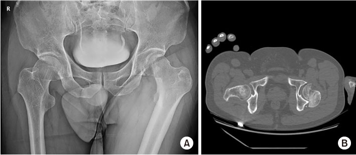
Fig. 1
(A) Anteroposterior Hip radiograph shows left femoral head fracture. (B) Axial computed tomography image shows a left hip posterior dislocation with Pipkin type II femoral head fracture.
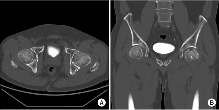
Fig. 2
(A) Postreduction axial computed tomography (CT) image shows a correct position of the femoral head. (B) Postreduction coronal CT scan shows a defect of the anterior-superior weight bearing dome of the femoral head.

Fig. 3
(A) An intraoperative image of the femoral head after a surgical hip dislocation. (B) An intraoperative image showing placement of Acutrak (Acuemd) for fixation of the main fragment after anatomical reduction. A large osteochondral defect in the weight-bearing surface of the femoral head is visible.
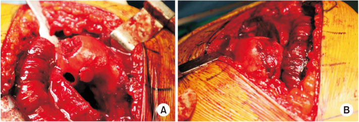
Fig. 4
(A) An intraoperative image showing harvesting 10-mm-diameter two osteochondral plugs from the inferior non-weight-bearing surface. (B) Final image showing a smooth contour after insertion of the donor plugs and greater trochanter autograft.
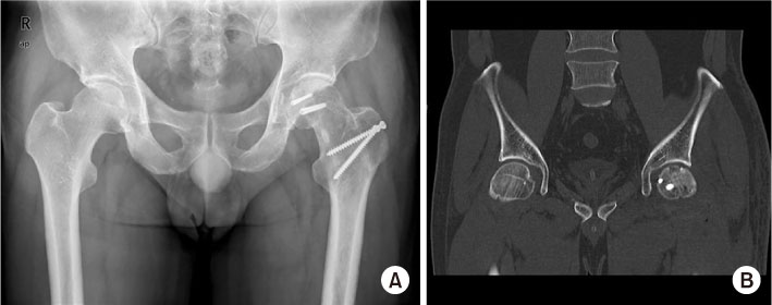
Fig. 5
(A) One year follow-up plain radiograph shows no evidence of the collapse of femoral head. (B) Coronal computed tomography image at 1 year postoperatively, showing no evidence of posttraumatic osteoarthritis or the collapse of femoral head.
Notes
Financial support:None.
Conflict of interests:None.
References
-
Epstein HC, Wiss DA, Cozen L. Posterior fracture dislocation of the hip with fractures of the femoral head. Clin Orthop Relat Res 1985;(201):9–17.
-
-
Kim JW, Kim JH. Updates on treatment of femoral head fractures. J Korean Orthop Assoc 2015;50:171–177.
-
-
Massè A, Aprato A, Alluto C, Favuto M, Ganz R. Surgical hip dislocation is a reliable approach for treatment of femoral head fractures. Clin Orthop Relat Res 2015;473:3744–3751.
-
-
Ganz R, Gill TJ, Gautier E, Ganz K, Krügel N, Berlemann U. Surgical dislocation of the adult hip a technique with full access to the femoral head and acetabulum without the risk of avascular necrosis. J Bone Joint Surg Br 2001;83:1119–1124.
-
-
Hangody L, Füles P. Autologous osteochondral mosaicplasty for the treatment of full-thickness defects of weight-bearing joints: ten years of experimental and clinical experience. J Bone Joint Surg Am 2003;85-A Suppl 2:25–32.
-
-
El Bitar YF, Lindner D, Jackson TJ, Domb BG. Joint-preserving surgical options for management of chondral injuries of the hip. J Am Acad Orthop Surg 2014;22:46–56.
-
-
Hart R, Janecek M, Visna P, Bucek P, Kocis J. Mosaicplasty for the treatment of femoral head defect after incorrect resorbable screw insertion. Arthroscopy 2003;19:E1–E5.
-
-
Nam D, Shindle MK, Buly RL, Kelly BT, Lorich DG. Traumatic osteochondral injury of the femoral head treated by mosaicplasty: a report of two cases. HSS J 2010;6:228–234.
-
-
Girard J, Roumazeille T, Sakr M, Migaud H. Osteochondral mosaicplasty of the femoral head. Hip Int 2011;21:542–548.
-
 Cite
Cite
