 E-submission
 TOTA
TOTA TOTS
TOTS
Articles
- Page Path
- HOME > J Musculoskelet Trauma > Volume 22(4); 2009 > Article
-
Original Article
- Treatment of Shatzker Type VI Tibia Plateau Fracture Using Lateral and Posteromedial Dual Incision Approach and Dual Plating
- In-Jung Chae, M.D., Sang-Won Park, M.D., Soon-Hyuck Lee, M.D., Won Noh, M.D., Ho-Joong Kim, M.D., Seung-Beom Hahn, M.D.
-
Journal of the Korean Fracture Society 2009;22(4):252-258.
DOI: https://doi.org/10.12671/jkfs.2009.22.4.252
Published online: October 30, 2009
Department of Orthopaedic Surgery, Korea University Anam Hospital, Seoul, Korea.
- Address reprint requests to: Seung-Beom Hahn, M.D. Department of Orthopaedic Surgery, Korea University Anam Hospital, 126-1, Anam-dong 5-ga, Seongbuk-gu, Seoul 136-705, Korea. Tel: 82-2-920-5924, Fax: 82-2-924-2471, oshan@korea.ac.kr
• Received: November 30, 2008 • Revised: March 9, 2009 • Accepted: September 21, 2009
Copyright © 2009 The Korean Fracture Society. All rights reserved.
This is an Open Access article distributed under the terms of the Creative Commons Attribution Non-Commercial License (http://creativecommons.org/licenses/by-nc/3.0/) which permits unrestricted non-commercial use, distribution, and reproduction in any medium, provided the original work is properly cited.
- 1,603 Views
- 21 Download
- 3 Crossref
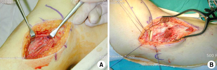
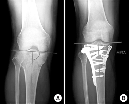
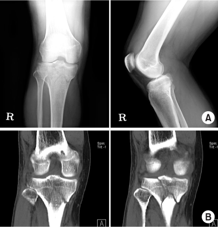
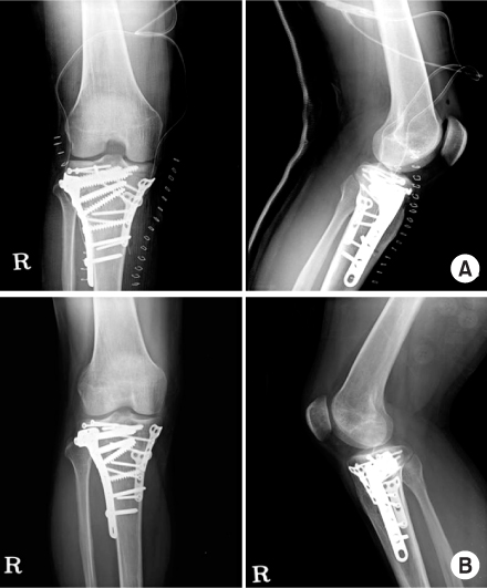
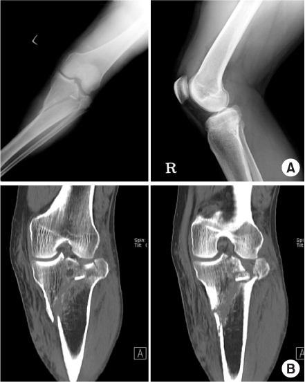
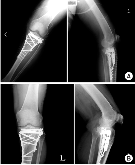
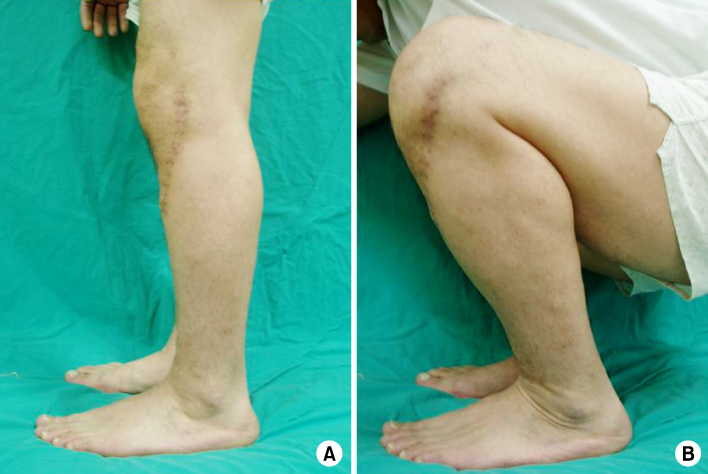
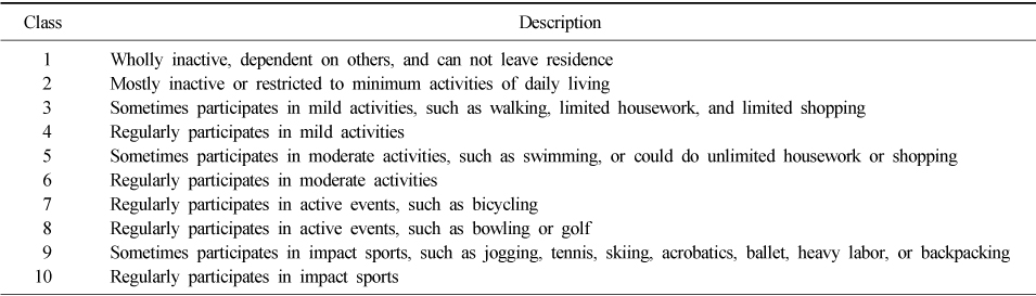
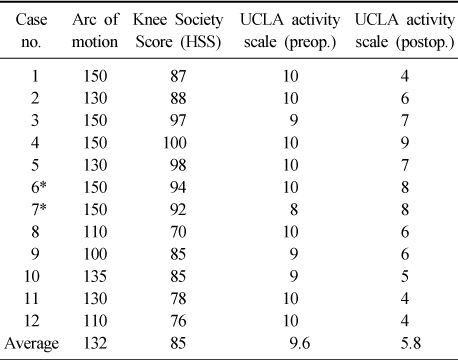
Figure & Data
REFERENCES
Citations
Citations to this article as recorded by 
- Staged Treatment of Bicondylar Tibial Plateau Fracture (Schatzker Type V or VI) Using Temporary External Fixator: Correlation between Clinical and Radiological Outcomes
Seung Min Ryu, Han Seok Yang, Oog Jin Shon
Knee Surgery and Related Research.2018; 30(3): 261. CrossRef - Medial Minimally Invasive Percutaneous Plate Osteosynthesis in Proximal Tibial Comminuted Fractures
Jae-Ang Sim, Kwang-Hui Kim, Yong-Seuk Lee, Sang-Jin Lee, Beom-Koo Lee
Journal of the Korean Orthopaedic Association.2014; 49(4): 278. CrossRef - Current Concepts in Management of Tibia Plateau Fracture
Sang Hak Lee, Kang-Il Kim
Journal of the Korean Fracture Society.2014; 27(3): 245. CrossRef
 Cite
CiteTreatment of Shatzker Type VI Tibia Plateau Fracture Using Lateral and Posteromedial Dual Incision Approach and Dual Plating


Fig. 1
(A) Posteromedial approach.
(B) Lateral submeniscal approach.
Fig. 2
(A) Preoperative MPTA.
(B) Postoperative MPTA.
Fig. 3
37 years old patient who sustained crushing injury.
(A) Plain radiograph showing complex tibial plateau fracture (Shatzker type VI).
(B) 3D-CT scan (coronal view).
Fig. 4
(A) Immediate postoperative plain radiographs showing dual plating.
(B) Postoperative 12 months follow-up showing complete union state and normal alignment of knee joint.
Fig. 5
57 years old male patient who sustained traffic accident.
(A) Plain radiograph showing complex tibial plateau fracture (Shatzker type VI).
(B) 3D-CT scan (coronal view).
Fig. 6
(A) Immediate postoperative plain radiographs showing dual plating.
(B) Postoperative 15 months follow-up showing complete union state and normal alignment of knee joint.
Fig. 7
In postoperative 18 months follow-up photographs, the patient recovered the full range of motion but UCLA activity scale was decreased significantly (A) the patient can extend the knee in full degrees (B) the patient can flex the knee in full degrees.
Fig. 1
Fig. 2
Fig. 3
Fig. 4
Fig. 5
Fig. 6
Fig. 7
Treatment of Shatzker Type VI Tibia Plateau Fracture Using Lateral and Posteromedial Dual Incision Approach and Dual Plating
Demographics
*The same patient who sustained the bilateral tibia plateau fracture, case no. 6 and 7 indicates right and left knee joint respectively.
UCLA scale
Clinical results
*The same patient who sustained the bilateral tibia plateau fracture, case no. 6 and 7 indicates right and left knee joint respectively.
Table 1
Demographics
*The same patient who sustained the bilateral tibia plateau fracture, case no. 6 and 7 indicates right and left knee joint respectively.
Table 2
UCLA scale
Table 3
Clinical results
*The same patient who sustained the bilateral tibia plateau fracture, case no. 6 and 7 indicates right and left knee joint respectively.

