 E-submission
 TOTA
TOTA TOTS
TOTS
Articles
- Page Path
- HOME > J Musculoskelet Trauma > Volume 22(4); 2009 > Article
-
Original Article
- Long Term Follow up Results of the Operative Treatment of the Acromioclavicular Joint Dislocation with a Wolter Plate
- Ki-Ser Kang, M.D., Han-Jun Lee, M.D., Jae-Sung Lee, M.D., Jae-Yoon Kim, M.D., Yong-Beom Park, M.D.
-
Journal of the Korean Fracture Society 2009;22(4):259-263.
DOI: https://doi.org/10.12671/jkfs.2009.22.4.259
Published online: October 30, 2009
Department of Orthopedic Surgery, Yong-San Hospital, Chung-Ang University College of Medicine, Seoul, Korea.
- Address reprint requests to: Jae-Sung Lee, M.D. Department of Orthopaedic Surgery, Yong-San Hospital, Chung-Ang University College of Medicine, 65-207, Hangangro-3ga, Yongsan-gu, Seoul 140-757, Korea. Tel: 82-2-748-9746, Fax: 82-2-793-6634, boneman@cau.ac.kr
• Received: April 11, 2009 • Revised: May 29, 2009 • Accepted: September 11, 2009
Copyright © 2009 The Korean Fracture Society. All rights reserved.
This is an Open Access article distributed under the terms of the Creative Commons Attribution Non-Commercial License (http://creativecommons.org/licenses/by-nc/3.0/) which permits unrestricted non-commercial use, distribution, and reproduction in any medium, provided the original work is properly cited.
- 1,039 Views
- 2 Download
- 2 Crossref
Abstract
-
Purpose
- To evaluate the long-term clinical and radiological results of the operative treatment of the acromioclavicular dislocation with a Wolter plate.
-
Materials and Methods
- We reviewed clinical and radiological data of twenty patients (mean age: 37 years) who underwent the operative treatment of acromioclavicular joint dislocation using a Wolter plate from September, 1999 to December, 2002 with minimum of five years follow-up (average 6 years 7 months). The clinical outcomes of twenty patients were evaluated by UCLA scoring and radiological results of fifteen patients with available radiograph were evaluated by Zanca view and stress view.
-
Results
- The mean UCLA score was mean 33 points (range, 27~35) at final follow up. By clinical evaluation, twelve cases (60%) were excellent, six cases (30%) were good and two cases were poor (10%). By radiological evaluation, eight cases (54%) were excellent (without displacement), five cases (33%) were good (displacement<5 mm) and two cases (13%) were poor (displacement>5 mm). Erosive change in acromioclavicular joint was seen in poor case.
-
Conclusion
- Wolter plate fixation may be a useful modality for treating acromioclavicular joint dislocation. Great care should be taken to make the hook hole at the appropriate position during operation for long-term prognosis.
- 1. Bannister GC, Wallace WA, Stableforth PG, Huston MA. The management of acute acromioclavicular dislocation. A randomised prospective controlled trial. J Bone Joint Surg Br, 1989;71:848-850.ArticlePDF
- 2. Bosworth BM. Acromioclavicular dislocation; end-results of screw suspension treatment. Ann Surg, 1948;127:98-111.
- 3. Calve E, Lópe-Franco M, Arribas IM. Clinical and radiologic outcomes of surgical and conservative treatment of type III acromioclavicular joint injury. J Shoulder Elbow Surg, 2006;15:300-305.Article
- 4. Chun JM, Kim SY, Choi JH, Kim TS, Kim KY. Surgical treatment of the acute acromioclavicular joint dislocation using a wolter plate. J Korean Orthop Assoc, 2002;37:185-190.ArticlePDF
- 5. Dewar FP, Barrington TW. The treatment of chronic acromioclavicular dislocation. J Bone Joint Surg Br, 1965;47:32-35.ArticlePDF
- 6. Ellman H, Hanker G, Bayer M. Repair of the rotator cuff. End-result study of factors influencing reconstruction. J Bone Joint Surg Am, 1986;68:1136-1144.Article
- 7. Gstettner C, Tauber M, Hitzl W, Resch H. Rockwood type III acromioclavicular dislocation: Surgical versus conservative treatment. J Shoulder Elbow Surg, 2008;17:220-225.Article
- 8. Guy DK, Wirth MA, Griffin JL, Rockwood CA Jr. Reconstruction of chronic and complete dislocation of the acromioclavicular joint. Clin Orthop Relat Res, 1998;347:138-149.
- 9. Habernek H, Weinstabl R, Schmid L, Fialka C. A crook plate for treatment of acromioclavicular joint separation: indication, technique, and results after one year. J Trauma, 1993;35:893-901.
- 10. Hessmann M, Gotzen L, Gehling H. Acromioclavicular reconstruction augmented with polydioxanonsulphate bands. Surgical technique and results. Am J Sports Med, 1995;23:552-556.ArticlePDF
- 11. Inman VT, Saunders JB, Abott LC. Observation on the function of the shoulder joint 1944. Clin Orthop Relat Res, 1996;330:3-12.
- 12. Kennedy JC, Cameron H. Complete dislocation of the acromioclaviclar joint. J Bone Joint Surg Br, 1954;36:202-208.
- 13. Larsen E, Hede A. Treatment of acute acromioclavicular dislocation. Three different methods of treatment prospectively studied. Acta Orthop Belg, 1987;53:480-484.
- 14. Lee HJ, Lee EW, Kang KS, et al. Operative treatment of complete dislocation of the acromioclavicular joint with a wolter plate. J Korean Orthop Assoc, 2002;37:459-463.ArticlePDF
- 15. Lin B, Lian KJ, Guo LX, et al. Comparative study on treating complete dislocation of acromioclavicular joint with three different methods. Chin J Traumatol, 2004;7:101-107.
- 16. MacDonald PB, Alexander MJ, Frejuk J, Johnson GE. Comprehensive functionctional analysis of shoulders following complete acromioclavicular separation. Am J Sports Med, 1988;16:475-480.ArticlePDF
- 17. Morrison DS, Lemos MJ. Acromioclavicular separation. Reconstruction using synthetic loop augmentation. Am J Sports Med, 1995;23:105-110.ArticlePDF
- 18. Phemister DB. The treatment of dislocation of the acromioclavicular joint by open reduction and threaded-wire fixation. J Bone Joint Surg, 1942;24:166-169.
- 19. Rockwood CA, Matsen FA. Disorders of the acromioclavicular joint. The shoulder. 2nd ed. Philadelphia: WE Saunders Co; 1998. p. 483-543.
- 20. Sim E, Schwarz N, Höcker K, Berzlanovich A. Repair of complete acromioclavicular separations using the acromioclavicular-hook plate. Clin Orthop Relat Res, 1995;314:134-142.Article
- 21. Taft TN, Wilson FC, Oglesby JW. Dislocation of the acromioclavicular joint. An end-result study. J Bone Joint Surg Am, 1987;69:1045-1051.Article
- 22. Weaver JK, Dunn HK. Treatment of acromioclavicular injuries, especially complete acromioclavicular separation. J Bone Joint Surg Am, 1972;54:1187-1194.Article
REFERENCES

Fig. 2
(A) Preoperative stress radiograph shows type V acromioclavicular dislocation.
(B) Postoperative 8 years stress radiograph of same patient shows no differences of coracoclavicular interval and degenerative changes of acromioclavicular joint.


Figure & Data
REFERENCES
Citations
Citations to this article as recorded by 
- Clinical outcomes of bending versus non-bending of the plate hook in acromioclavicular joint dislocation
Min Su Joo, Hoi Young Kwon, Jeong Woo Kim
Clinics in Shoulder and Elbow.2021; 24(4): 202. CrossRef - Clinical Comparison of Two Types of Hook Plate in Surgical Treatment of Acromioclavicular Dislocation - AO Hook Plate and Wolter Plate -
Jea-Yeol Choi, Eugene Kim, Haw-Jae Jeong, Jin Whan Ahn, Hun-Kyu Shin, Se-Jin Park, Seung-Hee Lee, Jae-Wook Lee, Kyu-Bo Choi
The Journal of the Korean Shoulder and Elbow Society.2012; 15(2): 123. CrossRef
 Cite
CiteLong Term Follow up Results of the Operative Treatment of the Acromioclavicular Joint Dislocation with a Wolter Plate
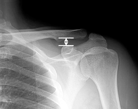
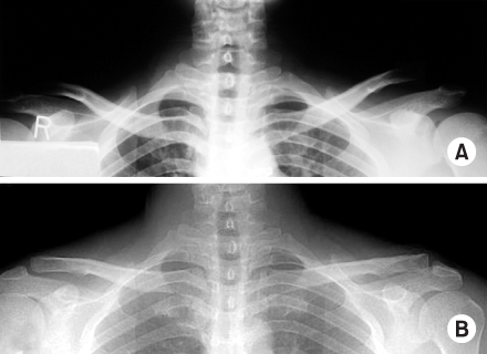
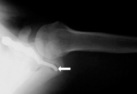
Fig. 1
X-ray shows the coracoclavicular distance (white arrow).
Fig. 2
(A) Preoperative stress radiograph shows type V acromioclavicular dislocation.
(B) Postoperative 8 years stress radiograph of same patient shows no differences of coracoclavicular interval and degenerative changes of acromioclavicular joint.
Fig. 3
X-ray of 45 year old male at postoperative 3months shows widening of the hook hole (white arrow).
Fig. 1
Fig. 2
Fig. 3
Long Term Follow up Results of the Operative Treatment of the Acromioclavicular Joint Dislocation with a Wolter Plate
Average scores of university of california at los angeles end-results
FF: Forward flexion.
Radiologic results of coracoclavicular distance (paired t-test)
Table 1
Average scores of university of california at los angeles end-results
FF: Forward flexion.
Table 2
Radiologic results of coracoclavicular distance (paired t-test)

