 E-submission
 TOTA
TOTA TOTS
TOTS
Articles
- Page Path
- HOME > J Musculoskelet Trauma > Volume 25(1); 2012 > Article
-
Case Report
- Deep Femoral Vessel Injury Following Subtrochanteric Hip Fracture: A Case Report
- Jae-Hyuk Yang, M.D., Jung-Ro Yoon, M.D., Kyu-Bok Kang, M.D., Ho-Hyun Yun, M.D., Young-Soo Shin, M.D., Yun-Ku Cho, M.D.
-
Journal of the Korean Fracture Society 2012;25(1):64-68.
DOI: https://doi.org/10.12671/jkfs.2012.25.1.64
Published online: January 31, 2012
Department of Orthopedic Surgery, Seoul Veterans Hospital, Seoul, Korea.
*Department of Radiology, Seoul Veterans Hospital, Seoul, Korea.
- Address reprint requests to: Jae-Hyuk Yang, M.D. Department of Orthopedic Surgery, Seoul Veterans Hospital, 6-2, Dunchon-dong, Kangdong-gu, Seoul 134-791, Korea. Tel: 82-2-2225-1351, Fax: 82-2-2225-2190, jaekorea@gmail.com
• Received: January 25, 2011 • Revised: July 3, 2011 • Accepted: October 31, 2011
Copyright © 2012 The Korean Fracture Society
- 1,919 Views
- 5 Download
- 2 Crossref
Abstract
- Arterial trauma associated with hip fracture treatment is still a rare complication. We present a case in which an arterial injury was discovered during closed reduction and intramedullary nail fixation of a subtrochanteric hip fracture. The preoperative thigh circumference was increased due to severe swelling, and the vascular injury was located substantially proximal to the fracture and the instrumentation area. An interventional angiogram revealed a damaged vessel originating from one of the minor proximal branches of the right deep femoral artery while filling a 2 cm-sized pseudoaneurysm. Embolization was performed without further complications.
CASE REPORT
DISCUSSION
- 1. Bartonícek J. Injuries to femoral vessels in fractures of the hip. Rozhl Chir, 2009;88:203-205.
- 2. Grimaldi M, Courvoisier A, Tonetti J, Vouaillat H, Merloz P. Superficial femoral artery injury resulting from intertrochanteric hip fracture fixation by a locked intramedullary nail. Orthop Traumatol Surg Res, 2009;95:380-382.Article
- 3. Keel JD, Eyres KS. Vascular injury by an intertrochanteric fracture fragment. Injury, 1993;24:350-352.Article
- 4. Kuzniec S, Kauffman P, Molnár LJ, Aun R, Puech-Leão P. Diagnosis of limbs and neck arterial trauma using duplex ultrasonography. Cardiovasc Surg, 1998;6:358-366.Article
- 5. Lazarides MK, Arvanitis DP, Dayantas JN. Iatrogenic arterial trauma associated with hip joint surgery: an overview. Eur J Vasc Surg, 1991;5:549-556.
- 6. Lazarides MK, Arvanitis DP, Liatas AC, Dayantas JN. Iatrogenic and noniatrogenic arterial trauma: a comparative study. Eur J Surg, 1991;157:17-20.
- 7. Miller-Thomas MM, West OC, Cohen AM. Diagnosing traumatic arterial injury in the extremities with CT angiography: pearls and pitfalls. Radiographics, 2005;25:Suppl 1. S133-S142.Article
- 8. Moreyra CE, Lavernia CJ, Cooke CC. Late vascular injury following intertrochanteric fracture reduction with sliding hip screw. J Surg Orthop Adv, 2004;13:170-173.
- 9. Ohki T, Veith FJ, Marin ML, Cynamon J, Sanchez LA. Endovascular approaches for traumatic arterial lesions. Semin Vasc Surg, 1997;10:272-285.
- 10. Panetta T, Sclafani SJ, Goldstein AS, Phillips TF, Shaftan GW. Percutaneous transcatheter embolization for massive bleeding from pelvic fractures. J Trauma, 1985;25:1021-1029.
- 11. Rajaesparan K, Amin A, Arora S, Walton NP. Pseudoaneurysm of a branch of the profunda femoris artery following distal locking of an intramedullary hip nail: an unusual anatomical location. Hip Int, 2008;18:231-235.Article
- 12. Ryzewicz M, Robinson M, McConnell J, Lindeque B. Vascular injury during fixation of an intertrochanteric hip fracture in a patient with severe atherosclerosis. A case report. J Bone Joint Surg Am, 2006;88:2483-2486.
- 13. Søballe K, Christensen F. Laceration of the superficial femoral artery by an intertrochanteric fracture fragment. A case report. J Bone Joint Surg Am, 1987;69:781-783.Article
- 14. Storm RK, Sing AK, de Graaf EJ, Tetteroo GW. Latrogenic arterial trauma associated with hip fracture treatment. J Trauma, 2000;48:957-959.
- 15. Whitehill R, Wang GJ, Edwards JR, Stamp WG. Late injuries to femoral vessels after fracture of the hip. Case report. J Bone Joint Surg Am, 1978;60:541-542.Article
REFERENCES
Fig. 1
(A, B) Preoperative radiographs; anteroposterior and translateral view showing subtrochanteric fracture with proximal migration of the femur shaft and deformed proximal fragment. Notice the anterior beak of the proximal fragment with severe angulation (arrow). Extensive calcifications are seen in the arteries.
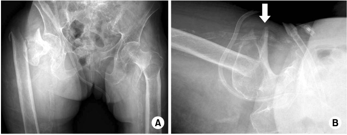
Fig. 2Clinical photograph of thigh swelling with knee flexion contracture. Notice the swelling of injured limb is more than twice the circumference of the contralateral limb.

Fig. 3Computed tomographic angiogram showing the extravasation of contrast material from branch of deep femoral artery.

Fig. 4
(A) Digital subtraction angiography showing contrast extravasations originating from one of the minor proximal branch of right deep femoral artery, while filling a 2 cm sized pseudoaneurysm (arrow).
(B) After embolization. Postembolization angiography demonstrated no additional extravasation of contrast medium.
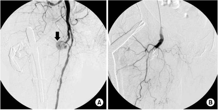
Figure & Data
REFERENCES
Citations
Citations to this article as recorded by 
- Proximal femoral fractures and vascular injuries in adults: Incidence, aetiology and outcomes
Antonio Barquet, Andrés Gelink, Peter V. Giannoudis
Injury.2015; 46(12): 2297. CrossRef - Pertrochanteric Hip Fracture: A “Routine” Fracture With a Potentially Devastating Vascular Complication
Matthew Patrick Sullivan, Mara Lynne Schenker, Samir Mehta
Orthopedics.2015;[Epub] CrossRef
 Cite
CiteDeep Femoral Vessel Injury Following Subtrochanteric Hip Fracture: A Case Report

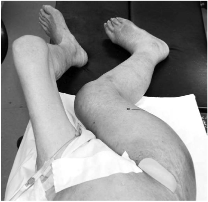
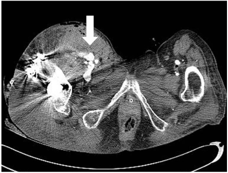

Fig. 1
(A, B) Preoperative radiographs; anteroposterior and translateral view showing subtrochanteric fracture with proximal migration of the femur shaft and deformed proximal fragment. Notice the anterior beak of the proximal fragment with severe angulation (arrow). Extensive calcifications are seen in the arteries.
Fig. 2
Clinical photograph of thigh swelling with knee flexion contracture. Notice the swelling of injured limb is more than twice the circumference of the contralateral limb.
Fig. 3
Computed tomographic angiogram showing the extravasation of contrast material from branch of deep femoral artery.
Fig. 4
(A) Digital subtraction angiography showing contrast extravasations originating from one of the minor proximal branch of right deep femoral artery, while filling a 2 cm sized pseudoaneurysm (arrow).
(B) After embolization. Postembolization angiography demonstrated no additional extravasation of contrast medium.
Fig. 1
Fig. 2
Fig. 3
Fig. 4
Deep Femoral Vessel Injury Following Subtrochanteric Hip Fracture: A Case Report

