 E-submission
 TOTA
TOTA TOTS
TOTS
Articles
- Page Path
- HOME > J Musculoskelet Trauma > Volume 25(1); 2012 > Article
-
Case Report
- Simultaneous Bilateral Proximal Femoral Fracture associated with Generalized Tonic-Clonic Seizure: A Case Report
- Sang-Hoo Lee, M.D., Kyeong-Seop Song, M.D., Seung-Joo Jeon, M.D., Seong-Hwan Hong, M.D.
-
Journal of the Korean Fracture Society 2012;25(1):69-72.
DOI: https://doi.org/10.12671/jkfs.2012.25.1.69
Published online: January 31, 2012
Department of Orthopedic Surgery, Kwangmyung Sung-Ae Hospital, Gwangmyeong, Korea.
*Department of Orthopedic Surgery, Sung-Ae General Hospital, Seoul, Korea.
- Address reprint requests to: Kyeong-Seop Song, M.D. Department of Orthopedic Surgery, Kwangmyung Sung-Ae Hospital, 389, Cheolsan 3-dong, Gwangmyeong 423-711, Korea. Tel: 82-2-2680-7236, Fax: 82-2-2617-8938, sksub@paran.com
• Received: September 24, 2011 • Revised: October 21, 2011 • Accepted: December 8, 2011
Copyright © 2012 The Korean Fracture Society
- 835 Views
- 0 Download
Abstract
- Simultaneous bilateral proximal femoral fractures are extremely rare, and a few have been reported in and outside the country. It may have various causes, and most cases were associated with major trauma, repetitive minor trauma, seizure, parathyroid or renal dysfunction, and anti-epileptic medications. We experienced a case of simultaneous bilateral proximal femoral fractures after generalized tonic-clonic seizure in a 70-year-old female. Herein, we report it with a review of the literature.
- 1. Chen CE, Kao CL, Wang CJ. Bilateral pathological femoral neck fractures secondary to ectopic parathyroid adenoma. Arch Orthop Trauma Surg, 1998;118:164-166.ArticlePDF
- 2. Finelli PF, Cardi JK. Seizure as a cause of fracture. Neurology, 1989;39:858-860.Article
- 3. Haronian E, Silver JW, Mesa J. Simultaneous bilateral femoral neck fracture and greater tuberosity shoulder fracture resulting from seizure. Orthopedics, 2002;25:757-758.Article
- 4. Madhok R, Rand JA. Ten-year follow-up study of missed, simultaneous, bilateral femoral-neck fractures treated by bipolar arthroplasties in a patient with chronic renal failure. Clin Orthop Relat Res, 1993;291:185-187.Article
- 5. Pearson JR, Hargadon EJ. Fractures of the pelvis involving the floor of the acetabulum. J Bone Joint Surg Br, 1962;44:550-561.ArticlePDF
- 6. Rahman MM, Awada A. Bilateral simultaneous hip fractures secondary to an epileptic seizure. Saudi Med J, 2003;24:1261-1263.Article
- 7. Remec PT, Evarts CM. Bilateral central dislocation of the hip. A case report. Clin Orthop Relat Res, 1983;181:118-120.
- 8. Shaheen MA, Sabet NA. Bilateral simultaneous fracture of the femoral neck following electrical shock. Injury, 1984;16:13-14.Article
- 9. Souverein PC, Webb DJ, Petri H, Weil J, Van Staa TP, Egberts T. Incidence of fractures among epilepsy patients: a population-based retrospective cohort study in the General Practice Research Database. Epilepsia, 2005;46:304-310.Article
- 10. Vasconcelos D. Compression fractures of the vertebrae during major epileptic seizures. Epilepsia, 1973;14:323-328.Article
REFERENCES
Fig. 1Preoperative radiograph of both hip shows neck fracture of the left femur and intertrochanteric fracture of the right femur.
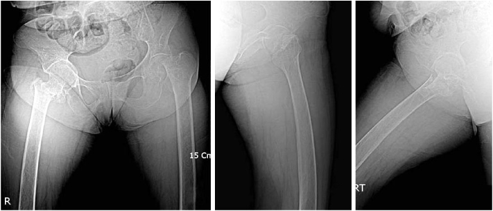


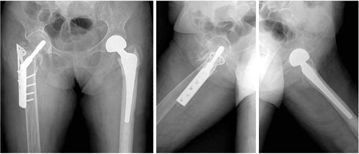
Figure & Data
REFERENCES
Citations
Citations to this article as recorded by 
 Cite
CiteSimultaneous Bilateral Proximal Femoral Fracture associated with Generalized Tonic-Clonic Seizure: A Case Report

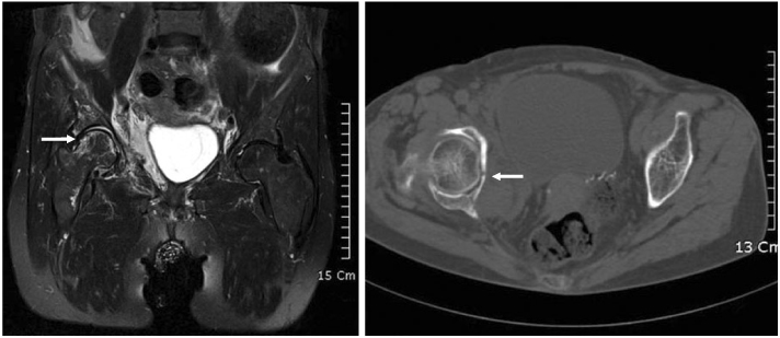
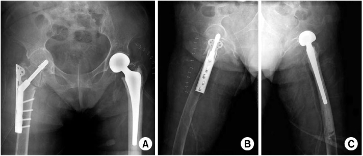

Fig. 1
Preoperative radiograph of both hip shows neck fracture of the left femur and intertrochanteric fracture of the right femur.
Fig. 2
MRI and CT shows fracture of the acetabulum and femoral head.
Fig. 3
(A~C) Immidiatly postoperative radiograph shows: The left femur neck fracture are replaced non-cemented bipolar hemiarthroplasty. The right femur intertrochanteric fracture are reduced and stabilized with compressive hip screw.
Fig. 4
Postoperative radiograph of the both hip 3 months after operation.
Fig. 1
Fig. 2
Fig. 3
Fig. 4
Simultaneous Bilateral Proximal Femoral Fracture associated with Generalized Tonic-Clonic Seizure: A Case Report

