 E-submission
 TOTA
TOTA TOTS
TOTS
Articles
- Page Path
- HOME > J Musculoskelet Trauma > Volume 29(2); 2016 > Article
-
Original Article
- Usefulness of Computed Tomography on Distal Tibia Intra-Articular Fracture Associated with Spiral Tibia Shaft Fracture
- Seong-Eun Byun, M.D., Sang-June Lee, M.D., Uk Kim, M.D., Young Rak Choi, M.D., Soo-Hong Han, M.D., Ph.D., Byong-Guk Kim, M.D., Ph.D.
-
Journal of the Korean Fracture Society 2016;29(2):114-120.
DOI: https://doi.org/10.12671/jkfs.2016.29.2.114
Published online: April 19, 2016
Department of Orthopaedic Surgery, CHA Bundang Medical Center, CHA University, Seongnam, Korea.
*Department of Orthopaedic Surgery, CHA Gumi Medical Center, CHA University, Gumi, Korea.
- Address reprint requests to: Byong-Guk Kim, M.D., Ph.D. Department of Orthopaedic Surgery, CHA Gumi Medical Center, 12 Sinsi-ro 10-gil, Gumi 39295, Korea. Tel: 82-31-780-5289, Fax: 82-31-708-3578, bgkimmd@gmail.com
• Received: July 6, 2015 • Revised: December 11, 2015 • Accepted: January 10, 2016
Copyright © 2016 The Korean Fracture Society. All rights reserved.
This is an Open Access article distributed under the terms of the Creative Commons Attribution Non-Commercial License (http://creativecommons.org/licenses/by-nc/4.0) which permits unrestricted non-commercial use, distribution, and reproduction in any medium, provided the original work is properly cited.
- 1,171 Views
- 8 Download
- 1 Crossref


Figure & Data
REFERENCES
Citations
Citations to this article as recorded by 
- Treatment of Distal Tibial Spiral Fractures Combined with Posterior Malleolar Fractures
Young Sung Kim, Ho Min Lee, Jong Pil Kim, Phil Hyun Chung, Soon Young Park
Journal of the Korean Orthopaedic Association.2021; 56(4): 317. CrossRef
 Cite
CiteUsefulness of Computed Tomography on Distal Tibia Intra-Articular Fracture Associated with Spiral Tibia Shaft Fracture
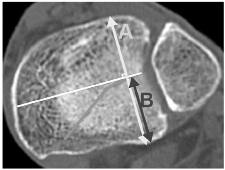
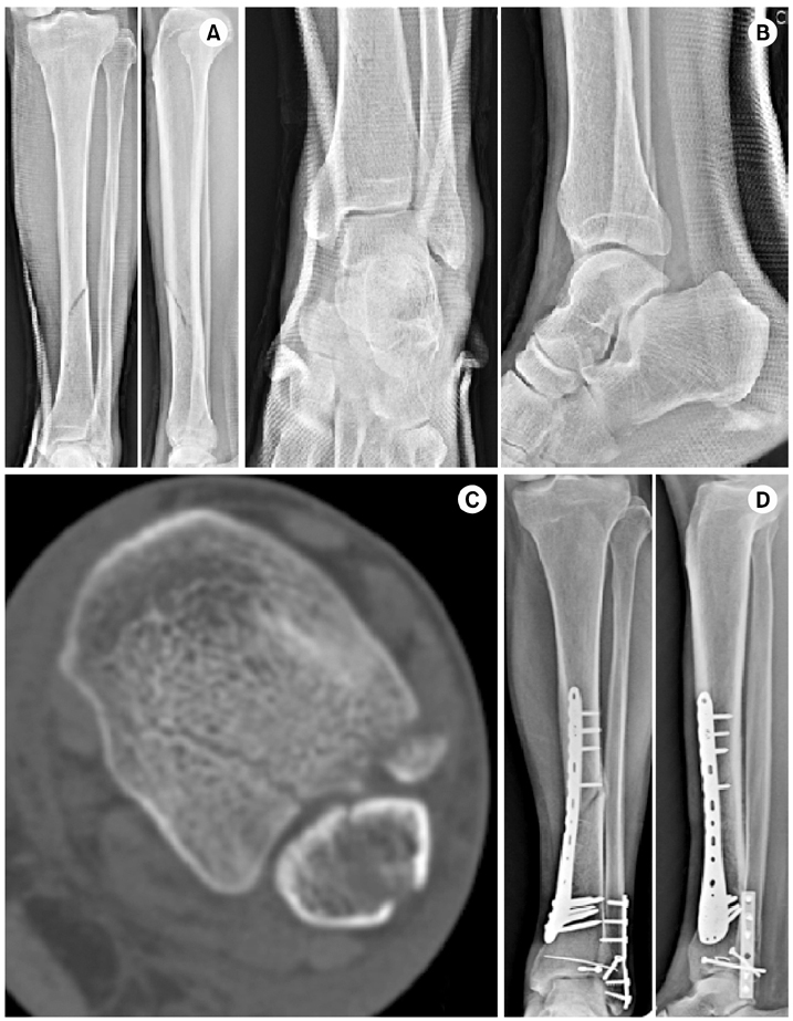
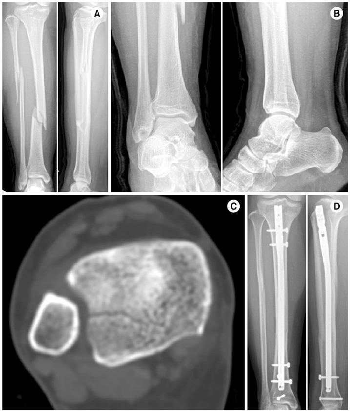
Fig. 1
Measurement of relative length of posterior malleolar fragment. A: anteroposterior length of articular surface, B: anteroposterior length of posterior malleolar fragment.
Fig. 2
(A) Preoperative tibia anteroposterior (AP) and lateral image of a 61-year-old male with an AO/OTA 42-A1 type fracture. (B) Preoperative ankle AP and lateral image showing a lateral malleolar fracture and no associated distal tibia intra-articular fracture. (C) Axial image of computed tomography showing a minimally displaced posterior malleolar fracture exceeding 25% of the articular surface and an avulsion fracture of the anterior tibiofibular ligament. (D) AP and lateral image after plating with screw fixation of the posterior malleolar fragment and Kirschner wire fixation of the avulsion fracture of the anterior tibiofibular ligament.
Fig. 3
(A) Preoperative tibia anteroposterior (AP) and lateral image of a 51-year-old female with an AO/OTA 42-A1 type fracture. (B) Preoperative ankle AP and lateral image showing no associated distal tibia intra-articular fracture. (C) Axial image of computed tomography showing a minimally displaced posterior malleolar fracture exceeding 25% of the articular surface. (D) AP and lateral image after intramedullary nailing with fixation of the posterior malleolar fragment.
Fig. 1
Fig. 2
Fig. 3
Usefulness of Computed Tomography on Distal Tibia Intra-Articular Fracture Associated with Spiral Tibia Shaft Fracture
Types of Associated Distal Tibia Intra-Articular Fracture in Tibia Shaft Fracture
| Variable | PM fracture | ATFL avulsion fracture | MM fracture | Combined fracture | Total |
|---|---|---|---|---|---|
| Plain radiograph | 15* | 5* | 5 | 5 | 30 |
| CT | 23 | 5 | 0 | 4 | 32 |
| Total | 37* | 5* | 5 | 15* | 62 |
Values are presented as number only. *One posterior malleolar fracture and 5 anterior tibio-fibular ligament avulsion fractures diagnosed by plain radiograph were revealed as combined injuries after reviewing computed tomography and classified as combined fracture in 'total' line. PM: Posterior malleolus, ATFL: Anterior tibio-fibular ligament, MM: Medial malleolus, CT: Computed tomography.
Component of Associated Combined Fractures
| PM & ATFL avulsion fracture | PM & MM fracture | MM & ATFL avulsion fracture | |
|---|---|---|---|
| No. of combined fracture | 12 | 3 | 0 |
PM: Posterior malleolus, ATFL: Anterior tibio-fibular ligament, MM: Medial malleolus.
Comparison between PM Diagnosed by Plain Radiograph and CT
| Variable | Diagnosed by radiograph (n=20) | Diagnosed by CT (n=32) |
|---|---|---|
| PM mets indication of surgical fixation | 16 | 26 |
| PM fragment size >25% | 16 | 26 |
| PM >2 mm of gap | 2 | 0 |
| PM fixed | 14 | 18 |
Values are presented in number only. PM: Posterior malleolar fracture, CT: Computed tomography.
Table 1
Types of Associated Distal Tibia Intra-Articular Fracture in Tibia Shaft Fracture
Values are presented as number only. *One posterior malleolar fracture and 5 anterior tibio-fibular ligament avulsion fractures diagnosed by plain radiograph were revealed as combined injuries after reviewing computed tomography and classified as combined fracture in 'total' line. PM: Posterior malleolus, ATFL: Anterior tibio-fibular ligament, MM: Medial malleolus, CT: Computed tomography.
Table 2
Component of Associated Combined Fractures
PM: Posterior malleolus, ATFL: Anterior tibio-fibular ligament, MM: Medial malleolus.
Table 3
Comparison between PM Diagnosed by Plain Radiograph and CT
Values are presented in number only. PM: Posterior malleolar fracture, CT: Computed tomography.

