 E-submission
 TOTA
TOTA TOTS
TOTS
Articles
- Page Path
- HOME > J Musculoskelet Trauma > Volume 20(3); 2007 > Article
-
Case Report
- Posterior Thigh Compartment Syndrome as a Result of Pseudoaneurysm of the Popliteal Artery in the Distal Femoral Fracture: A Case Report
- Seoung Jun Lee, M.D.
-
Journal of the Korean Fracture Society 2007;20(3):277-281.
DOI: https://doi.org/10.12671/jkfs.2007.20.3.277
Published online: June 14, 2016
Department of Orthopedic Surgery, Konkuk University College of Medicine, Seoul, Korea.
- Address reprint requests to: Seuong Jun Lee, M.D. Department of Orthopedic Surgery, Konkuk University Hospital, 4-12, Hwayang-dong, Gwangjin-gu, Seoul 143-701, Korea. Tel: 82-2-2030-7360, Fax: 82-2-2030-7369, lsjmd@kku.ac.kr
Copyright © The Korean Fracture Society. All rights reserved
- 1,038 Views
- 0 Download
- 2 Crossref
Abstract
- Compartment syndrome of the thigh is a rare condition and usually occurs in the anterior compartment. It is frequently caused by muscle injury, femur fracture, muscle overuse and vessel injury, but there have been few reports about posterior thigh compartment syndrome caused by pseudoaneurysm of the popliteal artery after fixation of distal femoral fracture with the retrograde intramedullary nail. We report a case of posterior thigh compartment syndrome caused by pseudoaneurysm of the popliteal artery, and report the clinical progression and result of our case.
- 1. Allen MJ, Stirling AJ, Crawshaw CV, Barnes MR. Intracompartmental pressure monitoring of leg injuries. An aid to management. J Bone Joint Surg Br, 1985;67:53-57.ArticlePDF
- 2. An HS, Simpson JM, Gale S, Jackson WT. Acute anterior compartment syndrome in the thigh: a case report and review of the literature. J Orthop Trauma, 1987;1:180-182.
- 3. Ebraheim NA, Hoeflinger MJ, Savolaine ER, Jackson WT. Anterior compartment syndrome of the thigh as a complication of blunt trauma in a patient on prolonged anticoagulation therapy. Clin Orthop Relat Res, 1991;263:180-184.Article
- 4. Gorman PW, McAndrew MP. Acute anterior compartmental syndrome of the thigh following contusion. A case report and review of the literature. J Orthop Trauma, 1987;1:68-70.Article
- 5. Karkos CD, Hughes R, Prasad V, D'Souza SP. Thigh compartment syndrome as a result of a false aneurysm of the profunda femoris artery complicating fixation of an intertrochanteric fracture. J Trauma, 1999;47:393-395.Article
- 6. Lindsay MB. Quadriceps compartment syndrome from minor trauma. Acad Emerg Med, 1999;6:860-861.Article
- 7. Mubarak SJ, Owen CA, Hargens AR, Garetto LP, Akeson WH. Acute compartment syndromes: diagnosis and treatment with the aid of the wick catheter. J Bone Joint Surg Am, 1978;60:1091-1095.
- 8. Rooser B, Bengtson S, Hagglund G. Acute compartment syndrome from anterior thigh muscle contusion: a report of eight cases. J Orthop Trauma, 1991;5:57-59.
- 9. Schwartz JT, Brumback RJ, Lakatos R, Poka A, Bathon GH, Burgess AR. Acute compartment syndrome of the thigh. A spectrum of injury. J Bone Joint Surg Am, 1989;71:392-400.Article
- 10. Suzuki T, Moirmura N, Kawai K, Sugiyama M. Arterial injury associated with acute compartment syndrome of the thigh following blunt trauma. Injury, 2005;36:151-159.Article
REFERENCES


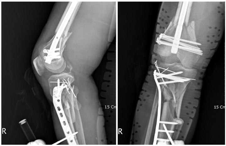

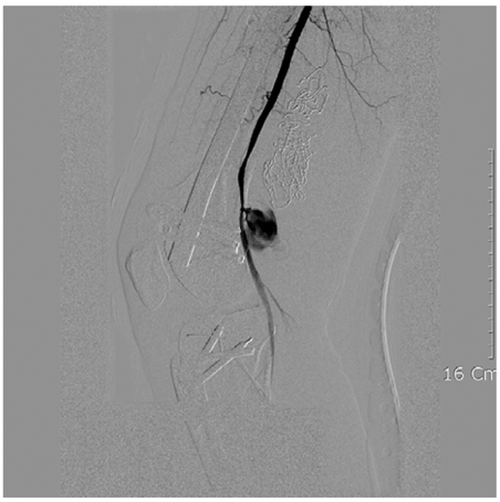
Figure & Data
REFERENCES
Citations
Citations to this article as recorded by 
- Huge Pseudoaneurysm of Popliteal Artery Following Conservative Treatment of a Distal Femur Fracture: A Case Report
Won-Chul Cho, Chong Bin Park, Young-Jun Choi, Hyun-Il Lee, Hee-Jae Won, Jae-Kwang Hwang
Journal of the Korean Fracture Society.2016; 29(2): 137. CrossRef - Is CT Angiography a Reliable Tool for Diagnosis of Traumatic Vessel Injury in the Lower Extremities?
Jong-Hyuk Park, Kwang-Bok Lee, Hyuk Park, Jun-Mo Lee
Journal of the Korean Fracture Society.2012; 25(1): 26. CrossRef
 Cite
CitePosterior Thigh Compartment Syndrome as a Result of Pseudoaneurysm of the Popliteal Artery in the Distal Femoral Fracture: A Case Report
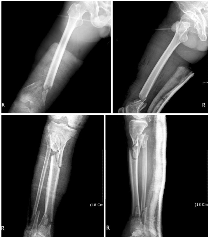
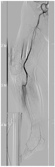


Fig. 1
Radiographies made on admission, showing comminuted fracture of the distal femur and proximal tibia.
Fig. 2
On the femoral angiogram, posterior tibial and peroneal arteries are not well visualized, but posterior tibial artery is faintly visualized through weak collateral flow.
Fig. 3
Postoperative radiographs of the knee joint.
Fig. 4
Sonogram shows 10×6 cm mass in the medial aspect of the thigh as like hematoma or abcess.
Fig. 5
Angiogram shows snowman appeared pseudoaneurysm in popliteal artery.
Fig. 1
Fig. 2
Fig. 3
Fig. 4
Fig. 5
Posterior Thigh Compartment Syndrome as a Result of Pseudoaneurysm of the Popliteal Artery in the Distal Femoral Fracture: A Case Report

