 E-submission
 TOTA
TOTA TOTS
TOTS
Articles
- Page Path
- HOME > J Musculoskelet Trauma > Volume 24(4); 2011 > Article
-
Original Article
- Analysis of Risk Factors for Nonunion after Intramedullary Nailing of Femoral Shaft Fracture in Adult
- Yong-Woon Shin, M.D., Yerl-Bo Sung, M.D., Jeong Yoon Choi, M.D., Minkyu Kim, M.D.
-
Journal of the Korean Fracture Society 2011;24(4):313-320.
DOI: https://doi.org/10.12671/jkfs.2011.24.4.313
Published online: October 30, 2011
Department of Orthopedic Surgery, Sanggye Paik Hospital, College of Medicine, Inje University, Seoul, Korea.
- Address reprint requests to: Yerl-Bo Sung, M.D. Department of Orthopedic Surgery, Sanggye Paik Hospital, College of Medicine, Inje University, 761-1, Sanggye 7-dong, Nowon-gu, Seoul 139-707, Korea. Tel: 82-2-950-1032, Fax: 82-2-934-6342, ybs58@paik.ac.kr
• Received: May 4, 2011 • Revised: May 24, 2011 • Accepted: September 5, 2011
Copyright © 2011 The Korean Fracture Society
- 1,450 Views
- 7 Download
- 3 Crossref
Abstract
-
Purpose
- To evaluate the union time and nonunion rate after intramedullary nailing of femoral shaft fracture in adult, we would like to analysis the operation techniques, comminution, contact surface and displacement.
-
Materials and Methods
- We reviewed retrospectively 53 patients undergoing femoral intramedullary nailing at least 2 years postoperatively and analysised the union time and nonunion rate by operation techniques, comminution, contact surface and displacement. Patients were operated by either antegrade or retrograde intramedullary nailing.
-
Results
- There were no differences in nonunion rate, the duration of bony union between antegrade and retrograde intramedullary nail groups. Significant differences were found in the duration of bony union between the Winquist and Hansen type I, II and the type III, IV (p<0.05). There were significant differences in the duration of bony union among simple, comminuted, and segmental fracture groups (p<0.05).
-
Conclusion
- The union time is affected by not operation techniques and fracture displacement, but Winquist-Hansen classification and number of fracture fragments in intramedullary nailing of adult femoral shaft fracture.
- 1. Brumback RJ, Uwagie-Ero S, Lakatos RP, Poka A, Bathon GH, Burgess AR. Intramedullary nailing of femoral shaft fractures. Part II: Fracture-healing with static interlocking fixation. J Bone Joint Surg Am, 1988;70:1453-1462.Article
- 2. Harley BJ, Beaupre LA, Jones CA, Dulai SK, Weber DW. The effect of time to definitive treatment on the rate of nonunion and infection in open fractures. J Orthop Trauma, 2002;16:484-490.Article
- 3. Hwang SK, Han JB. Treatment of femur shaft fracture by intrlocking intramedullary nailing. J Korean Orthop Assoc, 1995;30:395-402.ArticlePDF
- 4. Karadimas EJ, Papadimitriou G, Theodoratos G, Papanikolaou A, Maris J. The effectiveness of the antegrade reamed technique: the experience and complications from 415 traumatic femoral shaft fractures. Strategies Trauma Limb Reconstr, 2009;4:113-121.ArticlePDF
- 5. Kempf I, Grosse A, Beck G. Closed locked intramedullary nailing. Its application to comminuted fractures of the femur. J Bone Joint Surg Am, 1985;67:709-720.Article
- 6. Kim SS, Sohn SK, Kim CH, Lee MJ, Wang L. Cause and treatment of the nonunion of femoral shaft fracture after interlocking intramedullary nailing. J Korean Fract Soc, 2007;20:141-148.Article
- 7. Kuentscher G. Intramedullary surgical technique and its place in orthopedic surgery. J Bone Joint Surg Am, 1965;47:809-818.
- 8. Lee SH, Lee JY, Ha SH, Sohn HM, Lee KC. Treatment of Distal Femoral Shaft and Supracondylar Fracture with a Retrograde Intramedullary Nailing. J Korean Fract Soc, 2004;17:103-109.Article
- 9. Lee SW, Kwun KW, Kim SK, Choi CH, Chang HS. Closed interlocking nailing for femoral shaft fracture - comparison of results according to fracture comminution and site. J Korean Soc Fract, 1998;11:528-532.Article
- 10. Lee WS, Shin KH, Lim KS. Nonunion after intramedullary nailing of femoral shaft fracture. J Korean Soc Fract, 1999;12:577-583.Article
- 11. Meyer RW, Plaxton NA, Postak PD, Gilmore A, Froimson MI, Greenwald AS. Mechanical comparison of a distal femoral side plate and a retrograde intramedullary nail. J Orthop Trauma, 2000;14:398-404.Article
- 12. Moed BR, Watson JT. Retrograde nailing of the femoral shaft. J Am Acad Orthop Surg, 1999;7:209-216.Article
- 13. Morgan E, Ostrum RF, DiCicco J, McElroy J, Poka A. Effects of retrograde femoral intramedullary nailing on the patellofemoral articulation. J Orthop Trauma, 1999;13:13-16.Article
- 14. Murray P, Bergin P, Labropoulos P, Gunther S. Retrograde femoral nailing and knee function. Orthopedics, 2008;31.Article
- 15. Noumi T, Yokoyama K, Ohtsuka H, Nakamura K, Itoman M. Intramedullary nailing for open fractures of the femoral shaft: evaluation of contributing factors on deep infection and nonunion using multivariate analysis. Injury, 2005;36:1085-1093.Article
- 16. Ostrum RF, Agarwal A, Lakatos R, Poka A. Prospective comparison of retrograde and antegrade femoral intramedullary nailing. J Orthop Trauma, 2000;14:496-501.
- 17. Pritchett JW. Supracondylar fractures of the femur. Clin Orthop Relat Res, 1984;184:173-177.Article
- 18. Ricci WM, Bellabarba C, Evanoff B, Herscovici D, DiPasquale T, Sanders R. Retrograde versus antegrade nailing of femoral shaft fractures. J Orthop Trauma, 2001;15:161-169.Article
- 19. Sanders R, Koval KJ, DiPasquale T, Helfet DL, Frankle M. Retrograde reamed femoral nailing. J Orthop Trauma, 1993;7:293-302.Article
- 20. Scheerlinck T, Krallis P, Descamps PY, Hardy D, Delincé P. The femoral supracondylar nail: preliminary experience. Acta Orthop Belg, 1998;64:385-392.
- 21. Shon OJ, Lee WJ. Analysis of prognostic factors for union time after unreamed femoral nailing. J Korean Fract Soc, 2004;17:13-18.Article
- 22. Song KW, Lee SY, Shin SI, et al. Retrograde intamedullary nailing for femoral fracture. J Korean Fract Soc, 2006;19:314-318.Article
- 23. Sung YB, Park SC, Ahn JK, et al. Long term results of retrograde nailing in adult femoral shaft Fractures. J Korean Soc Fract, 2002;15:356-362.Article
- 24. Taitsman LA, Lynch JR, Agel J, Barei DP, Nork SE. Risk factors for femoral nonunion after femoral shaft fracture. J Trauma, 2009;67:1389-1392.Article
- 25. Tornetta P 3rd, Tiburzi D. Antegrade or retrograde reamed femoral nailing. A prospective, randomised trial. J Bone Joint Surg Br, 2000;82:652-654.
- 26. Winquist RA, Hansen ST Jr, Clawson DK. Closed intramedullary nailing of femoral fractures. A report of five hundred and twenty cases. J Bone Joint Surg Am, 1984;66:529-539.Article
- 27. Won CH, Kang SB, Shin K, Jeon KC, Yoo JS, Jang KH. Treatment of femoral shaft fractures with static interlocking intramedullary Nailing. J Korean Soc Fract, 1995;8:533-537.Article
- 28. Yoon JH, Ahn BW, Kim CK, et al. Retrograde intramedullary nailing or the treatment of segmental femoral shaft fracture including distal part. J Korean Fract Soc, 2009;22:145-151.Article
REFERENCES
Fig. 123 year-old female who sustained right femoral shaft fracture was treated by retrograde intramedullary nailing.
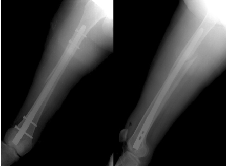
Fig. 267 year-old female who sustained left femoral shaft fracture was treated by antegrade intramedullary nailing.
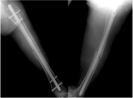
Fig. 3Graph showing duration of bony union according to type of Winquist and Hansen classification. Significant differences were found in the duration of bony union between the Winquist and Hansen type I, II and the type III, IV.

Fig. 4Graph showing duration of bony union according to the number of Fracture fragments. There were significant differences in the duration of bony union both between the simple and comminuted fracture groups, simple and segmental fracture.

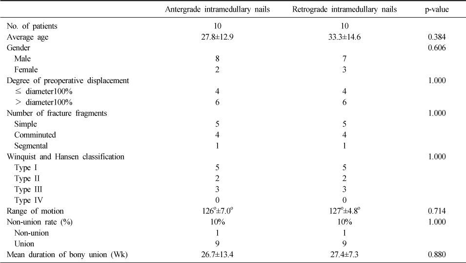
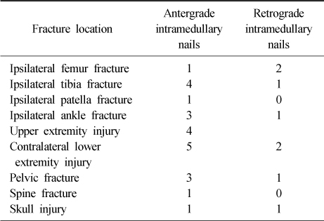
Table 3The differences of duration of bony union and non-union rate among Type I, Type II, Type III, and Type IV base on Winquist and Hansen classification (Results of a Kruskal-Wallis test and the Conover multiple comparison test as a post hoc analysis for comparison of Mean duration of bony union with the four groups)

Table 4Average duration for union of femoral shaft fracture and patient characteristics between Winquist and Hansen Type I, II and Type III, IV
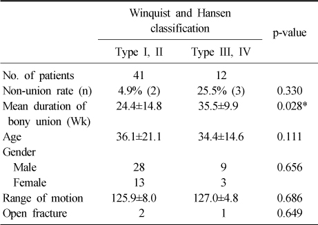
Table 5The differences of duration of bony union according to the degree of preoperative displacement
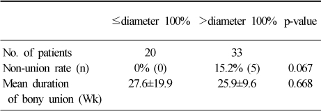
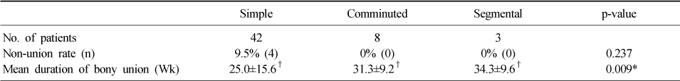
Figure & Data
REFERENCES
Citations
Citations to this article as recorded by 
- Extra-capsular proximal femoral fractures: a cohort comparison of union and complication rates after ballistic versus blunt trauma
Jordan Cook Serotte, Kevin Chen, Julia Nascimben, Jason Strelzow
European Journal of Orthopaedic Surgery & Traumatology.2025;[Epub] CrossRef - Factors Affecting Time to Bony Union of Femoral Subtrochanteric Fractures Treated with Intramedullary Devices
Jung-Yoon Choi, Yerl-Bo Sung, Jin-Hee Yoo, Sung-Jae Chung
Hip & Pelvis.2014; 26(2): 107. CrossRef - Augmentative Locking Plate Fixation for the Treatment of Femoral Nonunion after Intramedullary Nailing
Ki-Chul Park, Chul-Woong Kim, Kyu-Tae Hwang, Ye-Soo Park
Journal of the Korean Fracture Society.2013; 26(4): 268. CrossRef
 Cite
CiteAnalysis of Risk Factors for Nonunion after Intramedullary Nailing of Femoral Shaft Fracture in Adult


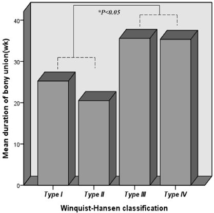
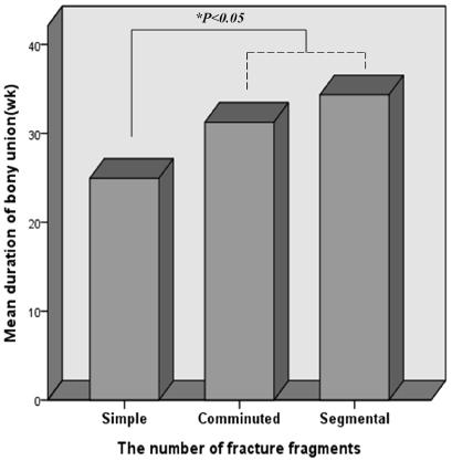
Fig. 1
23 year-old female who sustained right femoral shaft fracture was treated by retrograde intramedullary nailing.
Fig. 2
67 year-old female who sustained left femoral shaft fracture was treated by antegrade intramedullary nailing.
Fig. 3
Graph showing duration of bony union according to type of Winquist and Hansen classification. Significant differences were found in the duration of bony union between the Winquist and Hansen type I, II and the type III, IV.
Fig. 4
Graph showing duration of bony union according to the number of Fracture fragments. There were significant differences in the duration of bony union both between the simple and comminuted fracture groups, simple and segmental fracture.
Fig. 1
Fig. 2
Fig. 3
Fig. 4
Analysis of Risk Factors for Nonunion after Intramedullary Nailing of Femoral Shaft Fracture in Adult
Patients characteristics and the differences of duration of bony union between 2 groups
Associated fractures
The differences of duration of bony union and non-union rate among Type I, Type II, Type III, and Type IV base on Winquist and Hansen classification (Results of a Kruskal-Wallis test and the Conover multiple comparison test as a post hoc analysis for comparison of Mean duration of bony union with the four groups)
The values are given as the mean and the standard deviation. *Significant at p<0.05, †The differences in between the Winquist and Hansen type I, II and the type III, IV were significant (p<0.05).
Average duration for union of femoral shaft fracture and patient characteristics between Winquist and Hansen Type I, II and Type III, IV
The values are given as the mean and the standard deviation. *Significant at p<0.05.
The differences of duration of bony union according to the degree of preoperative displacement
The values are given as the mean and the standard deviation.
The differences of duration of bony union and non-union rate among simple, comminuted, and segmental fracture (Results of a Kruskal-Wallis test and the Conover multiple comparison test as a post hoc analysis for comparison of Mean duration of bony union with the three groups)
The values are given as the mean and the standard deviation. *Significant at p<0.05, †The differences in both between the simple and comminuted fracture groups, simple and segmental fracture groups were significant (p<0.05).
Table 1
Patients characteristics and the differences of duration of bony union between 2 groups
Table 2
Associated fractures
Table 3
The differences of duration of bony union and non-union rate among Type I, Type II, Type III, and Type IV base on Winquist and Hansen classification (Results of a Kruskal-Wallis test and the Conover multiple comparison test as a post hoc analysis for comparison of Mean duration of bony union with the four groups)
The values are given as the mean and the standard deviation. *Significant at p<0.05, †The differences in between the Winquist and Hansen type I, II and the type III, IV were significant (p<0.05).
Table 4
Average duration for union of femoral shaft fracture and patient characteristics between Winquist and Hansen Type I, II and Type III, IV
The values are given as the mean and the standard deviation. *Significant at p<0.05.
Table 5
The differences of duration of bony union according to the degree of preoperative displacement
The values are given as the mean and the standard deviation.
Table 6
The differences of duration of bony union and non-union rate among simple, comminuted, and segmental fracture (Results of a Kruskal-Wallis test and the Conover multiple comparison test as a post hoc analysis for comparison of Mean duration of bony union with the three groups)
The values are given as the mean and the standard deviation. *Significant at p<0.05, †The differences in both between the simple and comminuted fracture groups, simple and segmental fracture groups were significant (p<0.05).

