 E-submission
 TOTA
TOTA TOTS
TOTS
Articles
- Page Path
- HOME > J Musculoskelet Trauma > Volume 23(1); 2010 > Article
-
Case Report
- Nonunion of Humeral Intercondylar Comminuted Fracture Treated with Fibular Graft: A Case Report
- Jin Rok Oh, M.D., Chang Ho Lee, M.D., Ki Yeon Kwon, M.D., Hoi Jeong Chung, M.D.
-
Journal of the Korean Fracture Society 2010;23(1):118-121.
DOI: https://doi.org/10.12671/jkfs.2010.23.1.118
Published online: January 31, 2010
Department of Orthopedic Surgery, Wonju College of Medicine, Yonsei University, Wonju, Korea.
- Address reprint requests to: Jin Rok Oh, M.D. Department of Orthopedic Surgery, Wonju College of Medicine, Yonsei University, Ilsan-dong, Wonju 220-701, Korea. Tel: 82-33-741-1355, Fax: 82-33-746-7326, jroh@yonsei.ac.kr
• Received: August 31, 2009 • Revised: October 5, 2009 • Accepted: November 23, 2009
Copyright © 2010 The Korean Fracture Society
- 1,395 Views
- 5 Download
- 1 Crossref
Abstract
- Nonunion of comminuted distal humeral fracture is troublesome problem to orthopedic surgeon. We report a case of 59 years old woman, who suffered nonunion of comminuted distal humeral fracture previously treated by open reduction and internal fixation with plate and screws concomitantly autoiliac bone graft. We reconstructed humeral condyle with fibular inlay graft inside cortical shell of intercondylar bone fragment and obtained excellent result in radiological and functional outcome.
CASE REPORT
DISCUSSION
- 1. Ahn HS, Cho YH, Byun YS, Kwon DY, Nam SO, Kim DY. Elbow function and complications after internal fixation for fractures of the distal humerus. J Korean Fract Soc, 2006;19:56-61.Article
- 2. Azmi I, Razak M, Hyzan Y. The results of treatment of dislocation and fracture--dislocation of the elbow--a review of 41 patients. Med J Malaysia, 1998;53:Suppl A. 59-70.
- 3. El-Sayed M, El-Hadidi M, El-Adl W. Free non-vascularised fibular graft for treatment of post-traumatic bone defects. Acta Orthop Belg, 2007;73:70-76.
- 4. Fracture and dislocation compendium Orthopaedic trauma association committee for coding and classification. J Orthop Trauma, 1996;10:Suppl 1. v-ix. 1-154.
- 5. Helfet DL, Kloen P, Anand N, Rosen HS. ORIF of delayed unions and nonunions of distal humeral fractures Surgical technique. J Bone Joint Surg Am, 2004;86:Suppl 1. 18-29.
- 6. McKee MD, Veillette CJ, Hall JA, et al. A multicenter, prospective, randomized, controlled trial of open reduction--internal fixation versus total elbow arthroplasty for displaced intra-articular distal humeral fractures in elderly patients. J Shoulder Elbow Surg, 2009;18:3-12.Article
- 7. McKee MD, Wilson TL, Winston L, Schemitsch EH, Richards RR. Functional outcome following surgical treatment of intra-articular distal humeral fractures through a posterior approach. J Bone Joint Surg Am, 2000;82:1701-1707.Article
- 8. Ring D. Instability after total elbow arthroplasty. Hand Clin, 2008;24:105-112.
- 9. Rispoli DM, Athwal GS, Morrey BF. Neurolysis of the ulnar nerve for neuropathy following total elbow replacement. J Bone Joint Surg Br, 2008;90:1348-1351.PDF
- 10. Strauss EJ, Alaia M, Egol KA. Management of distal humeral fractures in the elderly. Injury, 2007;38:Suppl 3. S10-S16.
REFERENCES
Fig. 1
(A) Anterior-posterior and lateral view showed fracture with severe comminution on intercondylar and supracondylar area.
(B) The patient was treated by open reduction and internal fixation with plates and screws concomitantly autologous iliac bone graft.


Figure & Data
REFERENCES
Citations
Citations to this article as recorded by 
- Update 1 of: Destruction and Detection of Chemical Warfare Agents
Yoon Jeong Jang, Kibong Kim, Olga G. Tsay, David A. Atwood, David G. Churchill
Chemical Reviews.2015; 115(24): PR1. CrossRef
 Cite
CiteNonunion of Humeral Intercondylar Comminuted Fracture Treated with Fibular Graft: A Case Report
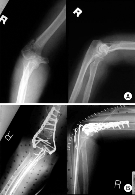
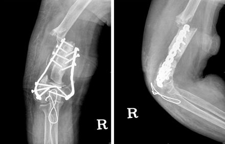
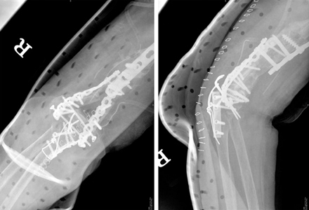
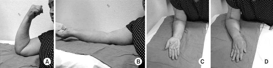
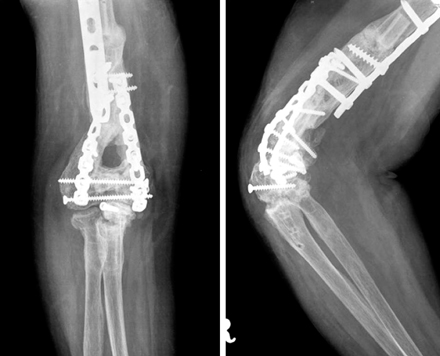
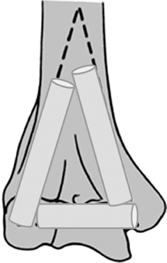
Fig. 1
(A) Anterior-posterior and lateral view showed fracture with severe comminution on intercondylar and supracondylar area.
(B) The patient was treated by open reduction and internal fixation with plates and screws concomitantly autologous iliac bone graft.
Fig. 2
Two years later, radiographs show the bone resorption of grafted bone and newly developed fracture near proximal portion of the plate.
Fig. 3
Anteriorposterior and translateral radiographs of the right elbow after re-operation.
Fig. 4
Excellent pain-free (A) flexion and (B) extension (C) supination (D) pronation resulted.
Fig. 5
Anteroposterior & translateral radiographs of the right elbow 10 months after the operation.
Fig. 6
Schematic view shows reconstructed condyle of distal humerus with three divided fibular graft.
Fig. 1
Fig. 2
Fig. 3
Fig. 4
Fig. 5
Fig. 6
Nonunion of Humeral Intercondylar Comminuted Fracture Treated with Fibular Graft: A Case Report

