 E-submission
 TOTA
TOTA TOTS
TOTS
Articles
- Page Path
- HOME > J Musculoskelet Trauma > Volume 23(1); 2010 > Article
-
Review Article
- Post-traumatic Osteomyelitis
- OokJin Shon, M.D.
-
Journal of the Korean Fracture Society 2010;23(1):122-136.
DOI: https://doi.org/10.12671/jkfs.2010.23.1.122
Published online: January 31, 2010
- Address reprint requests to: OokJin Shon, M.D. Department of Orthopedic Surgery, Yeung-Nam University Hospital, 317-1, Daemyung-5 dong, Nam-gu, Daegu 705-717, Korea. Tel: 82-53-620-3640, Fax: 82-53-628-4020, maestro-jin@hanmail.net
Copyright © 2010 The Korean Fracture Society
- 1,278 Views
- 22 Download
- 1. Barnes RW. Amputation; An illustrated Manual, 2000;1st ed. Philadelphia, Hanley & Belfus.
- 2. Bell SM. Further observations on the value of oral penicillins in chronic staphylococcal osteomyelitis. Med J Aust, 1976;2:591-593.ArticlePubMedPDF
- 3. Bhhaskar SN, Cutright DE, Runsnck EE, Gross A. Pulsating water jet devices in the debridement of combat wounds. Mil Med, 1971;136:264-266.PubMed
- 4. Böhm E, Jsten C. What's new in exogenous osteomyelitis? Pathol Res Pract, 1992;188:254-258.Article
- 5. Burgess EM. The below-knee amputation. Bull Prosthet Res, 1968;10:19.
- 6. Butt WP. The radiology of infection. Clin Orthop Relat Res, 1973;96:20-30.Article
- 7. Calhoun JH, Anger DM, Mader J, Ledbetter BR. The ilizarov technique in the treatment of osteomyelitis. Tex Med, 1991;87:56-59.
- 8. Carragee EJ, Kim D, van der Vlugt T, Vittum D. The clinical use of the erythrocyte sedimentation rate in pyogenic vertebral osteomyelitis. Spine (Phila Pa 1976), 1997;22:2089-2093.PubMed
- 9. Cho SH, Song HR, Koo KH, Jeong ST, Park YJ. Antibiotic-impregnated cement beads in the treatment of chronic osteomyelitis. Bull Hosp Jt Dis, 1997;56:140-144.
- 10. Cierny G 3rd. Chronic osteomyelitis: results of treatment. Instr Course Lect, 1990;39:495-508.
- 11. Cierny G 3rd. Infected tibial nonunions (1981-1995). The evolution of change. Clin Orthop Relat Res, 1999;360:97-105.
- 12. Cierny G 3rd, Mader JT. Approach to adult osteomyelitis. Orthop Rev, 1987;16:259-270.
- 13. Cierny G 3rd, Mader JT. Evarts CMC. The surgical treatment of adult osteomyelitis. In: Surgery of the musculoskeletal system, 1983;1st ed. New York, Churchill Livingstone. 15-35.
- 14. Cierny G 3rd, Mader JT, Penninck JJ. A clinical staging system for adult osteomyelitis. Clin Orthop Relat Res, 2003;414:7-24.
- 15. Cierny G 3rd, Mader JT, Penninck JJ. A clinical staging system for adult osteomyelitis. Contemp Orthop, 1985;10:17-37.
- 16. Couch L, Cierny G 3rd, Mader JT. Inpatient and outpatient use of the Hickman catheter for adults with osteomyelitis. Clin Orthop Relat Res, 1987;219:226-235.Article
- 17. Curtis MJ, Brown PR, Dick JD, Jinnah RH. Contaminated fracture of the tibia: a comparison of treatment modalities in an animal model. J Orthop Res, 1995;13:286-295.
- 18. Dendrinos GK, Kontos S, Lyritisis E. Use of the ilizarov technique for treatment of non-union of the tibia associated with infection. J Bone Joint Surg Am, 1995;77:835-846.Article
- 19. Deysine M, Rafkin H, Teicher I, et al. Diagnosis of chronic and postoperative osteomyelitis with gallium 67 citrate scans. Am J Surg, 1975;129:632-635.Article
- 20. Erdman WA, Tamburro F, Jayson HT, Wearherall PT, Ferry KB, Peshock RM. Osteomyelitis: characteristics and pitfalls of diagnosis with MR imaging. Radiology, 1991;180:533-539.Article
- 21. Fears RL, Gleis GE, Seligson D. Browner BD, Jupiter JB, Levine AM, Trafton PG. Diagnosis and treatment of complications. In: Skeletal Trauma, 1998;2nd ed. Philadelphia, PA, WB Saunders Co.
- 22. Gustilo RB. Evarts CM. Management of infected non-union. In: Surgery of the Musculoskeletal System, 1990;2nd ed. London, Churchill Livingstone. 44294455.
- 23. Gustilo RB, Anderson JT. Prevention of infection in the treatment of one thousand and twenty-five open fractures of long bones: retrospective and prospective analyses. J Bone Joint Surg Am, 1976;58:453-458.
- 24. Hedström SA. The prognosis of chronic staphylococcal osteomyelitis after long-term antibiotic treatment. Scand J Infect Dis, 1974;6:33-38.Article
- 25. Herscovici D Jr, Sanders RW, Scaduto JM, Infante A, DiPasquale T. Vacuum-assisted wound closure (VAC therapy) for the management of patients with high-energy soft tissue injuries. J Orthop Trauma, 2003;17:683-688.Article
- 26. Hickman RO, Buckner CD, Clift RA, Sanders JE, Stewart P, Thomas ED. A modified right atrial catheter for access to the venous system in marrow transplant recipients. Surg Gynecol Obstet, 1979;148:871-875.
- 27. Hong SW, Seah CS, Kuek LB, Tan KC. Soft tissue coverage in compound and complicated tibial fracture using microvascular flaps. Ann Acad Med Singapore, 1998;27:182-187.
- 28. Ilizarov GA. Transosseous osteosynthesis: theoretical and clinical aspects of the regeneration and growth of tissue, 1992;1st ed. Berlin, Springer-Verlag.
- 29. Jones AG, Francis MD, Davis MA. Bone scanning: radionuclidic reaction mechanisms. Semin Nucl Med, 1976;6:3-18.Article
- 30. Kindsfater K, Jonassen EA. Osteomyelitis in grade II and III open tibia fracture with late debridement. J Orthop Trauma, 1995;9:121-127.
- 31. Kuhn JP, Berger PE. Computed tomographic diagnosis of osteomyelitis. Radiology, 1979;130:503-506.Article
- 32. Lew DP, Waldvogel FA. Osteomyelitis. N Engl J Med, 1997;336:999-1007.Article
- 33. Mackowiak PA, Jones SR, Smith JW. Diagnostic value of sinus-tract cultures in chronic osteomyelitis. JAMA, 1978;239:2772-2775.Article
- 34. Mader JT, Ortiz M, Calhoun JH. Update on the diagnosis and management of osteomyelitis. Clin Podiatr Med Surg, 1996;13:701-724.Article
- 35. Mader JT, Shirtliff M, Calhoun JH. Staging and staging application in osteomyelitis. Clin Infect Dis, 1997;25:1303-1309.Article
- 36. Ma LD, Frassica FJ, Bluemke DA, Fishman EK. CT and MRI evaluation of musculoskeletal infection. Crit Rev Diagn Imaging, 1997;38:535-568.
- 37. Mathes SJ, Alpert BS, Chang N. Use of muscle flap in chronic osteomyelitis: experimental and clinical correlation. Plast Reconst Surg, 1982;69:815-829.
- 38. May JW Jr, Gallico GG 3rd, Lukash FN. Microvascular transfer of free tissue for closure of bone wounds of the distal lower extremity. N Engl J Med, 1982;306:253-257.Article
- 39. Merritt K. Factors increasing the risk of infection in patients with open fractures. J Trauma, 1988;28:823-827.Article
- 40. Meyer S, Weiland AJ, Willenegger H. The treatment of infected non-union of fractures of long bones: study of sixty four cases with a five to twenty-one-year follow-up. J Bone Joint Surg Am, 1975;57:836-842.Article
- 41. Minami A, Kaneda K, Itoga H. Treatment of infected segmental defect of long bone with vascularized bone transfer. J Reconstr Microsurg, 1992;8:75-82.Article
- 42. Modic MT, Pflanze W, Feiglin DH, Belhobek G. Magnetic resonance imaging of musculoskeletal infections. Radiol Clin North Am, 1986;24:247-258.Article
- 43. Modi SP, Eppes SC, Klein JD. Cat-scratch disease presenting as multifocal osteomyelitis with thoracic abscess. Pediatr Infect Dis J, 2001;20:1006-1007.Article
- 44. Mooney JF 3rd, Argenta LC, Marks MW, Morykwas MJ, DeFranzo AJ. Treatment of soft tissue defects in pediatric patients using the V.A.C. system. Clin Orthop Relat Res, 2000;376:26-31.Article
- 45. Norden CW, Dickens DR. Experimental osteomyelitis. 3. Treatment with cephaloridine. J Infect Dis, 1973;127:525-528.
- 46. Okike K, Bhattacharyya T. Trends in the management of open fractures. A critical analysis. J Bone Joint Surg Am, 2006;88:2739-2748.
- 47. Panda M, Ntungila N, Kalunda M, Hinsenkamp M. Treatment of chronic osteomyelitis using the Papineau technique. Int Orthop, 1998;22:37-40.ArticlePDF
- 48. Papineau LJ, Alfageme A, Dalcourt JP, Pilon L. Chronic osteomyelitis: open excision and grafting after saucerization (author's transl). Int Orthop, 1979;3:165-176.
- 49. Papineau LJ, Alfageme A, Dalcourt JP, Pilon L. Osteomyelite Chronique : Excision et greffe de spongieux a l'air libre apres mises a plat extensives. Int Orthop, 1979;3:165-176.
- 50. Patzakis MJ, Harrey JP Jr, Ivler D. The role of antibiotics in the management of open fracture. J Bone Joint Surg, 1974;56:532-541.
- 51. Perry M. Erythrocyte sedimentation rate and C reactive protein in the assessment of suspected bone infection--are they reliable indices? J R Coll Surg Edinb, 1996;41:116-118.
- 52. Rightmire E, Zurakowski D, Vrahas M. Acute infections after fracture repair: management with hardware in place. Clin Orthop Relat Res, 2008;466:466-472.
- 53. Roine I, Faingezicht I, Arguedas A, Herrera JF, Rodríguez F. Serial serum C-reactive protein to monitor recovery from acute hematogenous osteomyelitis in children. Pediatr Infect Dis J, 1995;14:40-44.Article
- 54. Rosen H. The treatment of nonunions and pseudarthroses of the humeral shaft. Orthop Clin North Am, 1990;21:725-742.Article
- 55. Russin LD, Staab EV. Unusual bone-scan findings in acute osteomyelitis: case report. J Nucl Med, 1976;17:617-619.
- 56. Schulak DJ, Rayhack JM, Lippert FG 3rd, Convery FR. The erythrocyte sedimentation rate in orthopaedic patients. Clin Orthop Relat Res, 1982;167:197-202.Article
- 57. Simpson AH, Deakin M, Latham JM. Chronic osteomyelitis. The effect of the extent of surgical resection on infection-free survival. J Bone Joint Surg Br, 2001;83:403-407.
- 58. Tehranzadeh J, Wang F, Mesgarzadeh M. Magnetic resonance imaging of osteomyelitis. Crit Rev Diagn Imaging, 1992;33:495-534.
- 59. Tetsworth K, Cierny G III. Osteomyelitis debridement techniques. Clin Orthop Relat Res, 1999;(360):87-96.Article
- 60. Unger E, Moldofsky P, Gatenby R, Hartz W, Broder G. Diagnosis of osteomyelitis by MR imaging. AJR Am J Roentgenol, 1988;150:605-610.Article
- 61. Waldvogel FA, Medoff G, Swartz MN. Osteomyelitis: a review of clinical features, therapeutic considerations and unusual aspects. N Engl J Med, 1970;282:198-206.Article
- 62. Waldvogel FA, Papageorgiou PS. Osteomyelitis: the past decade. N Engl J Med, 1980;303:360-370.Article
- 63. Weiland AJ, Moore JR, Daniel RK. The efficacy of free tissue transfer in the treatment of osteomyelitis. J Bone Joint Surg Am, 1984;66:181-193.Article
- 64. Zuluaga AF, Galvis W, Saldarriaga JG, Agudelo M, Salazar BE, Vesga O. Etiologic diagnosis of chronic osteomyelitis: a prospective study. Arch Intern Med, 2006;166:95-100.Article
REFERENCES
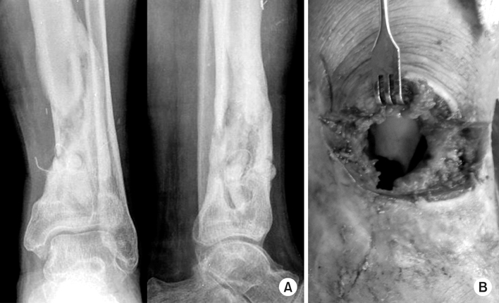
Fig. 2
(A) A 53 years old man with osteomyelitis on tibia.
(B) MRI showed osteomyelitis on mid shaft of tibia.
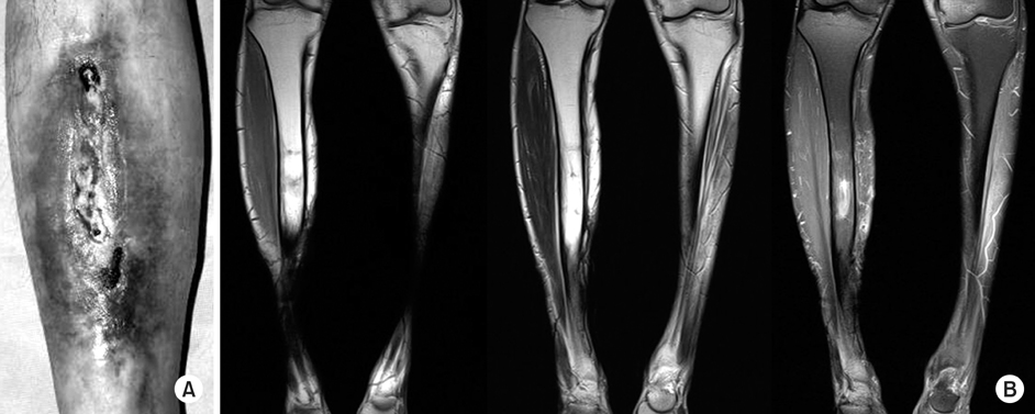
Fig. 3
(A) A 54 years old woman with left tibia open fracture.
(B) Irrigation and debridement and external fixator was applied, but (C) above knee amputation was done due to osteomyelitis.

Fig. 4
(A) A 49 years old female with left proximal femur osteomyelitis. Open debridement and irrigation, dead bone debridement with bead insertion was done. Bead was applied widely from intramedullary to subcutaneous space.
(B) After 4 weeks, bead was removed and internal fixation with locking plate by MIPO technique was done.
(C) X-ray at post-op 6 weeks.

Fig. 5
(A) A 71 years old man with left lower leg osteomyelitis
and compartment syndrome.
(B) Debridement and irrigation and bead insertion was done, but couldn't close the skin. This is not recommended.
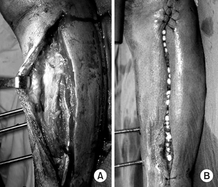
Fig. 6
(A) Left hand dorsum open wound with osteomyelitis.
(B) Both leg degloving injury.
(C) Forearm both bone fracture with degloving injury. In all case, after vacuum-assisted closure system was applied, granulation tissue was grown, and skin graft was done successfully.

Fig. 7
(A) OR/IF with tibia nail was done for right tibiofibular fracture.
(B) 6 month after operation, osteomyelitis was developed.
(C) All hardware remove and bead insertion was done.

Fig. 8
(A) A 57 years old female with left femur supracondylar open fracture.
(B) Temporary external fixator was applied.
(C) After open wound was healed, internal fixation with plate by MIPO technique was done.
(D, E) 3 months later, bone union was seen and full range of motion was acquired.

Fig. 9
(A) A 49 years old man with right distal tibia open fracture.
(B) OR/IF with skin graft was done but (C) osteomyelitis was developed.
(D, E) Bone transfer using ilizarov was done.
(F) OR/IF with plate using MIPO technique was done and
(G) last follow up x-ray shows bone union.


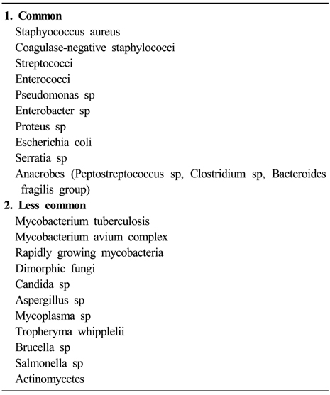
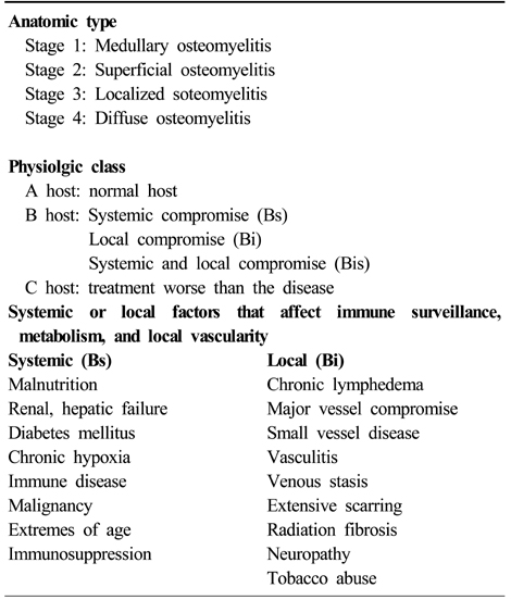
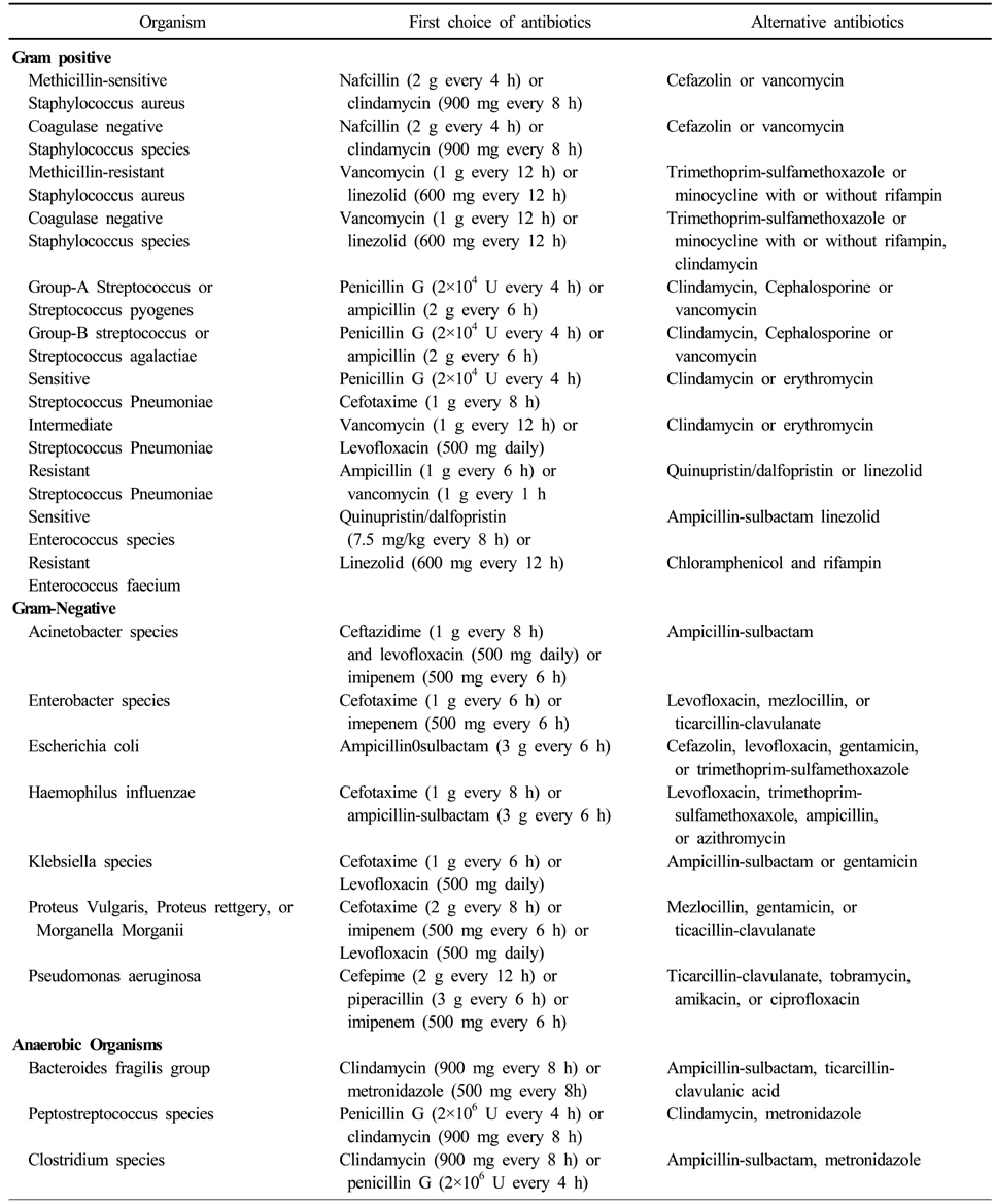
Figure & Data
REFERENCES
Citations
Citations to this article as recorded by 
 Cite
CitePost-traumatic Osteomyelitis


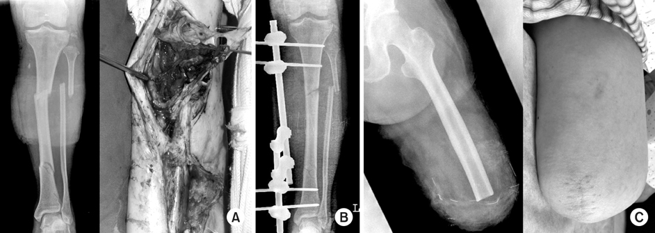
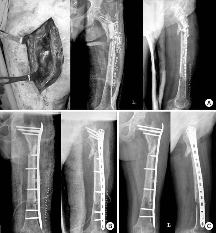

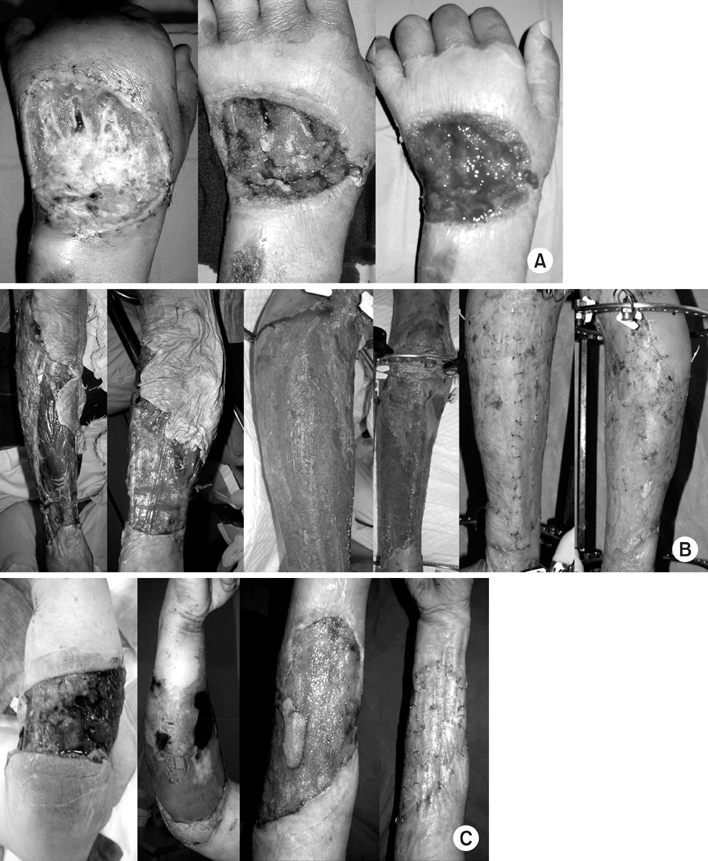
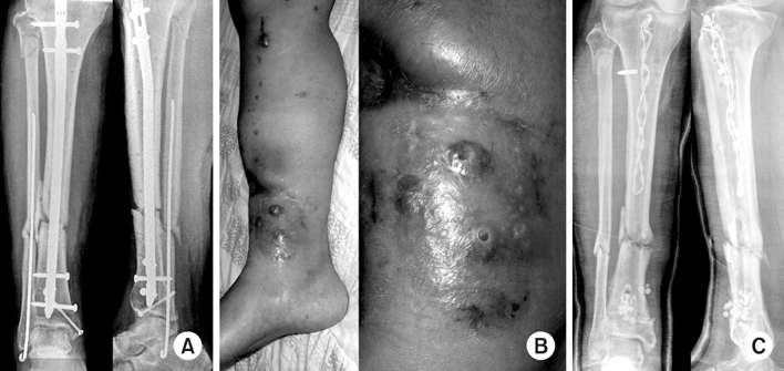
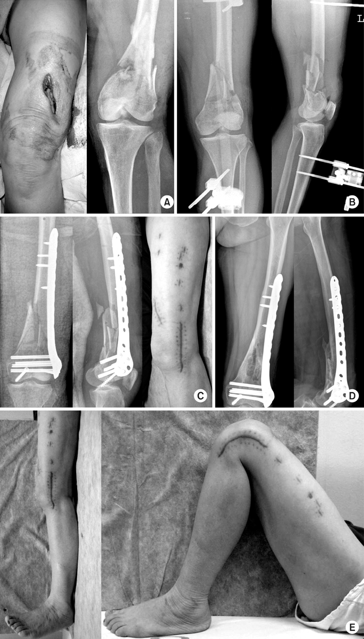
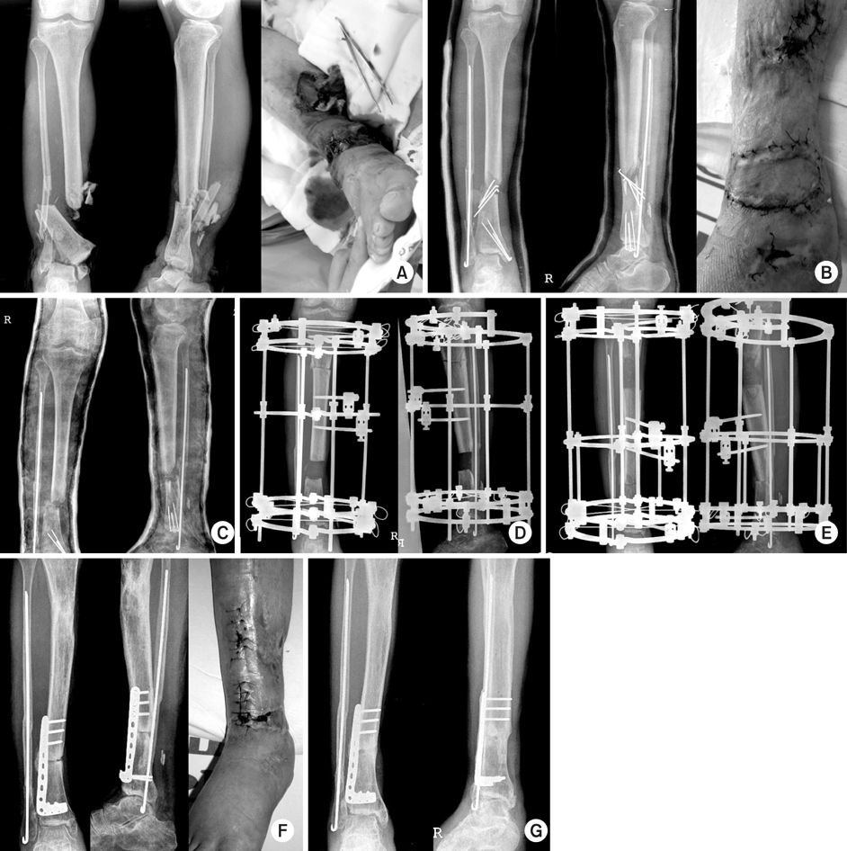
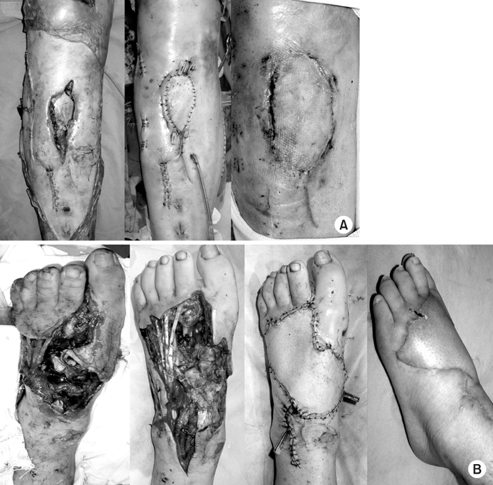
Fig. 1
(A, B) Non-union was seen at osteomyelitis after left distal tibiofibular open fx.
Fig. 2
(A) A 53 years old man with osteomyelitis on tibia.
(B) MRI showed osteomyelitis on mid shaft of tibia.
Fig. 3
(A) A 54 years old woman with left tibia open fracture.
(B) Irrigation and debridement and external fixator was applied, but (C) above knee amputation was done due to osteomyelitis.
Fig. 4
(A) A 49 years old female with left proximal femur osteomyelitis. Open debridement and irrigation, dead bone debridement with bead insertion was done. Bead was applied widely from intramedullary to subcutaneous space.
(B) After 4 weeks, bead was removed and internal fixation with locking plate by MIPO technique was done.
(C) X-ray at post-op 6 weeks.
Fig. 5
(A) A 71 years old man with left lower leg osteomyelitis
and compartment syndrome.
(B) Debridement and irrigation and bead insertion was done, but couldn't close the skin. This is not recommended.
Fig. 6
(A) Left hand dorsum open wound with osteomyelitis.
(B) Both leg degloving injury.
(C) Forearm both bone fracture with degloving injury. In all case, after vacuum-assisted closure system was applied, granulation tissue was grown, and skin graft was done successfully.
Fig. 7
(A) OR/IF with tibia nail was done for right tibiofibular fracture.
(B) 6 month after operation, osteomyelitis was developed.
(C) All hardware remove and bead insertion was done.
Fig. 8
(A) A 57 years old female with left femur supracondylar open fracture.
(B) Temporary external fixator was applied.
(C) After open wound was healed, internal fixation with plate by MIPO technique was done.
(D, E) 3 months later, bone union was seen and full range of motion was acquired.
Fig. 9
(A) A 49 years old man with right distal tibia open fracture.
(B) OR/IF with skin graft was done but (C) osteomyelitis was developed.
(D, E) Bone transfer using ilizarov was done.
(F) OR/IF with plate using MIPO technique was done and
(G) last follow up x-ray shows bone union.
Fig. 10
(A) A 55 years old man with right lower leg open wound. Gastrocnemius muscle head turn over flap was done.
(B) A 48 years old man with left foot open fracture. Medial sural artery perforator free flap was done.
Fig. 1
Fig. 2
Fig. 3
Fig. 4
Fig. 5
Fig. 6
Fig. 7
Fig. 8
Fig. 9
Fig. 10
Post-traumatic Osteomyelitis
Microbiology of osteomyelitis
Microbiology of osteomyelitis
Initial choice of antibiotics for therapy (Adult Doses)
Table 1
Microbiology of osteomyelitis
Table 2
Microbiology of osteomyelitis
Table 3
Initial choice of antibiotics for therapy (Adult Doses)

