 E-submission
 TOTA
TOTA TOTS
TOTS
Articles
- Page Path
- HOME > J Musculoskelet Trauma > Volume 26(4); 2013 > Article
-
Case Report
- Rupture of the Extensor Pollicis Longus Tendon at the Proximal Screw of Volar Plate Fixation for Distal Radius Fracture: A Case Report
- Dong-Ju Shin, M.D., Seung-Oh Nam, M.D., Hun-Sik Cho, M.D.
-
Journal of the Korean Fracture Society 2013;26(4):338-342.
DOI: https://doi.org/10.12671/jkfs.2013.26.4.338
Published online: October 18, 2013
Department of Orthopedic Surgery, Daegu Fatima Hospital, Daegu, Korea.
- Address reprint requests to: Seung-Oh Nam, M.D. Department of Orthopedic Surgery, Daegu Fatima Hospital, 99 Ayang-ro, Dong-gu, Daegu 701-724, Korea. Tel: 82-53-940-7324, Fax: 82-53-954-7417, nso1020@naver.com
• Received: May 20, 2013 • Revised: July 6, 2013 • Accepted: July 6, 2013
Copyright © 2013 The Korean Fracture Society. All rights reserved.
This is an Open Access article distributed under the terms of the Creative Commons Attribution Non-Commercial License (http://creativecommons.org/licenses/by-nc/3.0/) which permits unrestricted non-commercial use, distribution, and reproduction in any medium, provided the original work is properly cited.
- 830 Views
- 1 Download
Abstract
- As volar plate fixation of distal radius fracture becomes more common, reports of ruptured extensor pollicis longus tendon by a protruding distal screw tip are also increasing steadily. Authors have experienced a rare case of ruptured extensor pollicis longus tendon at the prominent proximal screw of fixed volar plate for distal radius fracture, and we report it herein with a review of the literature.
- 1. Al-Rashid M, Theivendran K, Craigen MA. Delayed ruptures of the extensor tendon secondary to the use of volar locking compression plates for distal radial fractures. J Bone Joint Surg Br, 2006;88:1610-1612.ArticlePubMedPDF
- 2. Arora R, Lutz M, Hennerbichler A, Krappinger D, Espen D, Gabl M. Complications following internal fixation of unstable distal radius fracture with a palmar locking-plate. J Orthop Trauma, 2007;21:316-322.ArticlePubMed
- 3. Augereau B, Lance D, Kerboul M. Plate osteosynthesis of unstable fractures of the wrist with anterior displacement. Int Orthop, 1983;7:55-59.PubMed
- 4. Benson EC, DeCarvalho A, Mikola EA, Veitch JM, Moneim MS. Two potential causes of EPL rupture after distal radius volar plate fixation. Clin Orthop Relat Res, 2006;451:218-222.ArticlePubMed
- 5. Cho NY, Seo CY, Kim MS, Kim HS, Lee KB. Extensor pollicis longus rupture after distal radius fracture. J Korean Fract Soc, 2012;25:52-57.Article
- 6. Christophe K. Rupture of the extensor pollicis longus tendon following colles fracture. J Bone Joint Surg Am, 1953;35:1003-1005.ArticlePubMed
- 7. Ozer K, Toker S. Dorsal tangential view of the wrist to detect screw penetration to the dorsal cortex of the distal radius after volar fixed-angle plating. Hand (N Y), 2011;6:190-193.ArticlePubMedPMCPDF
- 8. Perry DC, Machin DM, Casaletto JA, Brown DJ. Minimising the risk of extensor pollicis longus rupture following volar plate fixation of distal radius fractures: a cadaveric study. Ann R Coll Surg Engl, 2011;93:57-60.ArticlePubMed
- 9. White BD, Nydick JA, Karsky D, Williams BD, Hess AV, Stone JD. Incidence and clinical outcomes of tendon rupture following distal radius fracture. J Hand Surg Am, 2012;37:2035-2040.ArticlePubMed
- 10. Wong-Chung J, Quinlan W. Rupture of extensor pollicis longus following fixation of a distal radius fracture. Injury, 1989;20:375-376.ArticlePubMed
REFERENCES
Fig. 1Postoperative plain radiographs show that the proximal screw tips of fixed volar plate are protruded about 1-2 mm from the dorsal cortex of distal radius. The arrow marks the causative screw of ruptured extensor pollicis longus tendon.

Fig. 2Computerized tomographs show that any distal screws of fixed volar plate do not penetrate the dorsal cortex of distal radius, but the proximal screws are definitely protruded through the dorsal cortex. The middle one (arrow) is the most prominent screw in comparison of simple radiographs, measuring about 2.8 mm.
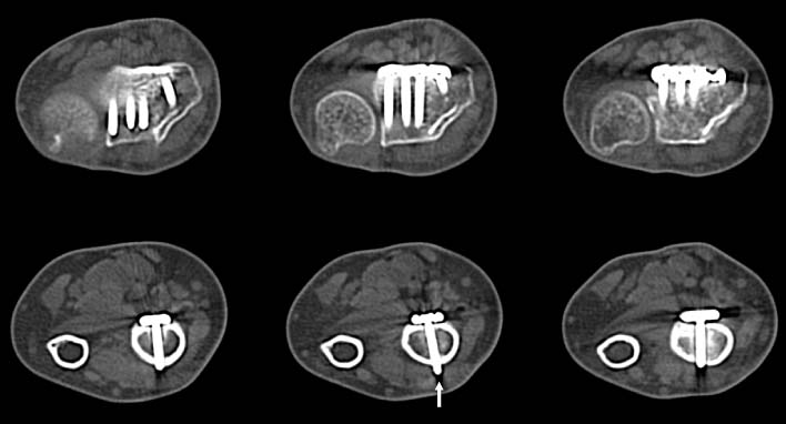
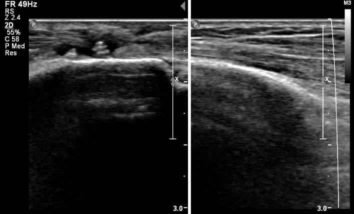

Figure & Data
REFERENCES
Citations
Citations to this article as recorded by 
 Cite
CiteRupture of the Extensor Pollicis Longus Tendon at the Proximal Screw of Volar Plate Fixation for Distal Radius Fracture: A Case Report
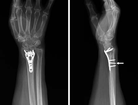


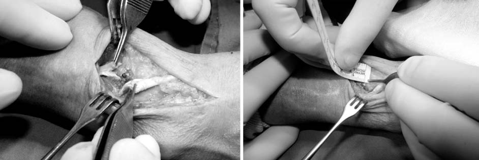
Fig. 1
Postoperative plain radiographs show that the proximal screw tips of fixed volar plate are protruded about 1-2 mm from the dorsal cortex of distal radius. The arrow marks the causative screw of ruptured extensor pollicis longus tendon.
Fig. 2
Computerized tomographs show that any distal screws of fixed volar plate do not penetrate the dorsal cortex of distal radius, but the proximal screws are definitely protruded through the dorsal cortex. The middle one (arrow) is the most prominent screw in comparison of simple radiographs, measuring about 2.8 mm.
Fig. 3
Preoperative ultrasonography shows protruding screw tips and discontinuity of extensor pollicis longus tendon at distal forearm region.
Fig. 4
Intraoperative photographs show a complete rupture of extensor pollicis longus tendon, where the ends are torn and fraying at prominent proximal screw tip, measuring about 2.5 mm.
Fig. 1
Fig. 2
Fig. 3
Fig. 4
Rupture of the Extensor Pollicis Longus Tendon at the Proximal Screw of Volar Plate Fixation for Distal Radius Fracture: A Case Report

