 E-submission
 TOTA
TOTA TOTS
TOTS
Articles
- Page Path
- HOME > J Musculoskelet Trauma > Volume 26(4); 2013 > Article
-
Review Article
- Reduction and Stabilization of Pelvic Ring Injury
- Ki-Chul Park, M.D.
-
Journal of the Korean Fracture Society 2013;26(4):343-347.
DOI: https://doi.org/10.12671/jkfs.2013.26.4.343
Published online: October 18, 2013
Department of Orthopedic Surgery, Hanyang University College of Medicine, Seoul, Korea.
- Address reprint requests to: Ki-Chul Park, M.D. Department of Orthopaedic Surgery, Hanyang University Guri Hospital, 153 Gyeongchun-ro, Guri 471-701, Korea. Tel: 82-31-560-2318, Fax: 82-31-557-8781, kcpark@hanyang.ac.kr
Copyright © 2013 The Korean Fracture Society. All rights reserved.
This is an Open Access article distributed under the terms of the Creative Commons Attribution Non-Commercial License (http://creativecommons.org/licenses/by-nc/3.0/) which permits unrestricted non-commercial use, distribution, and reproduction in any medium, provided the original work is properly cited.
- 610 Views
- 3 Download
- 1. Carlson DA, Scheid DK, Maar DC, Baele JR, Kaehr DM. Safe placement of S1 and S2 iliosacral screws: the "vestibule" concept. J Orthop Trauma, 2000;14:264-269.Article
- 2. Day AC, Kinmont C, Bircher MD, Kumar S. Crescent fracture-dislocation of the sacroiliac joint: a functional classification. J Bone Joint Surg Br, 2007;89:651-658.
- 3. Gänsslen A, Pohlemann T, Krettek C. A simple supraacetabular external fixation for pelvic ring fractures. Oper Orthop Traumatol, 2005;17:296-312.PubMed
- 4. Guthrie HC, Owens RW, Bircher MD. Fractures of the pelvis. J Bone Joint Surg Br, 2010;92:1481-1488.ArticlePubMedPDF
- 5. Lefaivre KA, Starr AJ, Reinert CM. Reduction of displaced pelvic ring disruptions using a pelvic reduction frame. J Orthop Trauma, 2009;23:299-308.ArticlePubMed
- 6. Letournel E. Surgical fixation of displaced pelvic fractures and dislocations of the symphysis pubis (excluding acetabular fractures) (author's transl). Rev Chir Orthop Reparatrice Appar Mot, 1981;67:771-782.PubMed
- 7. Metze M, Tiemann AH, Josten C. Male sexual dysfunction after pelvic fracture. J Trauma, 2007;63:394-401.ArticlePubMed
- 8. Nork SE, Jones CB, Harding SP, Mirza SK, Routt ML Jr. Percutaneous stabilization of U-shaped sacral fractures using iliosacral screws: technique and early results. J Orthop Trauma, 2001;15:238-246.ArticlePubMed
- 9. Poelstra KA, Kahler DM. Supra-acetabular placement of external fixator pins: a safe and expedient method of providing the injured pelvis with stability. Am J Orthop (Belle Mead NJ), 2005;34:148-151.PubMed
- 10. Routt ML Jr, Simonian PT, Grujic L. The retrograde medullary superior pubic ramus screw for the treatment of anterior pelvic ring disruptions: a new technique. J Orthop Trauma, 1995;9:35-44.PubMed
- 11. Schildhauer TA, Bellabarba C, Nork SE, Barei DP, Routt ML Jr, Chapman JR. Decompression and lumbopelvic fixation for sacral fracture-dislocations with spino-pelvic dissociation. J Orthop Trauma, 2006;20:447-457.ArticlePubMed
- 12. Simonian PT, Schwappach JR, Routt ML Jr, Agnew SG, Harrington RM, Tencer AF. Evaluation of new plate designs for symphysis pubis internal fixation. J Trauma, 1996;41:498-502.ArticlePubMed
- 13. Simpson LA, Waddell JP, Leighton RK, Kellam JF, Tile M. Anterior approach and stabilization of the disrupted sacroiliac joint. J Trauma, 1987;27:1332-1339.ArticlePubMed
- 14. The Korean Fracture Society. Principles of fracture management. 1st ed. Seoul: E-public Inc; 2013. p. 559-564.
- 15. Yinger K, Scalise J, Olson SA, Bay BK, Finkemeier CG. Biomechanical comparison of posterior pelvic ring fixation. J Orthop Trauma, 2003;17:481-487.ArticlePubMed
REFERENCES
Fig. 1
A 42-year-old man sustained a type B pelvic bone fracture with symphysis pubis dislocation.
(A) The symphysis pubis was displaced 2 cm initially.
(B) It was increased to 2.8 cm two days after injury.
(C) A 4.5 mm 4 hole limited contact-dynamic compression plate was fixed after reduction of symphysis pubis.
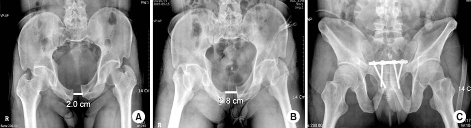
Fig. 2
(A) A 53-year-old man sustained a type B pelvic bone fracture with open wound in the inguinal area.
(B) Supra-acetabular external fixation was applied with reduction of the symphysis pubis.

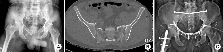
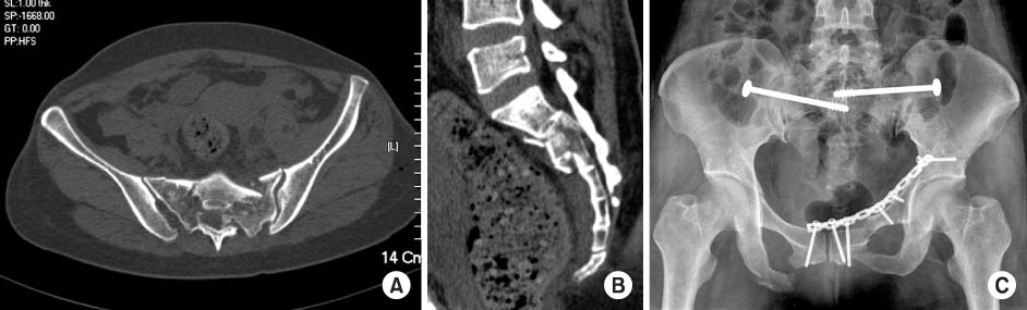
Figure & Data
REFERENCES
Citations
Citations to this article as recorded by 
 Cite
CiteReduction and Stabilization of Pelvic Ring Injury

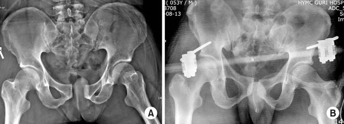


Fig. 1
A 42-year-old man sustained a type B pelvic bone fracture with symphysis pubis dislocation.
(A) The symphysis pubis was displaced 2 cm initially.
(B) It was increased to 2.8 cm two days after injury.
(C) A 4.5 mm 4 hole limited contact-dynamic compression plate was fixed after reduction of symphysis pubis.
Fig. 2
(A) A 53-year-old man sustained a type B pelvic bone fracture with open wound in the inguinal area.
(B) Supra-acetabular external fixation was applied with reduction of the symphysis pubis.
Fig. 3
(A) Pelvis anteroposterior radiograph and (B) axial computed tomography scan demonstrating both small superior crescent fractures.
(C) Both crescent fractures were fixed with iliosacral screws with anterior plating.
Fig. 4
(A) Axial and (B) sagittal reconstruction computed tomography scan, demonstrating spinal pelvic dissociation from a sacral fracture at S1/S2.
(C) A bilateral sacroiliac screw was fixed after closed reduction and anterior plating.
Fig. 1
Fig. 2
Fig. 3
Fig. 4
Reduction and Stabilization of Pelvic Ring Injury

