 E-submission
 TOTA
TOTA TOTS
TOTS
Articles
- Page Path
- HOME > J Musculoskelet Trauma > Volume 26(4); 2013 > Article
-
Original Article
- Analysis of Risk Factors for the Posterolateral Articular Depression and Status of Posterolateral Fragment in Lateral Condylar and Bicondylar Tibial Plateau Fractures with Joint Depression
- Jung-Yun Choi, M.D., Yong-Woon Shin, M.D., Beom-Jung Lee, M.D.
-
Journal of the Korean Fracture Society 2013;26(4):241-247.
DOI: https://doi.org/10.12671/jkfs.2013.26.4.241
Published online: October 18, 2013
Department of Orthopedic Surgery, Inje University Sanggye Paik Hospital, Seoul, Korea.
- Address reprint requests to: Yong-Woon Shin, M.D. Department of Orthopedic Surgery, Inje University Sanggye Paik Hospital, 1342 Dongil-ro, Nowon-gu, Seoul 139-707, Korea. Tel: 82-2-950-1032, Fax: 82-2-934-6342, woonysos@paik.ac.kr
• Received: April 24, 2013 • Revised: June 11, 2013 • Accepted: July 27, 2013
Copyright © 2013 The Korean Fracture Society. All rights reserved.
This is an Open Access article distributed under the terms of the Creative Commons Attribution Non-Commercial License (http://creativecommons.org/licenses/by-nc/3.0/) which permits unrestricted non-commercial use, distribution, and reproduction in any medium, provided the original work is properly cited.
- 1,046 Views
- 2 Download
- 1 Crossref
Abstract
-
Purpose
- To evaluate risk factors of posterolateral articular depression and characteristics of the posterolateral fragment in lateral condylar and bicondylar tibial plateau fractures with joint depression.
-
Materials and Methods
- We reviewed 48 patients of Schatzker type II and type V (type II 34, type V 14) and evaluated risk factors of posterolateral articular depression according to the posterolateral fragment, fibular fracture, and Schatzker classification. We evaluated the position of articular depression and anterolateral fracture line of the posterolateral fragment and measured anterior to posterior lengths of the posterolateral fragment.
-
Results
- Posterolateral articular depression was found in 20 of 34 cases (59%) with coexisting posterolateral fragment and in 16 of 21 cases (76%) with coexisting fibular fracture. There was a significant difference in the occurrence of posterolateral articular depression with the existence of the posterolateral fragment and fibular fracture (p<0.001). Multivariate regression analysis revealed that fibular fracture increased the occurrence of posterolateral articular depression (odds ratio 24.5, 95% confidence interval 2.2-267.2). Fifty-seven percentage of the anterolateral fracture line of the posterolateral fragment existed posterior to the anterior margin of the fibular head.
-
Conclusion
- This study showed that fibular fracture affects posterolateral articular depression in Schatzker type II and V tibial plateau fractures. Selecting a fixation device and performing fracture reduction requires a careful consideration since the anterolateral fracture line of the posterolateral fragment exists posterior to the anterior margin of the fibular head.
- 1. Barei DP, O'Mara TJ, Taitsman LA, Dunbar RP, Nork SE. Frequency and fracture morphology of the posteromedial fragment in bicondylar tibial plateau fracture patterns. J Orthop Trauma, 2008;22:176-182.Article
- 2. Bozkurt M, Turanli S, Doral MN, et al. The impact of proximal fibula fractures in the prognosis of tibial plateau fractures: a novel classification. Knee Surg Sports Traumatol Arthrosc, 2005;13:323-328.ArticlePDF
- 3. Brunner A, Honigmann P, Horisberger M, Babst R. Open reduction and fixation of medial Moore type II fractures of the tibial plateau by a direct dorsal approach. Arch Orthop Trauma Surg, 2009;129:1233-1238.ArticlePubMedPDF
- 4. Chan PS, Klimkiewicz JJ, Luchetti WT, et al. Impact of CT scan on treatment plan and fracture classification of tibial plateau fractures. J Orthop Trauma, 1997;11:484-489.ArticlePubMed
- 5. Chang SM, Zheng HP, Li HF, et al. Treatment of isolated posterior coronal fracture of the lateral tibial plateau through posterolateral approach for direct exposure and buttress plate fixation. Arch Orthop Trauma Surg, 2009;129:955-962.ArticlePubMedPDF
- 6. Fakler JK, Ryzewicz M, Hartshorn C, Morgan SJ, Stahel PF, Smith WR. Optimizing the management of Moore type I postero-medial split fracture dislocations of the tibial head: description of the Lobenhoffer approach. J Orthop Trauma, 2007;21:330-336.ArticlePubMed
- 7. Fernandez DL. Anterior approach to the knee with osteotomy of the tibial tubercle for bicondylar tibial fractures. J Bone Joint Surg Am, 1988;70:208-219.ArticlePubMed
- 8. Gosling T, Schandelmaier P, Muller M, Hankemeier S, Wagner M, Krettek C. Single lateral locked screw plating of bicondylar tibial plateau fractures. Clin Orthop Relat Res, 2005;439:207-214.ArticlePubMed
- 9. Huang YG, Chang SM. The posterolateral approach for plating tibial plateau fractures: problems in secondary hardware removal. Arch Orthop Trauma Surg, 2012;132:733-734.ArticlePubMedPDF
- 10. Kennedy JC, Bailey WH. Experimental tibial-plateau fractures. Studies of the mechanism and a classification. J Bone Joint Surg Am, 1968;50:1522-1534.PubMed
- 11. Lobenhoffer P. Posterolateral transfibular approach to tibial plateau fractures. J Orthop Trauma, 2011;25:e31. Article
- 12. Oh JK. Treatment of complex tibial plateau fractures. J Korean Fract Soc, 2005;18:349-358.Article
- 13. Partenheimer A, Gösling T, Müller M, et al. Management of bicondylar fractures of the tibial plateau with unilateral fixed-angle plate fixation. Unfallchirurg, 2007;110:675-683.ArticlePubMedPDF
- 14. Sarmiento A, Kinman PB, Latta LL, Eng P. Fracutres of the proximal tibia and tibial condyles: a clinical and laboratory comparative study. Clin Orthop Relat Res, 1979;(145):136-145.PubMed
- 15. Savolainen VT, Pajarinen J, Hirvensalo E, Lindahl J. Hybrid external fixation in treatment of proximal tibial fractures: a good outcome in AO/ASIF type-C fractures. Arch Orthop Trauma Surg, 2010;130:897-901.ArticlePDF
- 16. Tao J, Hang DH, Wang QG, et al. The posterolateral shearing tibial plateau fracture: treatment and results via a modified posterolateral approach. Knee, 2008;15:473-479.ArticlePubMed
- 17. Zhang W, Luo CF, Putnis S, Sun H, Zeng ZM, Zeng BF. Biomechanical analysis of four different fixations for the posterolateral shearing tibial plateau fracture. Knee, 2012;19:94-98.Article
REFERENCES
Fig. 1
Three demensional computed tomographys show the position of articular depression center in tibial plateau fractures. A line is the connecting line between the medial and lateral posterior cortex of the tibial plateau. B line is the parallel line to A line that crossing the anterior border of the lateral tibial condyle. C line is the bisecting line between lines A and B, which divide anterior and posterior depression. The position of joint depression is marked with an asterisk.
(A) Anterior depression of tibial plateau fracture.
(B) Posterior depression of tibial plateau fracture.

Fig. 2Photograph shows the anterior-posterior length of the posterolateral fragment with three dimensional computed tomography. A: lateral collateral ligament, B: biceps femoris muscle, C: distance between the posterior margin of the posterior condyle and the anterior margin of the posterolateral fragment, D: distance between the anterior margin of the fibular head and the anterior margin of the posterolateral fragment.
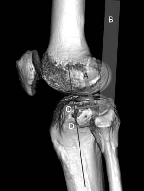
Table 1Analysis of Posterolateral Depression of Tibial Plateau according to Posterolateral Fragment, Fibular Fracture, Schatzker Type, Age, and Gender Differences
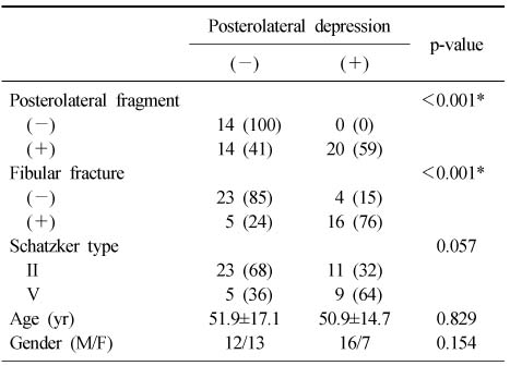
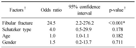
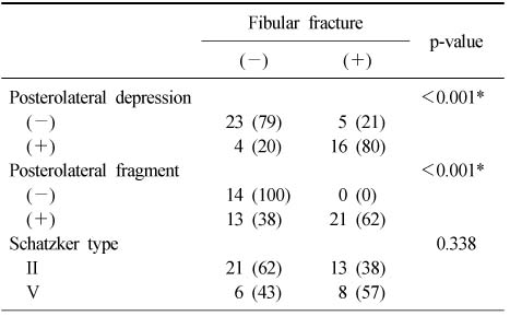
Figure & Data
REFERENCES
Citations
Citations to this article as recorded by 
- Current Concepts in Management of Tibia Plateau Fracture
Sang Hak Lee, Kang-Il Kim
Journal of the Korean Fracture Society.2014; 27(3): 245. CrossRef
 Cite
CiteAnalysis of Risk Factors for the Posterolateral Articular Depression and Status of Posterolateral Fragment in Lateral Condylar and Bicondylar Tibial Plateau Fractures with Joint Depression
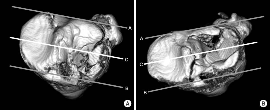

Fig. 1
Three demensional computed tomographys show the position of articular depression center in tibial plateau fractures. A line is the connecting line between the medial and lateral posterior cortex of the tibial plateau. B line is the parallel line to A line that crossing the anterior border of the lateral tibial condyle. C line is the bisecting line between lines A and B, which divide anterior and posterior depression. The position of joint depression is marked with an asterisk.
(A) Anterior depression of tibial plateau fracture.
(B) Posterior depression of tibial plateau fracture.
Fig. 2
Photograph shows the anterior-posterior length of the posterolateral fragment with three dimensional computed tomography. A: lateral collateral ligament, B: biceps femoris muscle, C: distance between the posterior margin of the posterior condyle and the anterior margin of the posterolateral fragment, D: distance between the anterior margin of the fibular head and the anterior margin of the posterolateral fragment.
Fig. 1
Fig. 2
Analysis of Risk Factors for the Posterolateral Articular Depression and Status of Posterolateral Fragment in Lateral Condylar and Bicondylar Tibial Plateau Fractures with Joint Depression
Analysis of Posterolateral Depression of Tibial Plateau according to Posterolateral Fragment, Fibular Fracture, Schatzker Type, Age, and Gender Differences
Values are presented as number (%) or mean±standard deviation. p-value using Fisher's exact test. M/F: Male/female. *Significant at p<0.05.
Multivariate Logistic Regression Analysis of Risk Factors for Posterolateral Articular Depression after Tibial Plateau Fractures
*Significant at p<0.05. †Factors were selected by stepwise method.
The Occurrence of Fibular Fracture according to Posterolateral Articular Depression of Tibial Plateau, Posterolateral Fragment, and Schatzker Types
Values are presented as number (%). p-value using Fisher's exact test. *Significant at p<0.05.
Table 1
Analysis of Posterolateral Depression of Tibial Plateau according to Posterolateral Fragment, Fibular Fracture, Schatzker Type, Age, and Gender Differences
Values are presented as number (%) or mean±standard deviation. p-value using Fisher's exact test. M/F: Male/female. *Significant at p<0.05.
Table 2
Multivariate Logistic Regression Analysis of Risk Factors for Posterolateral Articular Depression after Tibial Plateau Fractures
*Significant at p<0.05. †Factors were selected by stepwise method.
Table 3
The Occurrence of Fibular Fracture according to Posterolateral Articular Depression of Tibial Plateau, Posterolateral Fragment, and Schatzker Types
Values are presented as number (%). p-value using Fisher's exact test. *Significant at p<0.05.

