 E-submission
 TOTA
TOTA TOTS
TOTS
Articles
- Page Path
- HOME > J Musculoskelet Trauma > Volume 33(4); 2020 > Article
- Review Article Tendon Healing: A Review of Basic Science and Current Progress
- Young Woo Kwon, Pei Wei Wang, Jun-Ku Lee
-
Journal of Musculoskeletal Trauma 2020;33(4):227-237.
DOI: https://doi.org/10.12671/jkfs.2020.33.4.227
Published online: October 31, 2020
1Department of Orthopedic Surgery, Nowon Eulji Medical Center, Eulji University, Seoul, Korea
2Department of Orthopedic Surgery, Inje University Seoul Paik Hospital, Seoul, Korea
2Department of Orthopedic Surgery, Inje University Seoul Paik Hospital, Seoul, Korea
- 4,224 Views
- 140 Download
- 0 Crossref
- 0 Scopus
Abstract
The tendon connects the muscles to the bones and transmits the loads generated by the muscles to the bones to move the joints, support the joints, and provide stability to the joints. Approximately 30% of patients complaining of musculoskeletal pain are associated with tendon disease, and approximately 50% of musculoskeletal injuries are caused by a tendon injury. Despite this frequent treatment of tendon damage, studies on the basic biology that provide scientific evidence for treatment, such as development, tendon injury, and healing, are still very limited. This review first summarizes the classification and composition of the tendon identified so far, the surrounding tissue, and the blood supply to the tendon. The limitations of the tendon recovery process after a tendon injury are also discussed. Finally, this review examines ways to improve tendon recovery and the biological approaches and tissue engineering that have been currently studied. In conclusion, innovative progress in promoting tendon healing has not been achieved despite the many advances in the basic structure of the tendon, and the cell and regulatory molecular factors involved in tendon recovery. Biological approaches and tissue engineering, which have become a recent issue, have shown many possibilities for the recovery of damaged cases, but further research will be needed until clinical application.
J Korean Fract Soc. 2020 Oct;33(4):227-237. Korean.
Published online Oct 22, 2020.
https://doi.org/10.12671/jkfs.2020.33.4.227
Published online Oct 22, 2020.
https://doi.org/10.12671/jkfs.2020.33.4.227
Copyright © 2020 The Korean Fracture Society. All rights reserved.
Review
Tendon Healing: A Review of Basic Science and Current Progress

Abstract
The tendon connects the muscles to the bones and transmits the loads generated by the muscles to the bones to move the joints, support the joints, and provide stability to the joints. Approximately 30% of patients complaining of musculoskeletal pain are associated with tendon disease, and approximately 50% of musculoskeletal injuries are caused by a tendon injury. Despite this frequent treatment of tendon damage, studies on the basic biology that provide scientific evidence for treatment, such as development, tendon injury, and healing, are still very limited. This review first summarizes the classification and composition of the tendon identified so far, the surrounding tissue, and the blood supply to the tendon. The limitations of the tendon recovery process after a tendon injury are also discussed. Finally, this review examines ways to improve tendon recovery and the biological approaches and tissue engineering that have been currently studied. In conclusion, innovative progress in promoting tendon healing has not been achieved despite the many advances in the basic structure of the tendon, and the cell and regulatory molecular factors involved in tendon recovery. Biological approaches and tissue engineering, which have become a recent issue, have shown many possibilities for the recovery of damaged cases, but further research will be needed until clinical application.
Keywords
Tendon, Structure, Blood supply, Injury, Healing, Tissue engineering
Figures
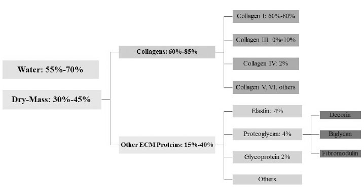
Fig. 1
Tendon composition. ECM: extracellular matrix.
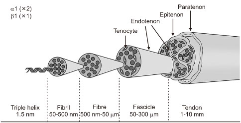
Fig. 2
Tendon structure.
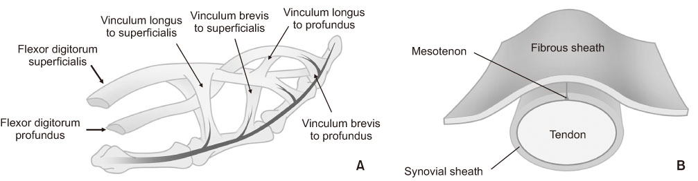
Fig. 3
Extrinsic direct blood supply through the Vincular system (A) and Mesotenon (B).
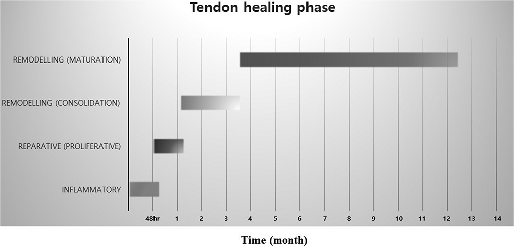
Fig. 4
Tendon repair process in humans. The healing of ruptured tendons passes through three main phases, inflammatory, proliferative, and remodeling phase, which contains the distinctive cell and molecular cascades. These phases overlap, and their duration depends upon the location and severity of the tendon injury.
Notes
Financial support:None.
Conflict of interests:None.
References
-
Sharma P, Maffulli N. Tendon injury and tendinopathy: healing and repair. J Bone Joint Surg Am 2005;87:187–202.
-
-
Bordoni B, Varacallo M. Anatomy, tendons. StatPearls [Internet]. Treasure Island (FL): StatPearls Publishing; 2020 Jan; [updated 2020 Jul 31]. [cited 2020 Jun 1].Available from: https://www.ncbi.nlm.nih.gov/books/NBK513237/.
-
-
Tozer S, Duprez D. Tendon and ligament: development, repair and disease. Birth Defects Res C Embryo Today 2005;75:226–236.
-
-
Wu F, Nerlich M, Docheva D. Tendon injuries: basic science and new repair proposals. EFORT Open Rev 2017;2:332–342.
-
-
Rees JD, Wilson AM, Wolman RL. Current concepts in the management of tendon disorders. Rheumatology (Oxford) 2006;45:508–521.
-
-
Bruns J, Kampen J, Kahrs J, Plitz W. Achilles tendon rupture: experimental results on spontaneous repair in a sheep-model. Knee Surg Sports Traumatol Arthrosc 2000;8:364–369.
-
-
Zafar MS, Mahmood A, Maffulli N. Basic science and clinical aspects of achilles tendinopathy. Sports Med Arthrosc Rev 2009;17:190–197.
-
-
Titan AL, Foster DS, Chang J, Longaker MT. Flexor tendon: development, healing, adhesion formation, and contributing growth factors. Plast Reconstr Surg 2019;144:639e–647e.
-
-
Benjamin M, Ralphs JR. The cell and developmental biology of tendons and ligaments. Int Rev Cytol 2000;196:85–130.
-
-
Schweitzer R, Chyung JH, Murtaugh LC, et al. Analysis of the tendon cell fate using Scleraxis, a specific marker for tendons and ligaments. Development 2001;128:3855–3866.
-
-
Lim WL, Liau LL, Ng MH, Chowdhury SR, Law JX. Current progress in tendon and ligament tissue engineering. Tissue Eng Regen Med 2019;16:549–571.
-
-
Leong NL, Kator JL, Clemens TL, James A, Enamoto-Iwamoto M, Jiang J. Tendon and ligament healing and current approaches to tendon and ligament regeneration. J Orthop Res 2020;38:7–12.
-
-
Lee WY, Lui PP, Rui YF. Hypoxia-mediated efficient expansion of human tendon-derived stem cells in vitro. Tissue Eng Part A 2012;18:484–498.
-
-
Mienaltowski MJ, Adams SM, Birk DE. Tendon proper- and peritenon-derived progenitor cells have unique tenogenic properties. Stem Cell Res Ther 2014;5:86
-
-
Mienaltowski MJ, Adams SM, Birk DE. Regional differences in stem cell/progenitor cell populations from the mouse achilles tendon. Tissue Eng Part A 2013;19:199–210.
-
-
Aparecida de Aro A, Vidal Bde C, Pimentel ER. Biochemical and anisotropical properties of tendons. Micron 2012;43:205–214.
-
-
Nelson E, Jones NEH, Brandt M. Biologics for tendon surgery. Clin Podiatr Med Surg 2020;37:601–608.
-
-
Sharma P, Maffulli N. Biology of tendon injury: healing, modeling and remodeling. J Musculoskelet Neuronal Interact 2006;6:181–190.
-
-
Taye N, Karoulias SZ, Hubmacher D. The “other” 15–40%: the role of non-collagenous extracellular matrix proteins and minor collagens in tendon. J Orthop Res 2020;38:23–35.
-
-
Frank CB, Shrive NG, Lo IKY, Hart DA. Form and function of tendon and ligament. In: Einhorn TA, O'Keefe RJ, Buckwalter JA, editors. Orthopaedic basic science: foundations of clinical practice. 3rd ed. Rosemont: American Academy of Orthopaedic Surgeons; 2007. pp. 191-222.
-
-
Ling SC, Chen CF, Wan RX. A study on the vascular supply of the supraspinatus tendon. Surg Radiol Anat 1990;12:161–165.
-
-
Schmidt-Rohlfing B, Graf J, Schneider U, Niethard FU. The blood supply of the Achilles tendon. Int Orthop 1992;16:29–31.
-
-
Clancy WG Jr, Narechania RG, Rosenberg TD, Gmeiner JG, Wisnefske DD, Lange TA. Anterior and posterior cruciate ligament reconstruction in rhesus monkeys. J Bone Joint Surg Am 1981;63:1270–1284.
-
-
Frey C, Shereff M, Greenidge N. Vascularity of the posterior tibial tendon. J Bone Joint Surg Am 1990;72:884–888.
-
-
Aström M. Laser Doppler flowmetry in the assessment of tendon blood flow. Scand J Med Sci Sports 2000;10:365–367.
-
-
Fenwick SA, Hazleman BL, Riley GP. The vasculature and its role in the damaged and healing tendon. Arthritis Res 2002;4:252–260.
-
-
Abrahamsson SO. Matrix metabolism and healing in the flexor tendon. Experimental studies on rabbit tendon. Scand J Plast Reconstr Surg Hand Surg Suppl 1991;23:1–51.
-
-
Molloy T, Wang Y, Murrell G. The roles of growth factors in tendon and ligament healing. Sports Med 2003;33:381–394.
-
-
Hogan MV, Bagayoko N, James R, Starnes T, Katz A, Chhabra AB. Tissue engineering solutions for tendon repair. J Am Acad Orthop Surg 2011;19:134–142.
-
-
Legrand A, Kaufman Y, Long C, Fox PM. Molecular biology of flexor tendon healing in relation to reduction of tendon adhesions. J Hand Surg Am 2017;42:722–726.
-
-
Takai S, Woo SL, Horibe S, Tung DK, Gelberman RH. The effects of frequency and duration of controlled passive mobilization on tendon healing. J Orthop Res 1991;9:705–713.
-
-
Maeda E, Shelton JC, Bader DL, Lee DA. Differential regulation of gene expression in isolated tendon fascicles exposed to cyclic tensile strain in vitro. J Appl Physiol 2009;106:506–512.
-
-
Boyer MI, Gelberman RH, Burns ME, Dinopoulos H, Hofem R, Silva MJ. Intrasynovial flexor tendon repair. An experimental study comparing low and high levels of in vivo force during rehabilitation in canines. J Bone Joint Surg Am 2001;83:891–899.
-
-
Diao E, Soejima O, Lotz J, Hariharan J. Randomized biomechanical study of zone II human flexor tendon repairs. J Hand Surg Am 1999;24:871–873.
-
-
Loutzenheiser TD, Harryman DT 2nd, Ziegler DW, Yung SW. Optimizing arthroscopic knots using braided or monofilament suture. Arthroscopy 1998;14:57–65.
-
-
Singer G, Ebramzadeh E, Jones NF, Meals R. Use of the Taguchi method for biomechanical comparison of flexor-tendon-repair techniques to allow immediate active flexion. A new method of analysis and optimization of technique to improve the quality of the repair. J Bone Joint Surg Am 1998;80:1498–1506.
-
-
Woo SL, Gelberman RH, Cobb NG, Amiel D, Lothringer K, Akeson WH. The importance of controlled passive mobilization on flexor tendon healing. A biomechanical study. Acta Orthop Scand 1981;52:615–622.
-
-
Boyer MI, Goldfarb CA, Gelberman RH. Recent progress in flexor tendon healing. The modulation of tendon healing with rehabilitation variables. J Hand Ther 2005;18:80–85.quiz 86.
-
-
Thomopoulos S, Williams GR, Soslowsky LJ. Tendon to bone healing: differences in biomechanical, structural, and compositional properties due to a range of activity levels. J Biomech Eng 2003;125:106–113.
-
-
Miller BS, Downie BK, Kohen RB, et al. When do rotator cuff repairs fail? Serial ultrasound examination after arthroscopic repair of large and massive rotator cuff tears. Am J Sports Med 2011;39:2064–2070.
-
-
Iannotti JP, Deutsch A, Green A, et al. Time to failure after rotator cuff repair: a prospective imaging study. J Bone Joint Surg Am 2013;95:965–971.
-
-
Liu CF, Aschbacher-Smith L, Barthelery NJ, Dyment N, Butler D, Wylie C. What we should know before using tissue engineering techniques to repair injured tendons: a developmental biology perspective. Tissue Eng Part B Rev 2011;17:165–176.
-
-
Rodriguez A, Anastassov GE, Lee H, Buchbinder D, Wettan H. Maxillary sinus augmentation with deproteinated bovine bone and platelet rich plasma with simultaneous insertion of endosseous implants. J Oral Maxillofac Surg 2003;61:157–163.
-
-
Hernigou P, Flouzat Lachaniette CH, Delambre J, et al. Biologic augmentation of rotator cuff repair with mesenchymal stem cells during arthroscopy improves healing and prevents further tears: a case-controlled study. Int Orthop 2014;38:1811–1818.
-
-
van den Boom NAC, Winters M, Haisma HJ, Moen MH. Efficacy of stem cell therapy for tendon disorders: a systematic review. Orthop J Sports Med 2020;8:2325967120915857
-
-
Barber FA, McGarry JE, Herbert MA, Anderson RB. A biomechanical study of Achilles tendon repair augmentation using GraftJacket matrix. Foot Ankle Int 2008;29:329–333.
-
-
Derwin KA, Baker AR, Spragg RK, Leigh DR, Iannotti JP. Commercial extracellular matrix scaffolds for rotator cuff tendon repair. Biomechanical, biochemical, and cellular properties. J Bone Joint Surg Am 2006;88:2665–2672.
-
-
Snyder SJ, Arnoczky SP, Bond JL, Dopirak R. Histologic evaluation of a biopsy specimen obtained 3 months after rotator cuff augmentation with GraftJacket Matrix. Arthroscopy 2009;25:329–333.
-
 Cite
Cite
