 E-submission
 TOTA
TOTA TOTS
TOTS
Articles
- Page Path
- HOME > J Musculoskelet Trauma > Volume 20(1); 2007 > Article
-
Case Report
- Old Atlantoaxial Rotary Subluxation Associated with High-riding Vertebral Arteries: Arthrodesis Using C1 Lateral Mass Screws and C2 Laminar Screws: A Case Report
- Kyeong Hwan Kim, M.D., Jin Sup Yeom, M.D., Kun Woo Park, M.D., Soon Woo Hong, M.D., Bong Soon Chang, M.D., Choon Ki Lee, M.D.
-
Journal of the Korean Fracture Society 2007;20(1):90-93.
DOI: https://doi.org/10.12671/jkfs.2007.20.1.90
Published online: June 14, 2016
Department of Orthopaedic Surgery, Seoul National University College of Medicine, Seoul, Korea.
- Address reprint requests to: Jin Sup Yeom, M.D. Department of Orthopaedic Surgery, Seoul National University Bundang Hospital, 300, Gumi-dong, Bundang-gu, Seongnam 463-707, Korea. Tel: 82-31-787-7190, Fax: 82-31-787-4056, ortho@hananet.net
Copyright © The Korean Fracture Society. All rights reserved
- 986 Views
- 0 Download
- 1 Crossref
Abstract
- To the best of our knowledge, there has been no domestic report on posterior atlantoaxial fusion with segmental screw fixation using C2 laminar screws and C1 lateral mass screws for atlantoaxial subluxation. We report the result of this operation performed in a patient with old atlantoaxial rotary subluxation who required posterior fusion. We chose this technique in this patient because wire fixation was not suitable due to osteoporosis, and transarticular screw fixation and use of C2 pedicle screws were not feasible due to the peculiar bony anatomy of the axis.
- 1. Fayyazi AH, An HS. Anterior/posterior cervical instrumentation. In: An HS, Jenis LG, editors. Complications of spine surgery. 1st ed. Philadelphia: Lippincott Williams & Wilkins; 2006. p. 65-66.
- 2. Goel A, Desai KI, Muzumdar DP. Atlantoaxial fixation using plate and screw method: a report of 160 treated patients. Neurosurgery, 2002;51:1351-1356.Article
- 3. Harms J, Melcher RP. Posterior C1-C2 fusion with polyaxial screw and rod fixation. Spine, 2001;26:2467-2471.Article
- 4. Leventhal MR. Fractures, dislocations, and fracture-dislocations of spine. In: Canale ST, editor. Campbell's operative orthopaedics. 10th ed. Philadelphia: Mosby Inc; 2003. p. 1617-1618.
- 5. Levine AM, Edwards CC. Traumatic lesions of the occipitoatlantoaxial complex. Clin Orthop Relat Res, 1989;239:53-68.Article
- 6. Wright NM. Posterior C2 fixation using bilateral, crossing C2 laminar screws: case series and technical note. J Spinal Disord Tech, 2004;17:158-162.
REFERENCES
Fig. 1
(A) Preoperative open mouth view shows obliteration of the right C1-2 facet joint (black arrowhead), which arouses suspicion of rotary subluxation or unilateral facet joint destruction.
(B) On lateral view, the C1-2 area does not seem cleanly, which might be a result of facet joint destruction.
(C) On bone scan, there is a hot uptake on the right C1-2 facet joint area.

Preoperative radiographs (A, B) and a bone scan image (C) are shown.

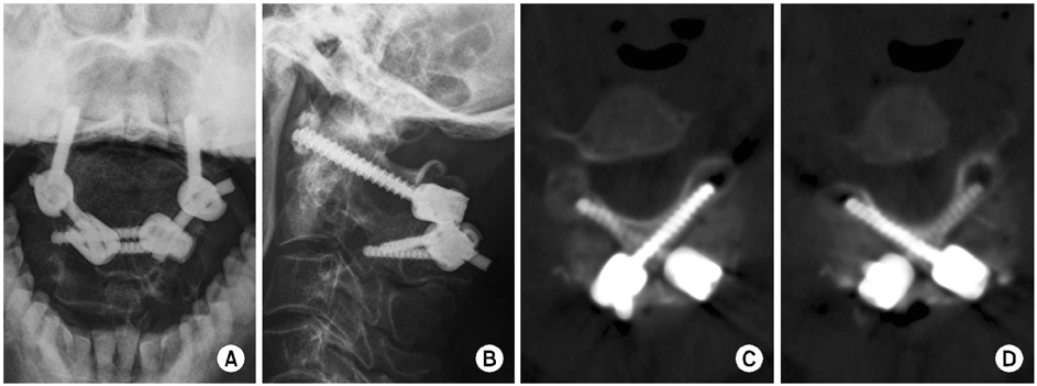
Figure & Data
REFERENCES
Citations
Citations to this article as recorded by 
- Indirect Decompression using Segmental Screw Fixation for Cervical Myelopathy Caused by C1-2 Subluxation - Technical Note -
Yoon Jong Kim, Kyeong Hwan Kim, Jong Hwa Won, Hak Jin Min, Ui Seong Yoon, Jin Sup Yeom
The Journal of the Korean Orthopaedic Association.2007; 42(6): 815. CrossRef
 Cite
CiteOld Atlantoaxial Rotary Subluxation Associated with High-riding Vertebral Arteries: Arthrodesis Using C1 Lateral Mass Screws and C2 Laminar Screws: A Case Report
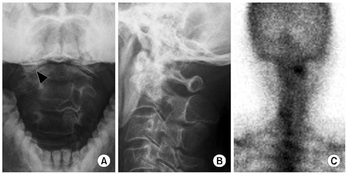
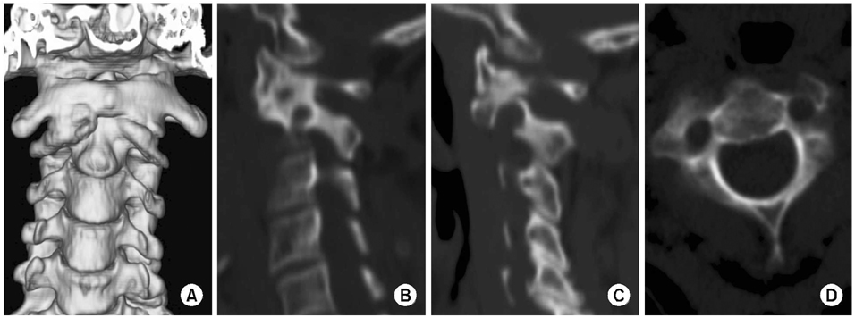

Fig. 1
Preoperative radiographs (A, B) and a bone scan image (C) are shown.
(A) Preoperative open mouth view shows obliteration of the right C1-2 facet joint (black arrowhead), which arouses suspicion of rotary subluxation or unilateral facet joint destruction.
(B) On lateral view, the C1-2 area does not seem cleanly, which might be a result of facet joint destruction.
(C) On bone scan, there is a hot uptake on the right C1-2 facet joint area.
Fig. 2
(A) Three-dimensional CT scan image shows rotary subluxation and severe osteophyte formation at the Rt C1-2 facet joint. (B, C) Sagittal reconstruction images show anomalous high-riding vertebral arteries on both sides. (D) An axial image shows small pedicles on both sides.
Fig. 3
Postoperative radiographs (A, B) and CT scan images (C, D) show satisfactory position of the screws.
Fig. 1
Fig. 2
Fig. 3
Old Atlantoaxial Rotary Subluxation Associated with High-riding Vertebral Arteries: Arthrodesis Using C1 Lateral Mass Screws and C2 Laminar Screws: A Case Report

