 E-submission
 TOTA
TOTA TOTS
TOTS
Articles
- Page Path
- HOME > J Musculoskelet Trauma > Volume 20(1); 2007 > Article
-
Case Report
- Hip Fracture-dislocation with Sciatic Nerve Palsy and Ipsilateral Femoral Shaft Open Fracture: A Case Report
- Kap Jung Kim, M.D., Ha Yong Kim, M.D., Sung Il Kang, M.D., Won Sik Choy, M.D.
-
Journal of the Korean Fracture Society 2007;20(1):94-98.
DOI: https://doi.org/10.12671/jkfs.2007.20.1.94
Published online: June 14, 2016
Department of Orthopedic Surgery, Eulji University College of Medicine, Daejeon, Korea.
- Address reprint requests to: Kap Jung Kim, M.D. Department of Orthopedic Surgery, Eulji University Hospital, 1306, Dunsan-dong, Seo-gu, Daejeon 302-120, Korea. Tel: 82-42-611-3278, Fax: 82-42-259-1289, oskkj@eulji.ac.kr
Copyright © The Korean Fracture Society. All rights reserved
- 1,312 Views
- 4 Download
Abstract
- The posterior dislocation of the hip accounts for about 85~90% of traumatic hip dislocations and high energy mechanisms such as traffic accidents may cause them. The traumatic dislocation and fracture-dislocation of the hip are true orthopedic emergencies and it should be considered that a femoral head has poor vascularity and the sciatic nerve is closely located to it. We report on one patient who went through the surgical treatment of the concomitant ipsilateral open fracture of the femoral shaft and hip fracture-dislocation accompanying sciatic nerve injury with the review of the literatures.
- 1. Brav EA. Traumatic dislocation of the hip. J Bone Joint Surg Am, 1962;44:1115-1134.Article
- 2. Carlsen AW, Lind J. Traumatic dislocation of the hip with ipsilateral fracture of the femur: a method of reduction. Injury, 1991;22:68-69.ArticlePubMed
- 3. Epstein HC. Posterior fracture-dislocations of the hip; longerm follow-up. J Bone Joint Surg Am, 1974;56:1103-1127.PubMed
- 4. Epstein HC, Wiss DA, Cozen L. Posterior fracture dislocation of the hip with fractures of the femoral head. Clin Orthop Relat Res, 1985;201:9-17.Article
- 5. Harper MC. Traumatic dislocation of the hip with ipsilateral femoral shaft fracture: a method of treatment. Injury, 1982;13:391-394.ArticlePubMed
- 6. Hillyard RF, Fox J. Sciatic nerve injuries associated with traumatic posterior hip dislocations. Am J Emerg Med, 2003;21:545-548.ArticlePubMed
- 7. Lyddon DW Jr, Hartman JT. Traumatic dislocation of the hip with ipsilateral femoral fracture. A case report. J Bone Joint Surg Am, 1971;53:1012-1016.PubMed
- 8. Schoenecker Pl, Manske PR, Sertl GO. Traumatic hip dislocation with ipsilateral femoral shaft fractures. Clin Orthop Relat Res, 1978;130:233-238.Article
- 9. Seddon H. Surgical disorders of the peripheral nerves. 1st ed. Baltimore: Williams and Wilkins Co; 1972. p. 299-314.
- 10. Watson Jones R. Fractures and joint injuries. 6th ed. New York: Churchill-Lvingstone; 1982. p. 885-934.
REFERENCES
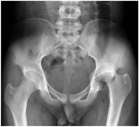
Fig. 2
(C) Medical photograph shows open wound of anterior thigh and tented soft tissue.

(A, B) Initial radiographs show femoral shaft fracture and displaced butterfly fragment.
Fig. 3

CT scan shows posterior hip dislocation and fracture fragment of posterior wall of acetabulum.
Fig. 4
(B) Intraoperative photograph shows reduced femoral head and posterior wall of acetabulaum.
(A) Intraoperative photograph shows dislocated femoral head, acetabular posterior wall fractured fragment and sciatic nerve. The continuity of sciatic nerve is intact but attenuated.
Fig. 5

Immediate postoperative radiograph shows reduction of hip dislocation and plate fixation of acetabular wall.
Fig. 6

Immediate postoperative radiographs show reduction of fractured femur and external fixator is applied.
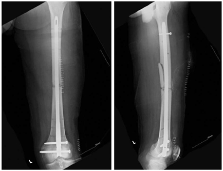
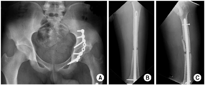
Figure & Data
REFERENCES
Citations
Citations to this article as recorded by 
 Cite
CiteHip Fracture-dislocation with Sciatic Nerve Palsy and Ipsilateral Femoral Shaft Open Fracture: A Case Report

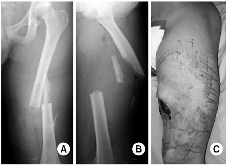
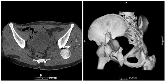

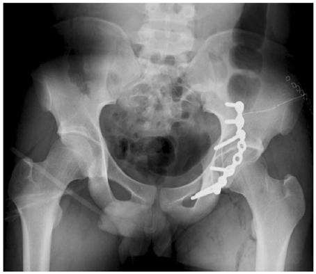
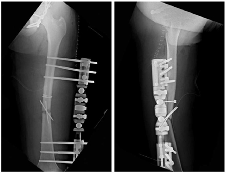


Fig. 1
Initial radiograph shows posterior hip dislocation and posterior wall fracture of acetabulum.
Fig. 2
(A, B) Initial radiographs show femoral shaft fracture and displaced butterfly fragment.
(C) Medical photograph shows open wound of anterior thigh and tented soft tissue.
Fig. 3
CT scan shows posterior hip dislocation and fracture fragment of posterior wall of acetabulum.
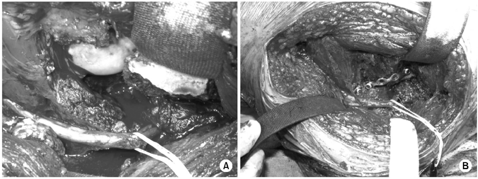
Fig. 4
(A) Intraoperative photograph shows dislocated femoral head, acetabular posterior wall fractured fragment and sciatic nerve. The continuity of sciatic nerve is intact but attenuated.
(B) Intraoperative photograph shows reduced femoral head and posterior wall of acetabulaum.
Fig. 5
Immediate postoperative radiograph shows reduction of hip dislocation and plate fixation of acetabular wall.
Fig. 6
Immediate postoperative radiographs show reduction of fractured femur and external fixator is applied.
Fig. 7
External fixation of femur is converted to internal fixation with retrograde intramedullary nail.
Fig. 8
Eight months follow up radiographs show complete union of posterior acetabular wall (A) but nonunion of femoral shaft (B, C).
Fig. 1
Fig. 2
Fig. 3
Fig. 4
Fig. 5
Fig. 6
Fig. 7
Fig. 8
Hip Fracture-dislocation with Sciatic Nerve Palsy and Ipsilateral Femoral Shaft Open Fracture: A Case Report

