 E-submission
 TOTA
TOTA TOTS
TOTS
Articles
- Page Path
- HOME > J Musculoskelet Trauma > Volume 25(3); 2012 > Article
-
Case Report
- Missed Variation of the Essex-Lopresti Injury Associated with Type-I Monteggia Equivalent Lesion: A Case Report
- Young Sung Kim, M.D., Phil Hyun Chung, M.D., Suk Kang, M.D., Ho Min Lee, M.D., Jong Pil Kim, M.D.
-
Journal of the Korean Fracture Society 2012;25(3):219-222.
DOI: https://doi.org/10.12671/jkfs.2012.25.3.219
Published online: July 16, 2012
Department of Orthopedic Surgery, College of Medicine, Dongguk University, Gyeongju, Korea.
- Address reprint requests to: Jong Pil Kim, M.D. Department of Orthopedic Surgery, Dongguk University Gyeongju Hospital, 87 Dongdae-ro, Gyeongju 780-350, Korea. Tel: 82-54-770-8225, Fax: 82-54-770-8378, kjpil@dongguk.ac.kr
• Received: August 16, 2011 • Revised: February 18, 2012 • Accepted: March 11, 2012
Copyright © 2012 The Korean Fracture Society
- 707 Views
- 3 Download
Abstract
- The authors report the case of a patient with the combination of a Type I Monteggia equivalent lesion and Essex-Lopresti injury. This combination of injury is very rare, and an associated distal radioulnar injury is often missed. We hope our experience illustrates the need to examine the wrist joint carefully and to be aware of the potential for distal radioulnar joint instability in all patients with type I Monteggia equivalent lesions.
- 1. Adams JE, Culp RW, Osterman AL. Interosseous membrane reconstruction for the Essex-Lopresti injury. J Hand Surg Am, 2010;35:129-136.ArticlePubMed
- 2. Cheung EV, Yao J. Monteggia fracture-dislocation associated with proximal and distal radioulnar joint instability. A case report. J Bone Joint Surg Am, 2009;91:950-954.PubMed
- 3. Edwards GS Jr, Jupiter JB. Radial head fractures with acute distal radioulnar dislocation. Essex-Lopresti revisited. Clin Orthop Relat Res, 1988;(234):61-69.
- 4. Eglseder WA, Hay M. Combined Essex-Lopresti and radial shaft fractures: case report. J Trauma, 1993;34:310-312.PubMed
- 5. Gong HS, Chung MS, Oh JH, Lee YH, Kim SH, Baek GH. Failure of the interosseous membrane to heal with immobilization, pinning of the distal radioulnar joint, and bipolar radial head replacement in a case of Essex-Lopresti injury: case report. J Hand Surg Am, 2010;35:976-980.ArticlePubMed
- 6. Jungbluth P, Frangen TM, Muhr G, Kälicke T. A primarily overlooked and incorrectly treated Essex-Lopresti injury: what can this lead to? Arch Orthop Trauma Surg, 2008;128:89-95.ArticlePubMedPDF
- 7. Lamas C, Castellanos J, Proubasta I, Dominguez E. Comminuted radial head fractures treated with pyrocarbon prosthetic replacement. Hand (N Y), 2011;6:27-33.ArticlePubMedPDF
- 8. Reckling FW. Unstable fracture-dislocations of the forearm (Monteggia and Galeazzi lesions). J Bone Joint Surg Am, 1982;64:857-863.ArticlePubMed
- 9. Rodriguez-Martin J, Pretell-Mazzini J, Vidal-Bujanda C. Unusual pattern of Essex-Lopresti injury with negative plain radiographs of the wrist: a case report and literature review. Hand Surg, 2010;15:41-45.Article
- 10. Smith AM, Urbanosky LR, Castle JA, Rushing JT, Ruch DS. Radius pull test: predictor of longitudinal forearm instability. J Bone Joint Surg Am, 2002;84-A:1970-1976.
REFERENCES
Fig. 1AP and lateral radiographs of the forearm show fracture of the ulnar shaft and displaced comminuted fracture of the radial neck.

Fig. 2Pre-operative AP and lateral radiographs of the forearm show a normal distal radioulnar joint at 6 weeks after injury.


Figure & Data
REFERENCES
Citations
Citations to this article as recorded by 
 Cite
CiteMissed Variation of the Essex-Lopresti Injury Associated with Type-I Monteggia Equivalent Lesion: A Case Report
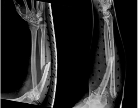
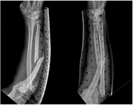

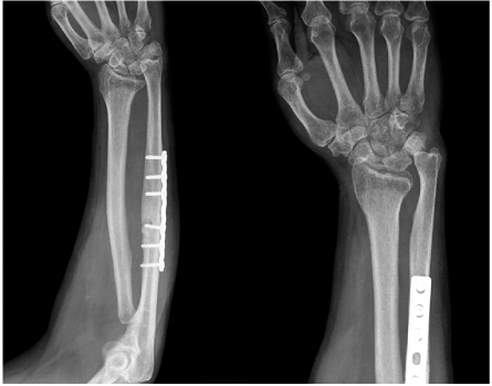
Fig. 1
AP and lateral radiographs of the forearm show fracture of the ulnar shaft and displaced comminuted fracture of the radial neck.
Fig. 2
Pre-operative AP and lateral radiographs of the forearm show a normal distal radioulnar joint at 6 weeks after injury.
Fig. 3
Immediate post-operative radiographs show a 6 mm migration of the radius proximally at the distal radioulnar joint.
Fig. 4
Post-operative one year radiographs show solid union of the ulnar shaft fracture but 15 mm migration of the radius proximally at the distal radioulnar joint and widening of the distal radioulnar gap.
Fig. 1
Fig. 2
Fig. 3
Fig. 4
Missed Variation of the Essex-Lopresti Injury Associated with Type-I Monteggia Equivalent Lesion: A Case Report

