 E-submission
 TOTA
TOTA TOTS
TOTS
Articles
- Page Path
- HOME > J Musculoskelet Trauma > Volume 26(4); 2013 > Article
-
Original Article
- The Comparison of Minimally Invasive Percutaneous Plate Osteosynthesis versus Open Plate Fixation in the Treatment of in the Distal Femur Fracture
- Seong-Jun Ahn, M.D., Suk-Woong Kang, M.D., Bu-Hwan Kim, M.D., Moo-Ho Song, M.D., Seong-Ho Yoo, M.D., Kwan-Taek Oh, M.D.
-
Journal of the Korean Fracture Society 2013;26(4):314-320.
DOI: https://doi.org/10.12671/jkfs.2013.26.4.314
Published online: October 18, 2013
Department of Orthopaedic Surgery, Daedong Hospital, Busan, Korea.
- Address reprint requests to: Suk-Woong Kang, M.D. Department of Orthopaedic Surgery, Daedong Hospital, 187 Chungnyeol-daero, Dongnae-gu, Busan 607-711, Korea. Tel: 82-51-554-8996, Fax: 82-51-553-7575, redmaniak@naver.com
• Received: May 2, 2013 • Revised: July 27, 2013 • Accepted: September 9, 2013
Copyright © 2013 The Korean Fracture Society. All rights reserved.
This is an Open Access article distributed under the terms of the Creative Commons Attribution Non-Commercial License (http://creativecommons.org/licenses/by-nc/3.0/) which permits unrestricted non-commercial use, distribution, and reproduction in any medium, provided the original work is properly cited.
- 1,278 Views
- 5 Download
- 1 Crossref
Abstract
-
Purpose
- To evaluate the efficacy of surgical treatment through retrospective comparison of minimally invasive percutaneous plate osteosynthesis (MIPPO) vs open plate fixation in the treatment of the distal femur fractures.
-
Materials and Methods
- Thirty-one patients with distal femur fractures from January 2002 to December 2010 were divided into two groups depending on the surgical method. Minimum follow up was 12 months. Group A consisted of 17 patients treated with MIPPO, and group B was comprised of 14 patients treated with open plate fixation. Clinical outcomes including operation time, transfusion rate, rehabilitation, range of motion, and interval change of postoperative C-reactive protein (CRP) were evaluated to assess postoperative inflammatory reaction, postoperative complications and clinical results with the use of Sanders criteria.
-
Results
- The operative time was 86/135 min and transfusion volume was 0.8/1.9 unit respectively. The postoperative 3-day and 7-day CRP were 7.4/1.5 mg% in group A and 10.3/2.4 mg% in group B, showing more minimal tissue injury and early recovery in group A. There were no significant differences in clinical results by Sanders criteria in both groups.
-
Conclusion
- Both MIPPO and open plate fixation for the treatment of distal femur fractures showed comparably good results. However, the MIPPO technique is superior to group B in view of minimal tissue injury and operation time and was proven to lessen the transfusion rate.
- 1. Arens S, Kraft C, Schlegel U, Printzen G, Perren SM, Hansis M. Susceptibility to local infection in biological internal fixation. Experimental study of open vs minimally invasive plate osteosynthesis in rabbits. Arch Orthop Trauma Surg, 1999;119:82-85.
- 2. Arneson TJ, Melton LJ 3rd, Lewallen DG, O'Fallon WM. Epidemiology of diaphyseal and distal femoral fractures in Rochester, Minnesota, 1965-1984. Clin Orthop Relat Res, 1988;(234):188-194.Article
- 3. Bradley GW, McKenna GB, Dunn HK, Daniels AU, Statton WO. Effects of flexural rigidity of plates on bone healing. J Bone Joint Surg Am, 1979;61:866-872.Article
- 4. Claes L, Heitemeyer U, Krischak G, Braun H, Hierholzer G. Fixation technique influences osteogenesis of comminuted fractures. Clin Orthop Relat Res, 1999;(365):221-229.Article
- 5. Colborn GL, Mattar SG, Taylor B, Skandalakis JE, Lumsden AB. The surgical anatomy of the deep femoral artery. Am Surg, 1995;61:336-346.
- 6. Court-Brown CM, Caesar B. Epidemiology of adult fractures: A review. Injury, 2006;37:691-697.Article
- 7. Danziger MB, Caucci D, Zecher SB, Segal D, Covall DJ. Treatment of intercondylar and supracondylar distal femur fractures using the GSH supracondylar nail. Am J Orthop (Belle Mead NJ), 1995;24:684-690.
- 8. Edwards CC. Staged reconstruction of complex open tibial fractures using Hoffmann external fixation. Clinical decisions and dilemmas. Clin Orthop Relat Res, 1983;(178):130-161.
- 9. Farouk O, Krettek C, Miclau T, Schandelmaier P, Guy P, Tscherne H. Minimally invasive plate osteosynthesis: does percutaneous plating disrupt femoral blood supply less than the traditional technique? J Orthop Trauma, 1999;13:401-406.Article
- 10. Farouk O, Krettek C, Miclau T, Schandelmaier P, Tscherne H. The topography of the perforating vessels of the deep femoral artery. Clin Orthop Relat Res, 1999;(368):255-259.Article
- 11. Gerber A, Ganz R. Combined internal and external osteosynthesis a biological approach to the treatment of complex fractures of the proximal tibia. Injury, 1998;29:Suppl 3. C22-C28.Article
- 12. Helfet DL, Shonnard PY, Levine D, Borrelli J Jr. Minimally invasive plate osteosynthesis of distal fractures of the tibia. Injury, 1997;28:Suppl 1. A42-A47.Article
- 13. Iannacone WM, Bennett FS, DeLong WG Jr, Born CT, Dalsey RM. Initial experience with the treatment of supracondylar femoral fractures using the supracondylar intramedullary nail: a preliminary report. J Orthop Trauma, 1994;8:322-327.Article
- 14. Johner R, Wruhs O. Classification of tibial shaft fractures and correlation with results after rigid internal fixation. Clin Orthop Relat Res, 1983;(178):7-25.Article
- 15. Kim SJ, Oh CW, Jeon IH, et al. Minimally invasive plate osteosynthesis for distal femoral fractures. J Korean Soc Fract, 2003;16:474-481.Article
- 16. Krettek C, Schandelmaier P, Miclau T, Tscherne H. Minimally invasive percutaneous plate osteosynthesis (MIPPO) using the DCS in proximal and distal femoral fractures. Injury, 1997;28:Suppl 1. A20-A30.Article
- 17. Lee HS, Kim JJ, Oh SK, Ahn HS. Treatment of distal tibial metaphyseal fracture using MIPPO technique. J Korean Foot Ankle Soc, 2004;8:166-170.
- 18. Martinet O, Cordey J, Harder Y, Maier A, Bühler M, Barraud GE. The epidemiology of fractures of the distal femur. Injury, 2000;31:Suppl 3. C62-C63.Article
- 19. McFerran MA, Smith SW, Boulas HJ, Schwartz HS. Complications encountered in the treatment of pilon fractures. J Orthop Trauma, 1992;6:195-200.Article
- 20. McKibbin B. The biology of fracture healing in long bones. J Bone Joint Surg Br, 1978;60:150-162.ArticlePDF
- 21. Ovadia DN, Beals RK. Fractures of the tibial plafond. J Bone Joint Surg Am, 1986;68:543-551.Article
- 22. Rhinelander FW. Tibial blood supply in relation to fracture healing. Clin Orthop Relat Res, 1974;(105):34-81.Article
- 23. Schütz M, Müller M, Krettek C, et al. Minimally invasive fracture stabilization of distal femoral fractures with the LISS: a prospective multicenter study. Results of a clinical study with special emphasis on difficult cases. Injury, 2001;32:Suppl 3. SC48-SC54.
- 24. Stoffel K, Dieter U, Stachowiak G, Gächter A, Kuster MS. Biomechanical testing of the LCP--how can stability in locked internal fixators be controlled? Injury, 2003;34:Suppl 2. B11-B19.Article
- 25. Tornetta P 3rd, Weiner L, Bergman M, et al. Pilon fractures: treatment with combined internal and external fixation. J Orthop Trauma, 1993;7:489-496.Article
- 26. Townsend CM, Beauchamp RD, Evers BM, Mattox KL. Sabiston textbook of surgery: the biological basis of modern surgical practice. 18th ed. Philadelphia: Saunders Elsevier; 2008. p. 130-135.
- 27. Weller S, Höntzsch D, Frigg R. Epiperiostal, percutaneous plate osteosynthesis. A new minimally invasive technique with reference to "biological osteosynthesis". Unfallchirurg, 1998;101:115-121.
- 28. Yang JY, Rhee KJ, Lee JK, Hwang DS, Shin HD, Lee HH. Minimally invasive percutaneous plate osteosynthesis for distal tibial shaft fracture. J Korean Soc Fract, 2002;15:286-291.Article
REFERENCES
Fig. 1
(A) A 66-year-old woman, AO/OTA classification 33-C1.
(B) Radigraphs show a postoperative state which was treated by the minimally invasive percutaneous plate osteosynthesis technique.
(C) The last follow up radiograph shows complete bone union.
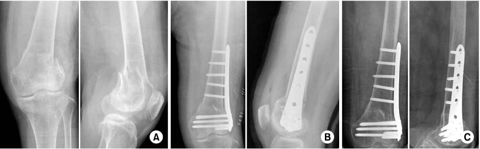
Fig. 2
(A) A 72-year-old woman, AO/OTA classification 33-A3.
(B) Radigraphs show a postoperative state which was treated by open reduced and internal fixation.
(C) The last follow up radiograph shows complete bone union.

Fig. 3The postoperative CRP changes in group A and B, showing more minimal tissue injury and early recovery. CRP: C-reactive protein, MIPPO: Minimally invasive percutaneous plate osteosynthesis.

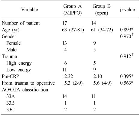
Figure & Data
REFERENCES
Citations
Citations to this article as recorded by 
- Usefulness of Reduction and Internal Fixation Using a 2.4 mm Hand Plating System in Type AO 33-A3 Distal Femur Fracture: Technical Note
Bong-Ju Lee, Ja-Yeong Yoon, Seungha Woo
Journal of the Korean Fracture Society.2023; 36(1): 25. CrossRef
 Cite
CiteThe Comparison of Minimally Invasive Percutaneous Plate Osteosynthesis versus Open Plate Fixation in the Treatment of in the Distal Femur Fracture

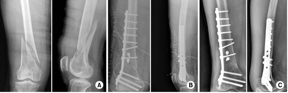
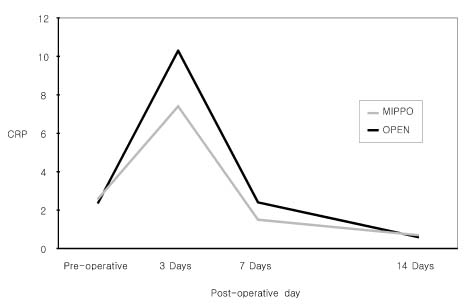
Fig. 1
(A) A 66-year-old woman, AO/OTA classification 33-C1.
(B) Radigraphs show a postoperative state which was treated by the minimally invasive percutaneous plate osteosynthesis technique.
(C) The last follow up radiograph shows complete bone union.
Fig. 2
(A) A 72-year-old woman, AO/OTA classification 33-A3.
(B) Radigraphs show a postoperative state which was treated by open reduced and internal fixation.
(C) The last follow up radiograph shows complete bone union.
Fig. 3
The postoperative CRP changes in group A and B, showing more minimal tissue injury and early recovery. CRP: C-reactive protein, MIPPO: Minimally invasive percutaneous plate osteosynthesis.
Fig. 1
Fig. 2
Fig. 3
The Comparison of Minimally Invasive Percutaneous Plate Osteosynthesis versus Open Plate Fixation in the Treatment of in the Distal Femur Fracture
Demographic Data, AO/OTA Classification
Values are presented as number or mean (range). MIPPO: Minimally invasive percutaneous plate osteosynthesis, Pre-CRP: Preoperative C-reactive protein. *Mann-Whitney U test, †Fisher's exact test.
Comparison of the Operative Time and Transfusion
Values are presented as mean (range) or % (number) or average unit. MIPPO: Minimally invasive percutaneous plate osteosynthesis. *Mann-Whitney U test, †Fisher's exact test.
Radiologic Result and Rehabilitation
Values are presented as mean (range). MIPPO: Minimally invasive percutaneous plate osteosynthesis, ROM: Range of motion. *Mann-Whitney U test.
Table 1
Demographic Data, AO/OTA Classification
Values are presented as number or mean (range). MIPPO: Minimally invasive percutaneous plate osteosynthesis, Pre-CRP: Preoperative C-reactive protein. *Mann-Whitney U test, †Fisher's exact test.
Table 2
Comparison of the Operative Time and Transfusion
Values are presented as mean (range) or % (number) or average unit. MIPPO: Minimally invasive percutaneous plate osteosynthesis. *Mann-Whitney U test, †Fisher's exact test.
Table 3
Radiologic Result and Rehabilitation
Values are presented as mean (range). MIPPO: Minimally invasive percutaneous plate osteosynthesis, ROM: Range of motion. *Mann-Whitney U test.

