 E-submission
 TOTA
TOTA TOTS
TOTS
Articles
- Page Path
- HOME > J Musculoskelet Trauma > Volume 20(2); 2007 > Article
-
Case Report
- Fatal Hemothorax Following Percutaneous Vertebroplasty: A Case Report
- Hee-Gon Park, M.D., Joo-Hong Lee, M.D.
-
Journal of the Korean Fracture Society 2007;20(2):202-205.
DOI: https://doi.org/10.12671/jkfs.2007.20.2.202
Published online: June 14, 2016
Department of Orthopaedic Surgery, College of Medicine, Dankook University, Cheonan, Korea.
- Address reprint requests to: Hee-Gon Park, M.D. Department of Orthopaedic Surgery, Dankook University Hospital, 16-5, Anseo-dong, Cheonan 330-715, Korea. Tel: 82-41-550-3950, Fax: 82-41-556-3238, heegon@chol.com
Copyright © The Korean Fracture Society. All rights reserved
- 642 Views
- 4 Download
Abstract
- Overall, the percutaneous vertebroplasty has low complication rate. Nevertheless, severe complications can occur. The majority of these are related to cement leakage. The cement migration through perivertebral venous system can lead to fatal complication. We present a case of death by hemothorax due to cement leakage following percutaneous vertebroplasty with literature review.
- 1. Bernhard J, Heini PF, Villiger PM. Asymptomatic diffuse pulmonary embolism caused by acrylic cement: an unusual complication of percutaneous vertebroplasty. Ann Rheum Dis, 2004;62:85-86.Article
- 2. Chen HL, Wong CS, Ho ST, Chang FL, Hsu CH, Wu CT. A lethal pulmonary embolism during percutaneous vertebroplasty. Anesth Analg, 2002;95:1060-1062.Article
- 3. Jensen ME, Evans AJ, Mathis JM, Kallmes DF, Cloft HJ, Dion JE. Percutaneous polymethylmethacrylate vertebroplasty in the treatment of osteoporotic vertebral body compression fracture: technical aspects. AJNR Am J neuroradiol, 1997;18:1897-1904.
- 4. Lee BJ, Lee SR, Yoo TY. Paraplegia as a complication of percutaneous vertebroplasty with polymethylm-ethacrylate: a case report. Spine (Phila Pa 1976), 2002;27:E419-E422.
- 5. Monticelli F, Meyer HJ, Tutsch-Bauer E. Fatal pulmonary cement embolism following percutaneous vertebroplasty (PVP). Forensic Sci Int, 2005;149:35-38.Article
- 6. Padovani B, Kasriel O, Brunner P, Peretti-Viton P. Pulmonary embolism caused by acrylic cement: a rare complication of percutaneous vertebroplasty. AJNR Am J Neuroradiol, 1999;20:375-377.
- 7. Pahuja K, Chand K. Fatal embolism associated with polymethyl methacrylate bone cement. Int Surg, 1976;61:19-22.
- 8. Tozzi P, Abdelmoumene Y, Corno AF, Gersbach PA, Hoogewoud HM, von Segesser LK. Management of pulmonary embolism during acrylic vertebroplasty. Ann Thorac Surg, 2002;74:1706-1708.Article
- 9. Wick MR, Ritter JH, Schuller D. Ruptured pulmonary infarction: a rare, fatal complication of thromboembolic disease. Mayo Clin Proc, 2000;75(6):639-642.
REFERENCES


Figure & Data
REFERENCES
Citations
Citations to this article as recorded by 
 Cite
CiteFatal Hemothorax Following Percutaneous Vertebroplasty: A Case Report
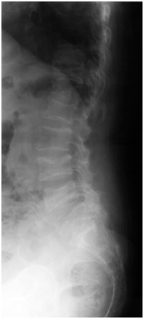
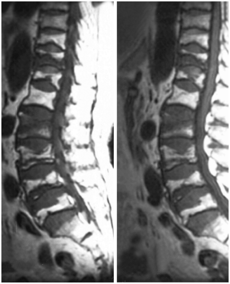
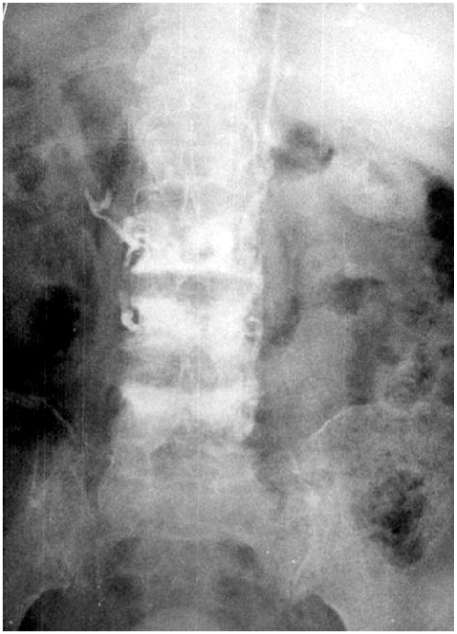
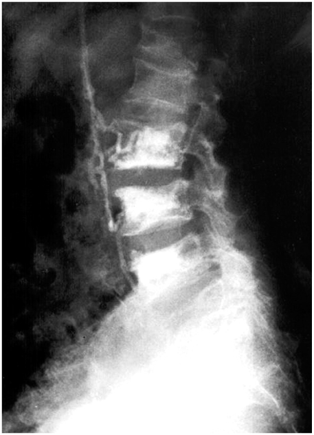
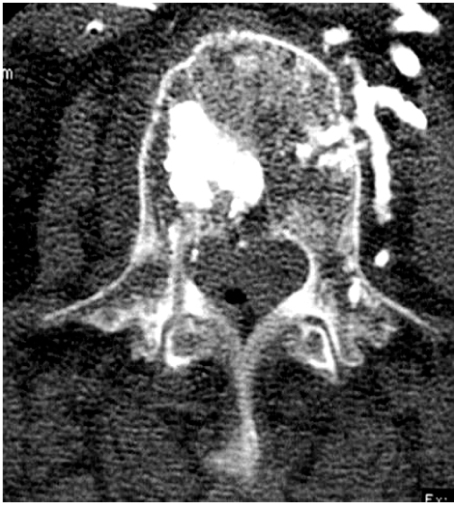
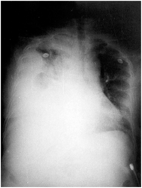
Fig. 1
An 80-year-old female had osteoporotic compression fracture of 11th and 12th thoracic vertebrae, 2nd, 3rd, 4th, and 5th lumbar vertebrae on the lateral. roenterogram.
Fig. 2
The T1WI magnetic resonance imaging (MRI) showing low signal on compression fracture.
Fig. 3
The anteroposterior roentgenogram showing leakage of PMMA along the vessel.
Fig. 4
The lateral roentgenogram showing leakage of PMMA along the vessel.
Fig. 5
Computed tomography (CT) showing leakage of PMMA.
Fig. 6
She had a hemothorax in 6th day after vertebroplasty.
Fig. 1
Fig. 2
Fig. 3
Fig. 4
Fig. 5
Fig. 6
Fatal Hemothorax Following Percutaneous Vertebroplasty: A Case Report

