 E-submission
 TOTA
TOTA TOTS
TOTS
Articles
- Page Path
- HOME > J Musculoskelet Trauma > Volume 21(2); 2008 > Article
-
Original Article
- The Comparison of LC-DCP versus LCP Fixation in the Plate Augmentation for the Nonunion of Femur Shaft Fractures after Intramedullary Nail Fixation
- Se Dong Kim, M.D., Oog Jin Sohn, M.D., Byung Hoon Kwack, M.D.
-
Journal of the Korean Fracture Society 2008;21(2):117-123.
DOI: https://doi.org/10.12671/jkfs.2008.21.2.117
Published online: April 30, 2008
Department of Orthopaedic Surgery, Yeungnam University Hospital, Daegu, Korea.
- Address reprint requests to: Oog Jin Sohn, M.D. Department of Orthopedic Surgery, Yeungnam University Hospital, 317-1, Daemyeong 5-dong, Nam-gu, Daegu 705-717, Korea. Tel: 82-53-620-3640, Fax: 82-53-628-4020, ossoj@med.yu.ac.kr
Copyright © 2008 The Korean Fracture Society. All rights reserved.
This is an Open Access article distributed under the terms of the Creative Commons Attribution Non-Commercial License (http://creativecommons.org/licenses/by-nc/3.0/) which permits unrestricted non-commercial use, distribution, and reproduction in any medium, provided the original work is properly cited.
- 2,219 Views
- 19 Download
- 3 Crossref
Abstract
-
Purpose
- The purpose of this study was to evaluate the efficacy of the surgical treatment through the comparison of LC-DCP (Limited Contact-Dynamic Compression Plate) versus LCP (Locking Compression Plate) fixation in the plate augmentation for the nonunion of femur shaft fractures after intramedullary nail fixation.
-
Materials and Methods
- Twenty-four patients with the nonunion of femur shaft fractures after intramedullary nail fixation who underwent plate augmentation were evaluated from Mar. 2001 to Sept. 2005. The group with LC-DCP augmentation was done bicortical screw fixation and the group with LCP was done monocortical fixation.
-
Results
- There was one case of nail breakage in LC-DCP group, but sound bony union were achieved uneventfully in all the cases of both group. LCP fixation was slightly superior to LC-DCP fixation in view of the bony union time, operating time, postoperative Hb down, amount of postoperative transfusion, but there was no statistical difference (p>0.05).
-
Conclusion
- We got the satisfactory results after monocortical LCP augmentation as well as bicortical LC-DCP fixation and have concluded that monocortical LCP fixation was an effective treatment option for nonunion of femur shaft fracture occurred after Intrmedullary nail fixation.
- 1. Bucholz RW, Heckman JD, Court-Brown C. Rockwood and Green's fractures in adults. 6th ed. Philadelphia: Lippincott-Williams & Wilkins Pb; 2006. p. 1845-1914.
- 2. Cho HO, Kwak KD, Kim JJ, Sohn SM, Kang CH, Lee HJ. Augmentative plate fixation for femoral nonunion after intramedullary nailing. J Korean Soc Fract, 2000;13:825-831.Article
- 3. Choi YS, Kim KS. Plate augmentation leaving the nail in situ and bone grafting for non-union of femoral shaft fractures. Int Orthop, 2005;29:287-290.ArticlePubMedPMCPDF
- 4. Gautier E, Sommer C. Guidelines for the clinical application of the LCP. Injury, 2003;34:Suppl 2. B63-B76.ArticlePubMed
- 5. Frigg R. Locking compression plate (LCP). An osteosynthesis plate based on the dynamic compression plate and the point contact fixator (PC-Fix). Injury, 2001;32:Suppl 2. 63-66.ArticlePubMed
- 6. Frigg R, Appenzeller A, Christensen R, Frenk A, Gilbert S, Schavan R. The development of the distal femur less invasive stabilization system (LISS). Injury, 2001;32:Suppl 3. SC24-SC31.ArticlePubMed
- 7. Gautier E, Sommer C. Guidelines for the clinical application of the LCP. Injury, 2003;34:Suppl 2. B63-B76.ArticlePubMed
- 9. Hak DJ, Lee SS, Goulet JA. Sucess of exchange reamed intramedullary nailing for femoral shaft nonunion or delayed union. J Orthop Trauma, 2000;14:178-182.PubMed
- 10. Helm AT, Karski MT, Parsons SJ, Sampath JS, Bale RS. A strategy for reducing blood-transfusion requirements in elective orthopaedic surgery. Audit of an algorithm for arthroplasty of the lower limb. J Bone Joint Surg Br, 2003;85:484-489.PubMed
- 11. Kim KS, Kim JO, Jung HG, Jung BO. Plate augmentation for the nonunion of femur shaft fractures after interlocking intramedullary nail fixation. J Korean Soc Fract, 1999;12:21-27.Article
- 12. Korner J, Lill H, Muller LP, Rommens PM, Schneider E, Linke B. The LCP-concept in the operative treatment of distal humerus fractures-biological, biomechanical and surgical aspects. Injury, 2003;34:Suppl 2. B20-B30.ArticlePubMed
- 13. Lee KH, Kim HM, Moon CW, Kim YS, Nam WS. Augmentation plate fixation for the management of long-bone nonunion after intramedullary nailing. J Korean Fract Soc, 2004;17:265-270.Article
- 14. Miller ME, Davis ML, MacClean CR, Davis JG, Smith BL, Humphries JR. Radiation exposure associated risks to operating-room personnel during use of Fluorscopic guidance for selected orthopaedic surgical procedures. J Bone Joint Surg Am, 1983;65:1-4.PubMed
- 15. Ring D, Jupiter JB, Sanders RA. Complex nonunion of fractures of the femoral shaft treated by wave-plate osteosynthesis. . J Bone Joint Surg Br, 1997;79:289-294.ArticlePubMedPDF
- 16. Schütz M, Sudkamp NP. Revolution in plate osteosythesis: new internal fixator systems. J Orthop Sci, 2003;8:252-258.PubMed
- 17. Sommer C. Locking compression plate. Injury, 2003;34:B4-B5.PubMed
- 18. Sommer C, Gautier E, Müller M, Helfet DL, Wagner M. First clinical results of the locking compression plate (LCP). Injury, 2003;34:Suppl 2. B43-B54.ArticlePubMed
- 19. Stoffel K, Dieter U, Stachowiak G, Gächter A, Kuster MS. Biomechanical testing of the LCP-how can stability in locked internal fixators be controlled? Injury, 2003;34:Suppl 2. B11-B19.ArticlePubMed
- 20. Taylor LW. Principles of treatment of fractures and nonunion of the shaft of femur. J Bone Joint Surg Am, 1963;45:191-198.
- 21. Tarr RR, Wiss DA. The mechanics and biology of intramedullary fracture fixation. Clin Orthop Relat Res, 1986;212:10-17.Article
- 22. Templeman D, Thomas M, Varecka T, Kyle R. Exchange reamed intramedullary nailing for delayed union and nonunion of the tibia. Clin Orthop Relat Res, 1995;315:169-175.Article
- 23. Ueng SW, Chao EK, Lee SS, Shih CH. Augmentative plate fixation for the management of femoral nonunion after intramedullary nailing. J Trauma, 1997;43:640-644.ArticlePubMed
- 24. Wagner M. General principles for the clinical use of the LCP. Injury, 2003;34:Suppl 2. B31-B42.ArticlePubMed
- 25. Weber BG, Brunner C. The treatment of nonunion without electrical stimulation. Clin Orthop Relat Res, 1981;161:24-32.
- 26. Winquist RA, Hansen ST Jr. Comminuted fractures of the femoral shaft treated by intramedullary nailing. Orthop Clin North Am, 1980;11:633-648.ArticlePubMed
- 27. Wu CC, Shih CH. Distal femoral nonunion treated with interlocking nailing. J Trauma, 1991;31:1659-1662.ArticlePubMed
- 28. Wu CC, Shih CH. Treatment of 84 cases of femoral nonunion. Acta Orthop Scand, 1992;63:57-60.ArticlePubMed
REFERENCES
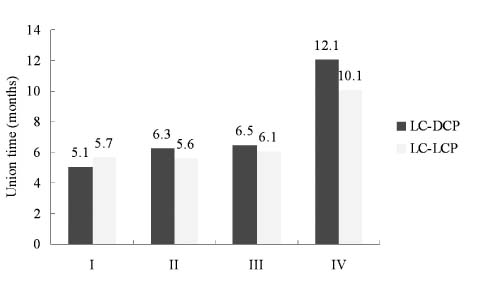
Fig. 2
(C, D) LC-DCP plate augmentation with autogenous iliac bone graft was done to the fracture site to counter the rotational instability leaving the intramedullary nail in situ.
(E, F) At 5.7 months after osteosynthesis, radiograph shows bone union.

(A, B) Radiograph taken at 10 months after intramedullary nailing for femoral shaft frature, shows hypertrophic nonunion.
Fig. 3
(C, D) LCP plate augmentation with autogenous iliac bone graft was done to the fracture site to counter the rotational instability leaving the intramedullary nail in situ.
(E, F) At 7.6 months after osteosynthesis, radiograph shows bone union.

(A, B) Radiograph taken at 12.5 months after intramedullary nailing for femoral shaft frature, shows oligotrophic nonunion.
Fig. 4
(C, D) LC-DCP plate augmentation with autogenous iliac bone graft was done to the fracture site to counter the rotational instability leaving the intramedullary nail in situ
(E, F) At 4 months after osteosynthesis, intramedullary nail was broken (arrow).
(G, H) At 8 months after osteosynthesis, radiograph shows bone union.
(A, B) Radiograph taken at 13.8 months after intramedullary nailing for femoral shaft frature, shows hypertrophic nonunion.
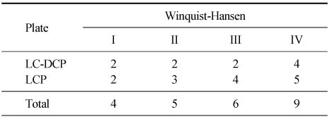
Figure & Data
REFERENCES
Citations
Citations to this article as recorded by 
- Delayed Union and Nonunion: Current Concepts, Prevention, and Correction: A Review
Kristin M. Bowers, David E. Anderson
Bioengineering.2024; 11(6): 525. CrossRef - RETRACTED ARTICLE: An experimental study on stress-shielding effects of locked compression plates in fixing intact dog femur
Xinwen Zhao, Wensen Jing, Zhe Yun, Xun Tong, Zhao Li, Jiajia Yu, Yaohui Zhang, Yabin Zhang, Zhixue Wang, Yanhua Wen, Heping Cai, Jun Wang, Baoan Ma, Haien Zhao
Journal of Orthopaedic Surgery and Research.2021;[Epub] CrossRef - The Treatment of IM Nailing of Femoral Shaft Fracture: Piriformis Fossa versus Trochanteric Entry Portal
Hyun Kook Youn, Oog Jin Shon, Dong Sung Han
Journal of the Korean Fracture Society.2008; 21(3): 200. CrossRef
 Cite
CiteThe Comparison of LC-DCP versus LCP Fixation in the Plate Augmentation for the Nonunion of Femur Shaft Fractures after Intramedullary Nail Fixation

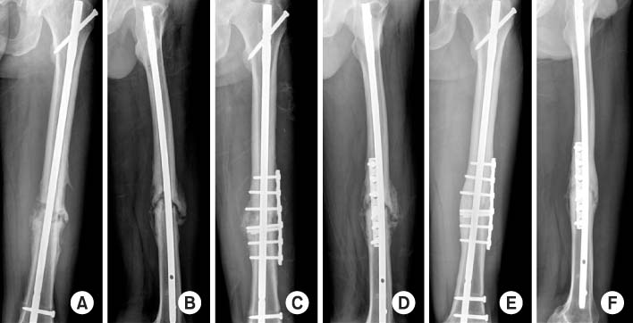
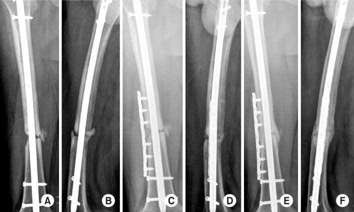

Fig. 1
Correlation with bony union time and initial fracture pattern.
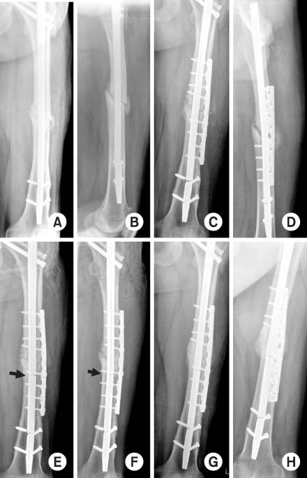
Fig. 2
(A, B) Radiograph taken at 10 months after intramedullary nailing for femoral shaft frature, shows hypertrophic nonunion.
(C, D) LC-DCP plate augmentation with autogenous iliac bone graft was done to the fracture site to counter the rotational instability leaving the intramedullary nail in situ.
(E, F) At 5.7 months after osteosynthesis, radiograph shows bone union.
Fig. 3
(A, B) Radiograph taken at 12.5 months after intramedullary nailing for femoral shaft frature, shows oligotrophic nonunion.
(C, D) LCP plate augmentation with autogenous iliac bone graft was done to the fracture site to counter the rotational instability leaving the intramedullary nail in situ.
(E, F) At 7.6 months after osteosynthesis, radiograph shows bone union.
Fig. 4
(A, B) Radiograph taken at 13.8 months after intramedullary nailing for femoral shaft frature, shows hypertrophic nonunion.
(C, D) LC-DCP plate augmentation with autogenous iliac bone graft was done to the fracture site to counter the rotational instability leaving the intramedullary nail in situ
(E, F) At 4 months after osteosynthesis, intramedullary nail was broken (arrow).
(G, H) At 8 months after osteosynthesis, radiograph shows bone union.
Fig. 1
Fig. 2
Fig. 3
Fig. 4
The Comparison of LC-DCP versus LCP Fixation in the Plate Augmentation for the Nonunion of Femur Shaft Fractures after Intramedullary Nail Fixation
Initial fracture pattern by Winquist-Hansen classification
Table 1
Initial fracture pattern by Winquist-Hansen classification

