 E-submission
 TOTA
TOTA TOTS
TOTS
Articles
- Page Path
- HOME > J Musculoskelet Trauma > Volume 21(2); 2008 > Article
-
Original Article
- Cerclage Wiring in Internal Fixation of Displaced Acetabular Fractures
- Chong-Kwan Kim, M.D., Jin-Woo Jin, M.D., Jong-Ho Yoon, M.D., Sung-Won Jung, M.D., Jung-Wook Peang, M.D.
-
Journal of the Korean Fracture Society 2008;21(2):95-102.
DOI: https://doi.org/10.12671/jkfs.2008.21.2.95
Published online: April 30, 2008
Department of Orthopedic Surgery, Masan Samsung Hospital, Sungkyunkwan University School of Medicine, Masan, Korea.
- Address reprint requests to: Jin-Woo Jin, M.D. Department of Orthopedic Surgery, Masan Samsung Hospital, 50, Hapseong 2-dong, Masan 630-723, Korea. Tel: 82-55-290-6036, Fax: 82-55-290-6888, ortho_ss@yahoo.co.kr
Copyright © 2008 The Korean Fracture Society. All rights reserved.
This is an Open Access article distributed under the terms of the Creative Commons Attribution Non-Commercial License (http://creativecommons.org/licenses/by-nc/3.0/) which permits unrestricted non-commercial use, distribution, and reproduction in any medium, provided the original work is properly cited.
- 800 Views
- 5 Download
- 2 Crossref
Abstract
-
Purpose
- To evaluate the usefulness of wire fixation in displaced acetabular fractures.
-
Materials and Methods
- From January 2000 to December 2005, 19 cases of displaced acetabular fracture were treated with wire fixation. According to Letournel's classification there were 9 both column fracture, 5 transverse fracture, 3 anterior column with posterior hemitransverse and 2 T-type fracture. Only wire fixation in 13 cases and wire with plate or wire with screw fixation in 6 cases.
-
Results
- We evaluate the accuracy of reduction by Matta' criteria, anatomical reduction in 12 cases, incomplete reduction in 4 cases, poor reduction in 2 cases and surgical secondary congruence in 1 case. The clinical results showed excellent in 12 cases, good in 4 cases, fair in 2 cases and poor in 1 case. The radiological results showed excellent in 10 cases, good in 4 cases, fair in 3 cases and poor in 2 cases. There were 4 cases of complication; wound infection in 1case, post-traumatic arthritis in 1 case and heterotopic ossification in 2 cases.
-
Conclusion
- The cerclage wiring is a preferable method in internal fixation of displaced acetabular fractures that can facilitate reduction and achieve stable fixation.
- 1. Chang JK, Gill SS, Zura RD, Krause WR, Wang GJ. Comparative strength of three methods of transverse acetabular fractures. Clin Orthop Relat Res, 2001;392:433-441.
- 2. Epstein HC. Posterior fracture-dislocation of the hip; long-term follow-up. J Bone Joint Surg Am, 1974;79:1103-1127.
- 3. Goulet JA, Bray TJ. Complex acetabular fractures. Clin Orthop Relat Res, 1989;240:9-20.Article
- 4. Judet R, Judet J, Letournel E. Fracture of acetabulum: classification and surgical approach for open reduction. Preliminary report. J Bone Joint Surg Am, 1964;46:1615-1646.
- 5. Kang CS, Min BW. Cable fixation in displaced fracture of the acetabulum: 21 patients followed for 2-8 years. Acta Orthop Scand, 2002;73:619-624.
- 6. Kang CS, Min BW, Song KS, Kang CH, Park JW. Cable fixation method for displaced acetabular fracture. J Korean Soc Fract, 1996;9:574-582.Article
- 7. Kim SK, Park JH, Park JW, Hong JS, Kim JH. Significance of anatomic reduction in acetabular fracture. J Korean Soc Fract, 2000;13:724-732.Article
- 8. Letournel E. The results of acetabular fractures treated surgically: 21 years experience, In: Proceedings of the seventh open scientific meeting for the hip society; St Louis, CV Mosby Co; 1979. p. 42-85.
- 9. Letournel E. Long term results in the surgical treatment of acetabular fractures, In: Presented at the annual meeting of AAOS; San Francisco, CA. 1993.
- 10. Letournel E, Judet R. Fracture of the acetabulum. 2th ed. Berlin, Germany: Springer-Verlag; 1993.
- 11. Matta JM, Merrit PO. Displaced acetabular fractures. Clin Orthop Relat Res, 1988;230:83-97.Article
- 12. Matta JM. Fracture of the acetabulum. Accuraly of reduction and clinical results in patients managed operatively within three weeks after injury. J Bone Joint Surg Am, 1996;78:1632-1645.
- 13. Mayo KA. Open reduction and internal fixation of fractures of the acetabulum: results in 163 fractures. Clin Orthop Relat Res, 1997;305:31-37.Article
- 14. Rockwood and Green. Fracture of the acetabulum. Fracture in adult. 5th ed. Lippincott Williams & Wilkins; 2001. p. 1513-1545.
- 15. Rowe CR, Lowell TD. Prognosis of fractures of the acetabulum. J Bone Joint Surg Am, 1961;43:30-59.Article
- 16. Stöckle U, Hoffman R, Nittinger M, Südkamp NP, Hass NP. Screw fixation of acetabular fractures. Int Orthop, 2000;24:143-147.ArticlePDF
- 17. Tile M, Helfet DL, Kellam JF. Fractures of the pelvis and acetabulum. 3rd ed. Philadelphia: Williams & Wilkins; p. 419-495.
- 18. Tornetta P 3rd. Non-operative management of acetabular fractures. The use of dynamic stress views. J Bone Joint Surg Br, 1999;81:67-70.
- 19. Wright R, Barrett K, Christe MJ, Johnson KD. Acetabular fractures: long term follow-up of open reduction and internal fixation. J Orthop Trauma, 1994;8:397-403.Article
- 20. Rim YT, Lee KB, Rowe SM, Chung JY, Song EK. Wire fixation for acetabular fracture - indication, advantage and technique -. J Korean Orthop Assoc, 1999;34:373-381.
REFERENCES
Fig. 1
(B) 3-Dimensional CT scan shows displaced fracture at the weight bearing dome.
(C) Immediate postoperative radiograph shows anatomical reduction with dual loop cerclage wiring and plate fixation.
(D) Radiograph after 19 months after operation shows well union of the fracture site and good joint congruency.

(A) Initial radiograph of a 37 years old male shows transverse and posterior wall fractue.
Fig. 2
(B) 3-Dimensional CT scan shows comminuted fracture at the medial wall and anterior column.
(C) Immediate postoperative radiograph shows anatomical reduction with dual loop cerclage wiring.
(D) Radiograph after 21 months after operation shows well union of the fracture site and good joint congruency.

(A) Initial radiograph of a 66 years old male shows both column fracture with spur sign.
Table 1

Patient data
A: Case number; B: Fracture type, BC: Both columns, ACPH: Anterior column & posterior hemitransverse, TPW: Transverse & posterior wall fracture, TS: T-shaped; C: Age; D: Sex, M: Male, F: Female; E: Operative approach, IL: Ilioinguinal, KL: Kocher-Langenbeck, KL+LIL: Kocher-Langenbeck+Limited Ilioinguinal, TR: Triradiate transtrochanteric; F: Fixation method, SW: Single loop cerclage wire, DW: Dual loop cerclage wire, CW+P: Cerclage wire+plate, CS: Cerclage wire+screw; G: Radiologic result; H: Clinical result, E: Excellent, G: Good, F: Fair, P: Poor; I: Complication, N: None, OA: Osteoarthritis, HO: Heterotopic ossifiaction, I: Infection.
Figure & Data
REFERENCES
Citations
Citations to this article as recorded by 
- Cerclage Clamping Using Cerclage Passer for Reduction of Anterior and Posterior Column Fracture
Ki Chul Park, Hyun Joong Cho, Hun Chul Kim, Kyung-Sik Min, Hae Won Jeong
Journal of the Korean Orthopaedic Association.2016; 51(6): 486. CrossRef - Comparative Results of Acetabular Both Column Fracture According to the Fixation Method
Kyung-Jae Lee, Byung-Woo Min, Eun-Seok Son, Hyuk-Jun Seo, Jin-Hyun Park
Hip & Pelvis.2011; 23(2): 131. CrossRef
 Cite
CiteCerclage Wiring in Internal Fixation of Displaced Acetabular Fractures
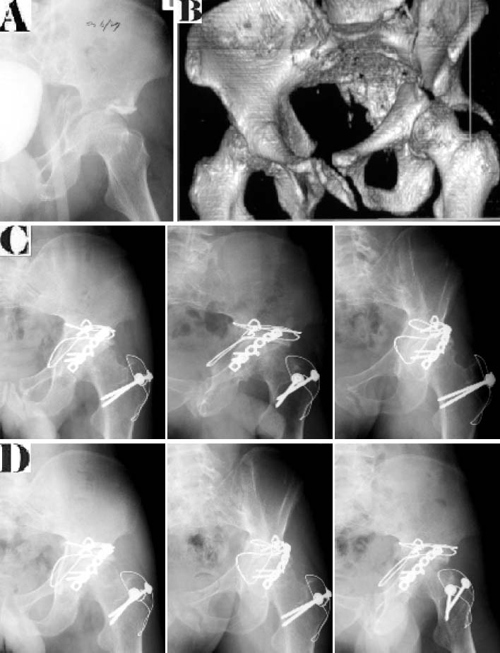
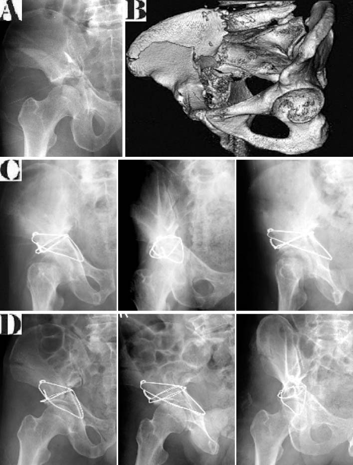
Fig. 1
(A) Initial radiograph of a 37 years old male shows transverse and posterior wall fractue.
(B) 3-Dimensional CT scan shows displaced fracture at the weight bearing dome.
(C) Immediate postoperative radiograph shows anatomical reduction with dual loop cerclage wiring and plate fixation.
(D) Radiograph after 19 months after operation shows well union of the fracture site and good joint congruency.
Fig. 2
(A) Initial radiograph of a 66 years old male shows both column fracture with spur sign.
(B) 3-Dimensional CT scan shows comminuted fracture at the medial wall and anterior column.
(C) Immediate postoperative radiograph shows anatomical reduction with dual loop cerclage wiring.
(D) Radiograph after 21 months after operation shows well union of the fracture site and good joint congruency.
Fig. 1
Fig. 2
Cerclage Wiring in Internal Fixation of Displaced Acetabular Fractures
Patient data
A: Case number; B: Fracture type, BC: Both columns, ACPH: Anterior column & posterior hemitransverse, TPW: Transverse & posterior wall fracture, TS: T-shaped; C: Age; D: Sex, M: Male, F: Female; E: Operative approach, IL: Ilioinguinal, KL: Kocher-Langenbeck, KL+LIL: Kocher-Langenbeck+Limited Ilioinguinal, TR: Triradiate transtrochanteric; F: Fixation method, SW: Single loop cerclage wire, DW: Dual loop cerclage wire, CW+P: Cerclage wire+plate, CS: Cerclage wire+screw; G: Radiologic result; H: Clinical result, E: Excellent, G: Good, F: Fair, P: Poor; I: Complication, N: None, OA: Osteoarthritis, HO: Heterotopic ossifiaction, I: Infection.
Table 1
Patient data
A: Case number; B: Fracture type, BC: Both columns, ACPH: Anterior column & posterior hemitransverse, TPW: Transverse & posterior wall fracture, TS: T-shaped; C: Age; D: Sex, M: Male, F: Female; E: Operative approach, IL: Ilioinguinal, KL: Kocher-Langenbeck, KL+LIL: Kocher-Langenbeck+Limited Ilioinguinal, TR: Triradiate transtrochanteric; F: Fixation method, SW: Single loop cerclage wire, DW: Dual loop cerclage wire, CW+P: Cerclage wire+plate, CS: Cerclage wire+screw; G: Radiologic result; H: Clinical result, E: Excellent, G: Good, F: Fair, P: Poor; I: Complication, N: None, OA: Osteoarthritis, HO: Heterotopic ossifiaction, I: Infection.

