 E-submission
 TOTA
TOTA TOTS
TOTS
Articles
- Page Path
- HOME > J Musculoskelet Trauma > Volume 23(1); 2010 > Article
-
Case Report
- Repetitive Insufficiency Fractures of the Femoral Shaft: A Case Report
- Ji-Hwan Kim, M.D., Young-Ho Cho, M.D., Young-Soo Byun, M.D., Jung-Hoon Shin, M.D., Chung-Yeol Lee, M.D., Tae-Gyun Kim, M.D.
-
Journal of the Korean Fracture Society 2010;23(1):109-112.
DOI: https://doi.org/10.12671/jkfs.2010.23.1.109
Published online: January 31, 2010
Department of Orthopedic Surgery, Daegu Fatima Hospital, Daegu, Korea.
- Address reprint requests to: Young-Ho Cho, M.D. Department of Orthopedic Surgery, Daegu Fatima Hosiptal, 576-31, Sinam-dong, Dong-gu, Daegu 701-600, Korea. Tel: 82-53-940-7320, Fax: 82-53-954-7417, fatimaos@unitel.co.kr
• Received: July 6, 2009 • Revised: September 7, 2009 • Accepted: October 29, 2009
Copyright © 2010 The Korean Fracture Society
- 769 Views
- 2 Download
Abstract
- Stress fractures occur when the loads applied to a bone exceed the mechanical resistance and fall into two groups. Fatigue fractures, in which abnormal mechanical stress is applied to a normal bone, and insufficiency fractures, in which fracture occurs when stress of normal activity is applied to a bone that has decreased elastic resistance. Femoral shaft insufficiency fractures are reported rarely in patients with postmenopausal osteoporosis. We report a case of repetitive insufficiency fractures of the femoral shaft in 70 year-old female with marked osteoporosis.
- 1. Clamp JA, King RJ, O'Hara JT, Hahn DM. Osteoporotic pelvic insufficiency fracture with gross instability. J Trauma, 2008;64:1380-1382.Article
- 2. Cooper KL, Beabout JW, Swee RG. Insufficiency fractures of the sacrum. Radiology, 1985;156:15-20.Article
- 3. Daffner RH, Pavlov H. Stress fractures: current concepts. AJR Am J Roentgenol, 1992;159:245-252.Article
- 4. De Smet AA, Neff JR. Pubic and sacral insufficiency fractures: clinical course and radiologic findings. AJR Am J Roentgenol, 1985;145:601-606.Article
- 5. Koh HS, Kang YK, Lee HY, et al. Insufficiency fractures of the femoral shaft associated with osteoporosis. J Korean Fract Soc, 2004;17:19-24.Article
- 6. Maraval A, Grados F, Royand V, Damade R, Boulu G, Fardellon P. Longitudinal femoral shaft fracture due to bone insufficiency. A review of three cases. Joint Bone Spine, 2003;70:526-531.Article
- 7. Ries T. Detection of osteoporotic sacral fracture with radionuclides. Radiology, 1983;146:783-785.Article
- 8. Roub LW, Gumerman LW, Hanley EN Jr, Clark MW, Goodman M, Herbert DL. Bone stress: a radionuclide imaging perspective. Radiology, 1979;132:431-438.Article
- 9. Soubrier M, Dubost JJ, Boisgard S, et al. Insufficiency fracture. A survey of 60 cases and review of the literature. Joint Bone Spine, 2003;70:209-218.Article
- 10. Yang KH, Sim DS. Clinical consideration on insufficiency fracture of femur. Korean J Bone Metab, 2009;16:37-41.
REFERENCES
Fig. 1Femur anteroposterior (A) and lateral (B) radiographs shows 3 radiolucent lines of lateral femoral cortex with endosteal and periosteal new bone formation.

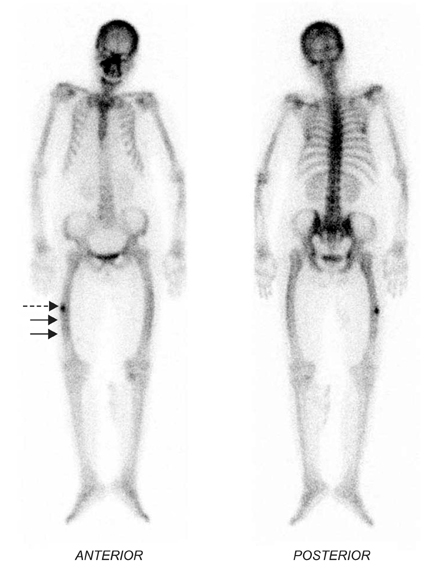
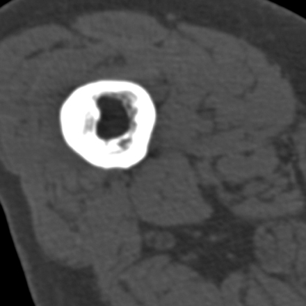
Figure & Data
REFERENCES
Citations
Citations to this article as recorded by 
 Cite
CiteRepetitive Insufficiency Fractures of the Femoral Shaft: A Case Report
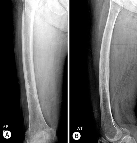


Fig. 1
Femur anteroposterior (A) and lateral (B) radiographs shows 3 radiolucent lines of lateral femoral cortex with endosteal and periosteal new bone formation.
Fig. 2
Whole body radionuclide scan shows a focal uptake (dotted arrow) and two tiny transverse linear uptakes (linear arrows) of right femur shaft.
Fig. 3
CT scan of distal femoral shaft shows thickened lateral femoral cortex and endosteal new bone formation.
Fig. 1
Fig. 2
Fig. 3
Repetitive Insufficiency Fractures of the Femoral Shaft: A Case Report

