 E-submission
 TOTA
TOTA TOTS
TOTS
Articles
- Page Path
- HOME > J Musculoskelet Trauma > Volume 23(1); 2010 > Article
-
Case Report
- Posterior Hip Dislocation with Ipsilateral Fractures of the Femoral Head and Intertrochanter: A Case Report
- Jai-Hyung Park, M.D., Hyung-Soo Kim, M.D., Soo-Tae Chung, M.D., Jeong-Hyun Yoo, M.D., Joo-Hak Kim, M.D., Seung-Do Cha, M.D., Tae-Woo Lee, M.D.
-
Journal of the Korean Fracture Society 2010;23(1):113-117.
DOI: https://doi.org/10.12671/jkfs.2010.23.1.113
Published online: January 31, 2010
Department of Orthopedic Surgery, Myongji Hospital, Kwandong University College of Medicine, Goyang, Korea.
- Address reprint requests to: Jai-Hyung Park, M.D. Department of Orthopedic Surgery, Myongji Hospital, Kwandong University College of Medicine, 697-24, Hwajeong-dong, Deogyang-gu, Goyang 412-270, Korea. Tel: 82-31-810-5520, Fax: 82-31-810-6537, wonnypia@kd.ac.kr
• Received: July 10, 2009 • Revised: August 24, 2009 • Accepted: October 19, 2009
Copyright © 2010 The Korean Fracture Society
- 1,009 Views
- 3 Download
- 1 Crossref
Abstract
- High-energy injury, as traffic accident or fall down, can cause fracture of femur head and posterior dislocation of hip joint which is accompanied with ipsilateral acetabulum fracture or femur neck fracture. But the case that femur head fracture and posterior dislocation of the hip joint coincide with ipsilateral intertrochanteric fracture of proximal femur is so uncommon that reports of the case is very rare. We hereby are to report the experienced and treated-cases of femur head fracture and posterior dislocation of the hip joint that is accompanied with ipsilateral intertrochanteric fracture.
- 1. Ganz R, Gill TJ, Gautier E, Ganz K, Krügel N, Berlemann U. Surgical dislocation of the adult hip a technique with full access to the femoral head and acetabulum without the risk of avascular necrosis. J Bone Joint Surg Br, 2001;83:1119-1124.
- 2. Henle P, Kloen P, Siebenrock KA. Femoral head injuries: which treatment strategy can be recommended? Injury, 2007;38:478-488.
- 3. Hunter GA. Posterior dislocation and fracture-dislocation of the hip. A review of fifty-seven patients. J Bone Joint Surg Br, 1969;51:38-44.
- 4. Jaskulka RA, Fischer G, Fenzl G. Dislocation and fracture-dislocation of the hip. J Bone Joint Surg Br, 1991;73:465-469.
- 5. Khan MH, Wright VJ, Prayson MJ. Ipsilateral intertrochanteric and pipkin fractures: an unusual case. Am J Orthop (Belle Mead NJ), 2007;36:E53-E55.
- 6. Klasen HJ, Binnendijk B. Fracture of the neck of the femur associated with posterior dislocation of the hip. J Bone Joint Surg Br, 1984;66:45-48.
- 7. Mostafa MM. Femoral head fractures. Int Orthop, 2001;25:51-54.
- 8. Paul Tornetta III. Bucholz R, Heckman J, Charles CB. Hip Dislocations and fracture of the femoral head. In: Rockwood and Green's Fractures in adults, 2005;6th. ed. Philadelphia, Lippincott Williams & Wilkins Co. 1715-1752.
- 9. Roeder LF Jr, DeLee JC. Femoral head fractures associated with posterior hip dislocations. Clin Orthop Relat Res, 1980;147:121-130.
- 10. Stannard JP, Harris HW, Volgas DA, Alonso JE. Functional outcome of patients with femoral head fractures associated with hip dislocations. Clin Orthop Relat Res, 2000;377:44-56.
REFERENCES
Fig. 1
(A) Anteroposterior Hip radiograph shows left femoral head fracture and combined ipsilateral intertrochanteric fracture. (B) Axial CT image shows large fracture fragment of femoral head and posterior hip dislocation in the left hip joint.
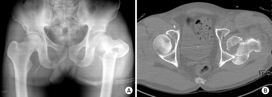
Fig. 2
(A, B) Immediate postoperative anteroposterior and axial Hip radiographs show that left femoral head fracture was fixated with standard Acutrak screw and ipsilateral intertrochanteric fracture was fixated with LCP-DF.

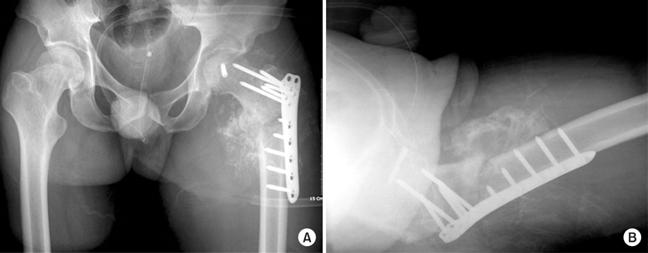
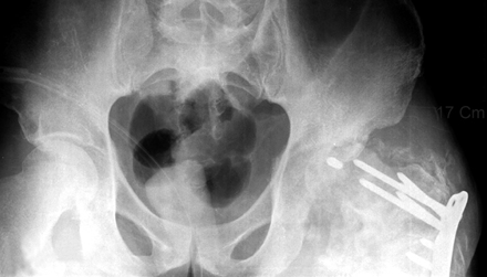
Figure & Data
REFERENCES
Citations
Citations to this article as recorded by 
- Decoding the behaviour of extracapsular proximal femur fracture- dislocation - A systematic review of a rare fracture pattern
Keyur B. Desai
Journal of Clinical Orthopaedics and Trauma.2021; 18: 157. CrossRef
 Cite
CitePosterior Hip Dislocation with Ipsilateral Fractures of the Femoral Head and Intertrochanter: A Case Report

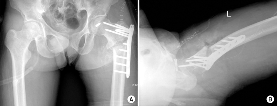


Fig. 1
(A) Anteroposterior Hip radiograph shows left femoral head fracture and combined ipsilateral intertrochanteric fracture. (B) Axial CT image shows large fracture fragment of femoral head and posterior hip dislocation in the left hip joint.
Fig. 2
(A, B) Immediate postoperative anteroposterior and axial Hip radiographs show that left femoral head fracture was fixated with standard Acutrak screw and ipsilateral intertrochanteric fracture was fixated with LCP-DF.
Fig. 3
(A, B) Postoperative 3 months later follow-up anteroposterior and axial Hip radiographs show bridging callus formation around previous intertrochanteric fracture area. Avascular necrosis of femur head or displacement does not observed.
Fig. 4
Postoperative 5 months later follow-up Abdomen radiograph shows erosion or avascular necrosis of femoral head and posterior dislocation.
Fig. 1
Fig. 2
Fig. 3
Fig. 4
Posterior Hip Dislocation with Ipsilateral Fractures of the Femoral Head and Intertrochanter: A Case Report

