 E-submission
 TOTA
TOTA TOTS
TOTS
Articles
- Page Path
- HOME > J Musculoskelet Trauma > Volume 34(2); 2021 > Article
- Case Report Delayed Pseudoaneurysm of Deep Femoral Artery Caused by Migration of Lesser Trochanter, Subsequent to an Intertrochanteric Fracture Surgery - A Case Report -
- Bum-Soo Kim, Seong-Tae Kim, Seungyup Shin, Chang Geun Yu
-
Journal of Musculoskeletal Trauma 2021;34(2):76-79.
DOI: https://doi.org/10.12671/jkfs.2021.34.2.76
Published online: April 30, 2021
Department of Orthopedic Surgery, Cheju Halla General Hospital, Jeju, Korea
- 988 Views
- 10 Download
- 0 Crossref
- 0 Scopus
Abstract
The lesser trochanteric fracture is commonly found amongst intertrochanteric fractures, where pseudoaneurysm of the femoral artery is a rare complication. A pseudoaneurysm could develop due to the penetration injury of the artery by the bone fragment during occurrence of the fracture, or by the insertion of screws during the surgical procedure. Minimal complication is seen when the lesser trochanter is not fixed during the intertrochanteric fracture surgery. However, in the current case, the authors experienced appearance of a delayed pseudoaneurysm of the deep femoral artery caused by migration of the lesser trochanter, which was successfully treated by excision.
J Korean Fract Soc. 2021 Apr;34(2):76-79. Korean.
Published online Apr 23, 2021.
https://doi.org/10.12671/jkfs.2021.34.2.76
Published online Apr 23, 2021.
https://doi.org/10.12671/jkfs.2021.34.2.76
Copyright © 2021 The Korean Fracture Society. All rights reserved.
Case Report
Delayed Pseudoaneurysm of Deep Femoral Artery Caused by Migration of Lesser Trochanter, Subsequent to an Intertrochanteric Fracture Surgery: A Case Report

Abstract
The lesser trochanteric fracture is commonly found amongst intertrochanteric fractures, where pseudoaneurysm of the femoral artery is a rare complication. A pseudoaneurysm could develop due to the penetration injury of the artery by the bone fragment during occurrence of the fracture, or by the insertion of screws during the surgical procedure. Minimal complication is seen when the lesser trochanter is not fixed during the intertrochanteric fracture surgery. However, in the current case, the uthors experienced appearance of a delayed pseudoaneurysm of the deep femoral artery caused by migration of the lesser trochanter, which was successfully treated by excision.
Keywords
Deep femoral artery, Lesser trochanter fracture, Delayed pseudoaneurysm, Peudoaneurysmectomy
Figures
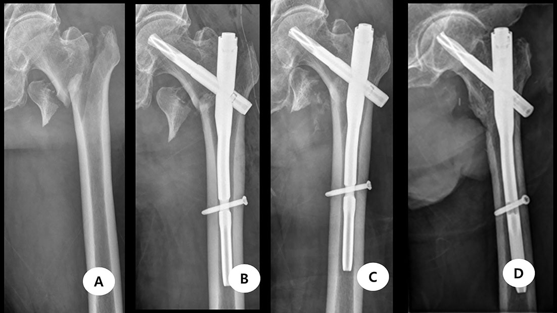
Fig. 1
(A) The fracture type was AO/OTA 31 A1.3. (B) Intramedullary fixation was performed. (C) The lesser trochater fragment migrated proximally. (D) Lesser trochnter excision was performed.
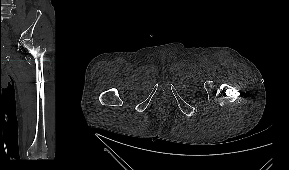
Fig. 2
The computed tomography taken 3 days postoperatively. Around the lesser trochaner there was neither hematoma nor pseudoaneurysm.

Fig. 3
Left lower extremity swelling was observed.
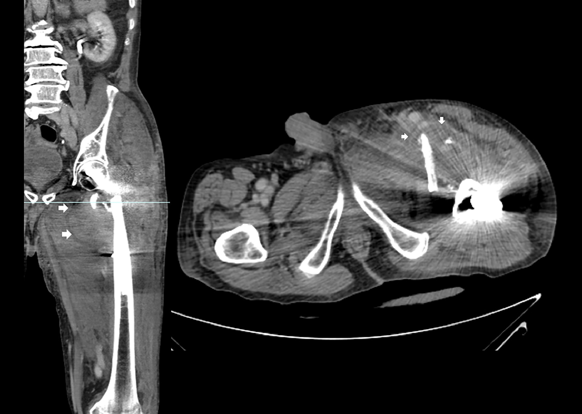
Fig. 4
On the computed tomography, there was pseudoaneurysm which was formed by the penetrating injury of the artery by the lesser trochanter bone fragment. White arrows indicate pseudoaneurysm.
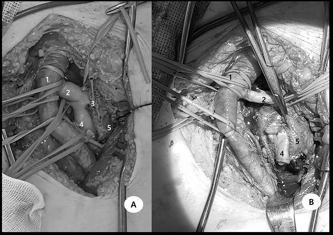
Fig. 5
(A) Through exploration pseudoaneurysm was found. (B) Descending branch of the deep fomoral artery was excised to remove pseudoaneurysm. 1: femoral artery, 2: deep femoral artery, 3: lateral circumflex femoral artery, 4: descending branch, 5: pseudoaneurysm.
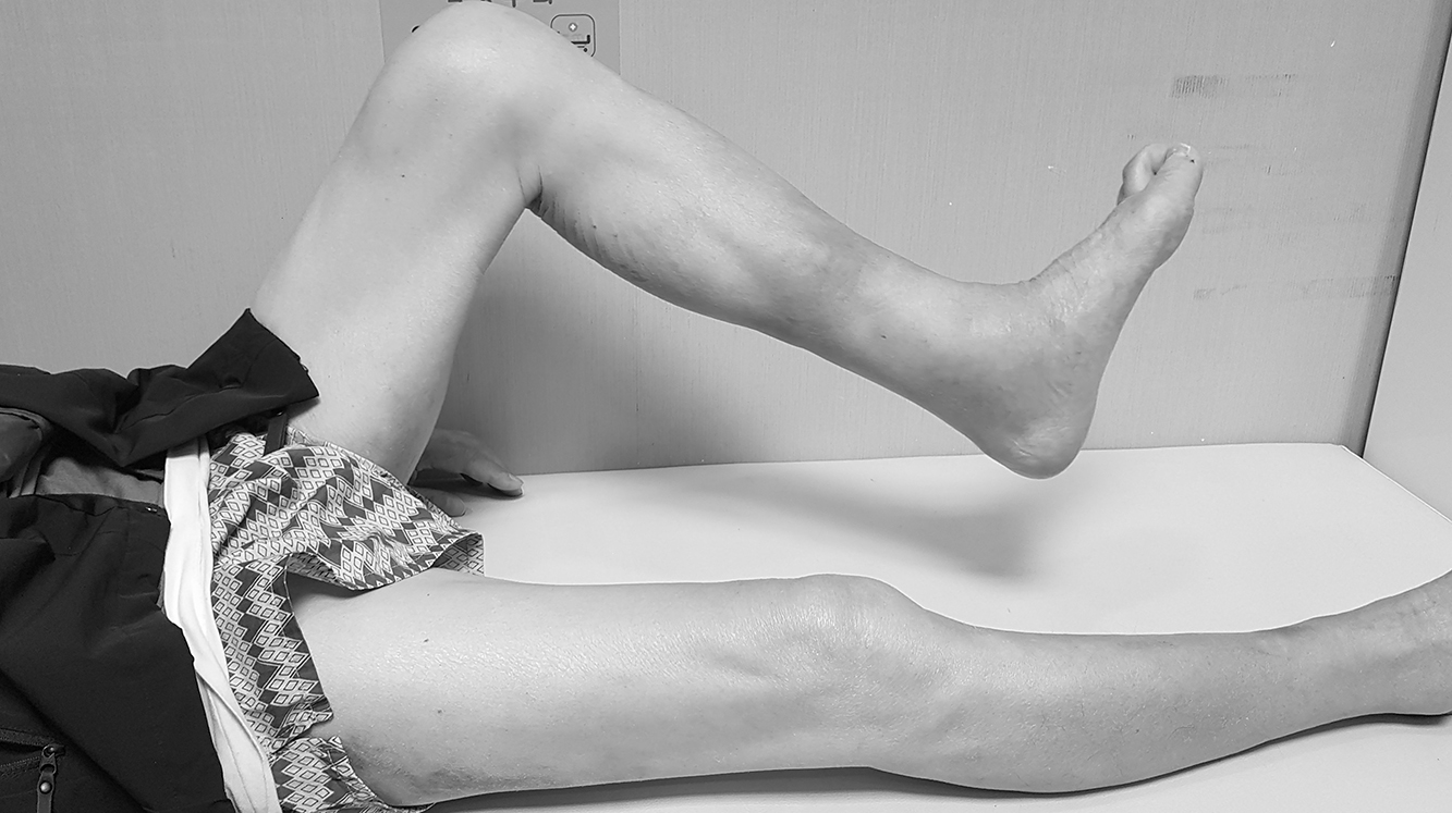
Fig. 6
The patient can flex the knee against resistance.
Notes
Financial support:None.
Conflict of interests:None.
References
-
Lindskog DM, Baumgaertner MR. Unstable intertrochanteric hip fractures in the elderly. J Am Acad Orthop Surg 2004;12:179–190.
-
-
Keel JD, Eyres KS. Vascular injury by an intertrochanteric fracture fragment. Injury 1993;24:350–352.
-
-
Karanikas I, Lazarides M, Arvanitis D, Papayanopoulos G, Exarchou E, Dayantas J. Iatrogenic arterial trauma associated with hip fracture surgery. Acta Chir Belg 1993;93:284–286.
-
-
Sharma G, Singh R, Kumar A, Sharma V, Farooque K. Acute femoral artery pseudoaneurysm due to lesser trochanter fragment: an unusual complication of an intertrochanteric fracture. Chin J Traumatol 2013;16:301–303.
-
-
Lazarides MK, Arvanitis DP, Dayantas JN. Iatrogenic arterial trauma associated with hip joint surgery: an overview. Eur J Vasc Surg 1991;5:549–556.
-
-
Yang KH, Park HW, Park SJ. Pseudoaneurysm of the superficial femoral artery after closed hip nailing with a Gamma nail: report of a case. J Orthop Trauma 2002;16:124–127.
-
-
Dhal A, Chadha M, Lal H, Singh T, Tyagi S. Encounters with pseudoaneurysms in orthopaedic practice. Injury 2001;32:771–778.
-
-
Karkos CD, Hughes R, Prasad V, D'Souza SP. Thigh compartment syndrome as a result of a false aneurysm of the profunda femoris artery complicating fixation of an intertrochanteric fracture. J Trauma 1999;47:393–395.
-
-
Aprato A, Lo Baido R, Crosio A, Matteotti R, Grosso E, Massè A. Does lesser trochanter implication affect hip flexion strength in proximal femur fracture? Eur J Trauma Emerg Surg 2015;41:523–529.
-
 Cite
Cite
