 E-submission
 TOTA
TOTA TOTS
TOTS
Articles
- Page Path
- HOME > J Musculoskelet Trauma > Volume 24(3); 2011 > Article
-
Case Report
- Multiple Non-contiguous Spine Fractures with Concomitant Injuries: A Case Report
- Soo Uk Chae, M.D., Yeung Jin Kim, M.D., Jung Hwan Yang, M.D., Ji Wan Lee, M.D., Jae In Park, M.D.
-
Journal of the Korean Fracture Society 2011;24(3):267-270.
DOI: https://doi.org/10.12671/jkfs.2011.24.3.267
Published online: July 15, 2011
Department of Orthopaedic Surgery, School of Medicine, Wonkwang University, Iksan, Korea.
- Address reprint requests to: Ji Wan Lee, M.D. Department of Orthopaedic Surgery, School of Medicine, Wonkwang University, 344-1, Shinyong-dong, Iksan 570-711, Korea. Tel: 82-63-472-5100, Fax: 82-63-472-5688, oschae@naver.com
• Received: February 4, 2010 • Revised: April 18, 2011 • Accepted: May 4, 2011
Copyright © 2011 The Korean Fracture Society
- 869 Views
- 10 Download
Abstract
- Multiple non-contiguous spinal fracture is a special type of multi-level spinal injury, which is rare but most frequently occur in motor vehicle accident or a falling from a height. We report five patients of multiple non-contiguous spinal fractures. All patients underwent segmental pedicle screws fixation without fusion for preserving facet joints and minimizing blood loss and operation time. We performed necessary operation for any concomitant injuries at the same day.
- 1. Chang HG, Kim YW, Kim YC, Kwon DJ, Seo KN, Lee KB. Multiple spine fracture of young adult (Over 3 Vertebrae). J Korean Soc Spine Surg, 2005;12:206-213.Article
- 2. Dai LY, Jia LS. Multiple non-contiguous injuries of the spine. Injury, 1996;27:573-575.Article
- 3. Gardner VO, Amstrong GW. Long-term lumbar facet joint change in spinal fracture patients treated with Harrington rods. Spine (Phila Pa 1976), 1990;15:479-484.
- 4. Huler RJ. Frymoyer JW. Thoracolumbar Spine Fracture. In: The adult spine-principles and practice, 1997;2nd ed. Philadelphia, Lippincott-Raven. 1473.
- 5. Jorgensen DR, Joseph J Jr. Multiple noncontiguous spine fractures at four levels in a neurologically intact patient. J Trauma, 1996;41:750-753.Article
- 6. Kim YM, Kim DS, Choi ES, et al. Results of non-fusion methods in thoracolumbar and lumbar spinal fractures. J Korean Soc Spine Surg, 2005;12:132-139.Article
- 7. Knop C, Fabian HF, Bastian L, Blauth M. Late results of thoracolumbar fractures after posterior instrumentation and transpedicular bone grafting. Spine (Phila Pa 1976), 2001;26:88-99.Article
- 8. Lian XF, Zhao J, Hou TS, Yuan JD, Jin GY, Li ZH. The treatment for multilevel noncontiguous spinal fractures. Int Orthop, 2007;31:647-652.ArticlePDF
- 9. Powell JN, Waddell JP, Tucker WS, Transfeldt EE. Multiple-level noncontiguous spinal fractures. J Trauma, 1989;28:1146-1150.Article
- 10. Wittenberg RH, Hargus S, Steffen R, Muhr G, Bötel U. Noncontiguous unstable spine fractures. Spine (Phila Pa 1976), 2002;27:254-257.Article
REFERENCES
Fig. 1
52-years old male who fell off ladder had T12, L2 & L4 spine fractures and concomitant distal radius and calcaneal fractures underwent operation on the day of the injury.
(A) Plain L-spine lateral image shows L2 and L4 bursting fracture and subluxation of L4/5 facet joint (black arrow).
(B) Axial CT images of L2 and L4 shows minimal invasion of spinal fracture fragment in vertebral canal. There were no neurologic abnormalities.
(C) Sagittal T1-weighted images of lumbar spine shows signal change and cortical irregularity.
(D) Postoperative sagittal CT image shows short segment fixation of the L3-5. T12 vertebral body fracture (white arrow) was newly found, which was not visible in preoperative CT scans.
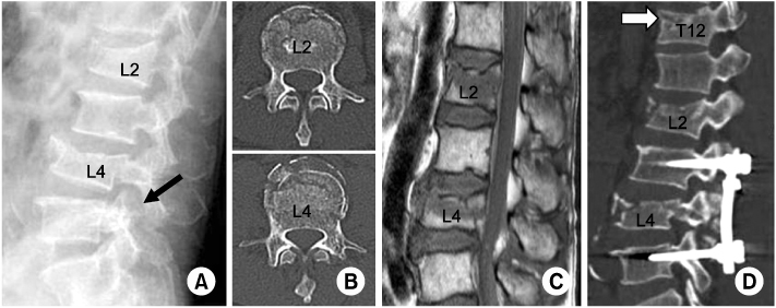
Fig. 2
(A) Axial and sagittal CT images shows T12 and L4 vertebral body fractures of 38-years old female who fell from a 6 m height.
(B) Postoperative T-L spine X-ray shows short segmental fixation of L3-5 without bone graft.
(C) Axial and sagittal CT images shows L1 and L4 vertebral body fractures of 39-years old female who fell from a 3m height.
(D) Postoperative T-L spine X-ray shows short segmental fixation of L3-5 without bone graft.


Figure & Data
REFERENCES
Citations
Citations to this article as recorded by 
 Cite
CiteMultiple Non-contiguous Spine Fractures with Concomitant Injuries: A Case Report

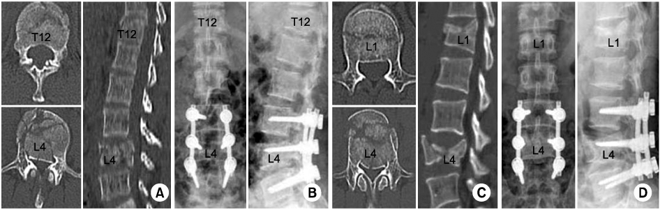
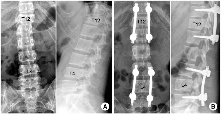
Fig. 1
52-years old male who fell off ladder had T12, L2 & L4 spine fractures and concomitant distal radius and calcaneal fractures underwent operation on the day of the injury.
(A) Plain L-spine lateral image shows L2 and L4 bursting fracture and subluxation of L4/5 facet joint (black arrow).
(B) Axial CT images of L2 and L4 shows minimal invasion of spinal fracture fragment in vertebral canal. There were no neurologic abnormalities.
(C) Sagittal T1-weighted images of lumbar spine shows signal change and cortical irregularity.
(D) Postoperative sagittal CT image shows short segment fixation of the L3-5. T12 vertebral body fracture (white arrow) was newly found, which was not visible in preoperative CT scans.
Fig. 2
(A) Axial and sagittal CT images shows T12 and L4 vertebral body fractures of 38-years old female who fell from a 6 m height.
(B) Postoperative T-L spine X-ray shows short segmental fixation of L3-5 without bone graft.
(C) Axial and sagittal CT images shows L1 and L4 vertebral body fractures of 39-years old female who fell from a 3m height.
(D) Postoperative T-L spine X-ray shows short segmental fixation of L3-5 without bone graft.
Fig. 3
(A) Preoperative L-spine X-ray shows T12, L4, and sacrum fractures that T12, L4 was diagnosed major fractures of 24-years old female who fell from a 4 m height.
(B) Postoperative L spine X-ray shows pedicular screw fixation on the upper and lower vertebrae of T12 and L4 without bone graft.
Fig. 1
Fig. 2
Fig. 3
Multiple Non-contiguous Spine Fractures with Concomitant Injuries: A Case Report
Summary of cases
*Death after POD 6 months, †Fx: fracture, IF: internal fixation, EF: external fixation.
Table 1
Summary of cases
*Death after POD 6 months, †Fx: fracture, IF: internal fixation, EF: external fixation.

