 E-submission
 TOTA
TOTA TOTS
TOTS
Articles
- Page Path
- HOME > J Musculoskelet Trauma > Volume 26(4); 2013 > Article
-
Review Article
- Complications of Pelvic Ring Injury
- Byung-Woo Min, M.D., Kyung-Jae Lee, M.D., Gyo-Wook Kim, M.D., Doohyun Kwon, M.D.
-
Journal of the Korean Fracture Society 2013;26(4):348-353.
DOI: https://doi.org/10.12671/jkfs.2013.26.4.348
Published online: October 18, 2013
Department of Orthopedic Surgery, Keimyung University School of Medicine, Daegu, Korea.
- Address reprint requests to: Byung-Woo Min, M.D. Department of Orthopedic Surgery, Keimyung University School of Medicine, 56 Dalseong-ro, Jung-gu, Daegu 700-712, Korea. Tel: 82-53-250-7267, Fax: 82-53-250-7205, min@dsmc.or.kr
Copyright © 2013 The Korean Fracture Society. All rights reserved.
This is an Open Access article distributed under the terms of the Creative Commons Attribution Non-Commercial License (http://creativecommons.org/licenses/by-nc/3.0/) which permits unrestricted non-commercial use, distribution, and reproduction in any medium, provided the original work is properly cited.
- 1,082 Views
- 7 Download
- 2 Crossref

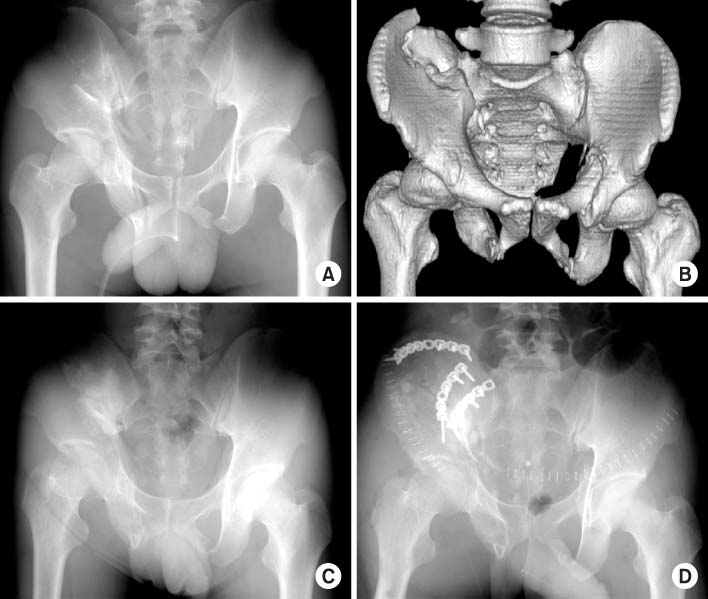
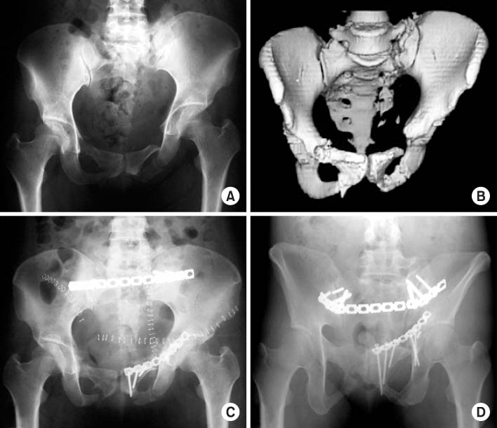
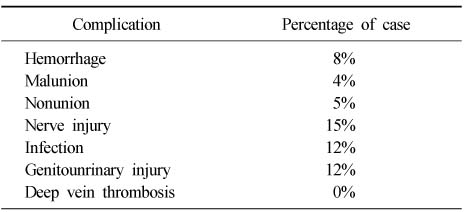
Figure & Data
REFERENCES
Citations
Citations to this article as recorded by 
- Young–Burgess Classification of Pelvic Ring Fractures as a Diagnostic Tool to Predict Vascular Injury Patterns and Targeted Embolization: A 10-Year Retrospective Study of Patients at a Single Regional Trauma Center in South Korea
Dae Hee Lee, Seong Wook Kim, Ki-Choul Kim
Hip & Pelvis.2025; 37(4): 321. CrossRef - Simultaneous Surgery on Jejunum perforation with Pelvic Ring Fracture: A Case Report
HoeJeong Chung, Keum-Seok Bae, Seong-yup Kim, Doosup Kim
Journal of Trauma and Injury.2016; 29(2): 56. CrossRef
 Cite
CiteComplications of Pelvic Ring Injury
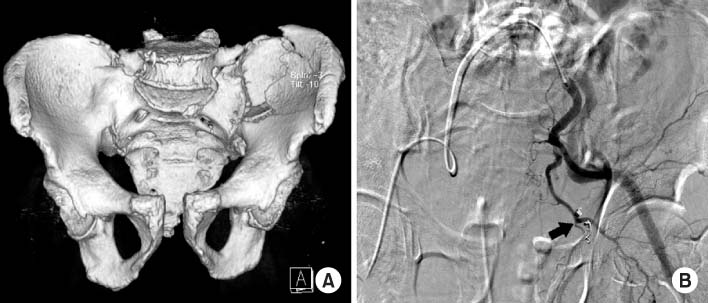


Fig. 1
(A) A 79-year-old male patient with Tile type C1 unstable pelvic fractures which were determined as left crescent fracture, sacroiliac joint disruption and symphysis pubis diastasis on 3-dimensional computed tomographys.
(B) Embolization with gelfoam and microcoil about the inferior epigastric artery (arrow).
Fig. 2
(A, B) A 28-year-old male patient with Tile type C1 unstable pelvic fractures which were determined as right crescent fracture including the ipsilateral superior and inferior rami on X-ray and 3-dimensional computed tomography.
(C) Conservative therapy with skeletal traction about 2 months due to liver injury.
(D) Osteotomy and multiple plate fixation about the malunion site of the ilium and ramus. Postoperative X-ray shows stable fixation but the right hemipelvis was rotated internally and migrated about 1 cm upward.
Fig. 3
(A, B) A 39-year-old female patient with Tile type C3 unstable pelvic fracture with left sacral fracture, which were both superior and inferior rami fractures on X-ray and 3-dimensional computed tomographys.
(C) Delayed operation due to problems with the patient's general condition. postoperative x-ray shows plate fixation on the left ramus, posterior sacral fracture.
(D) At 1 year, the left ramus and posterior sacral fracture site were nonunion and metal breakage on follow X-ray.
Fig. 1
Fig. 2
Fig. 3
Complications of Pelvic Ring Injury
Associated Complications of Pelvic Ring Fracture
Table 1
Associated Complications of Pelvic Ring Fracture

