 E-submission
 TOTA
TOTA TOTS
TOTS
Articles
- Page Path
- HOME > J Musculoskelet Trauma > Volume 25(1); 2012 > Article
-
Original Article
- Complications of Hook-Plate Fixation for Distal Clavicle Fractures
- Su-Han An, M.D., Hyung-Chun Kim, M.D., Kwang-Yeol Kim, M.D., Ji-Hoon Lee, M.D., Seung-Hyun Yoon, M.D.
-
Journal of the Korean Fracture Society 2012;25(1):38-45.
DOI: https://doi.org/10.12671/jkfs.2012.25.1.38
Published online: January 31, 2012
Department of Orthopedic Surgery, Wallace Memorial Baptist Hospital, Busan, Korea.
- Address reprint requests to: Hyung-Chun Kim, M.D. Department of Orthopedic Surgery, Wallace Memorial Baptist Hospital, 374-75, Namsan-dong, Heumjung-gu, Busan 609-728, Korea. Tel: 82-51-580-1422, Fax: 82-51-583-2568, dizziku@naver.com
• Received: December 13, 2011 • Accepted: December 16, 2011
Copyright © 2012 The Korean Fracture Society
- 3,167 Views
- 31 Download
- 2 Crossref
Abstract
-
Purpose
- To report on the complications of hook-plate fixation for distal clavicle fractures.
-
Materials and Methods
- Eighteen patients who underwent surgery for distal clavicle fracture with a hook-plate from April 2008 to April 2011 were enrolled with a minimum of 4 months follow-up. The reduction was qualified and evaluated according to the radiologic findings. We analyzed the results by UCLA score, Kona's functional evaluation, and VAS pain score.
-
Results
- By radiologic evaluation, 17 of 18 cases showed anatomical reduction and solid unions. Although satisfactory results were found in the clinical study as shown by the UCLA score, Kona's functional evaluation, and VAS pain score, complications arose in 7 cases, including osteolysis of the acromion in 2 cases, nonunion in 1 case, periprosthetic fracture in 2 cases, subacromial pain in 1 case, and skin irritation in 1 case. 2 cases of all required reoperation.
-
Conclusion
- To reduce the complications of the hook-plate, a precise surgical technique and the choice of an appropriate size for the hook-plate are needed. We suggest that early removal of the plate is necessary to decrease the risk of subacromial impingement and erosion in hook-plate fixation.
- 1. Ballmer FT, Gerber C. Coracoclavicular screw fixation for unstable fractures of the distal clavicle. A report of five cases. J Bone Joint Surg Br, 1991;73:291-294.ArticlePDF
- 2. Bottlang M, Doornink J, Byrd GD, Fitzpatrick DC, Madey SM. A nonlocking end screw can decrease fracture risk caused by locked plating in the osteoporotic diaphysis. J Bone Joint Surg Am, 2009;91:620-627.Article
- 3. Charity RM, Haidar SG, Ghosh S, Tillu AB. Fixation failure of the clavicular hook plate: a report of three cases. J Orthop Surg (Hong Kong), 2006;14:333-335.ArticlePDF
- 4. Chen CH, Chen WJ, Shih CH. Surgical treatment for distal clavicle fracture with coracoclavicular ligament disruption. J Trauma, 2002;52:72-78.Article
- 5. Chun JM, Kim SY, Lee KW, Shin SJ, Kim EG. Modified tenson band fixation for unstable fracture of the distal clavicle. J Korean Orthop Assoc, 2002;37:416-420.ArticlePDF
- 6. Constant CR, Murley AH. A clinical method of functional assessment of the shoulder. Clin Orthop Relat Res, 1987;(214):160-164.Article
- 7. Craig EV. Rock-wood CA, Green DP, Bucholz RW, Heckman JD. Fracture of the clavicle. In: Fractures in adults, 1996;4th ed. Philadelphia, Lippincott-Raven. 1109-1161.
- 8. Edwards DJ, Kavanagh TG, Flannery MC. Fractures of the distal clavicle: a case for fixation. Injury, 1992;23:44-46.Article
- 9. Ellman H, Hanker G, Bayer M. Repair of the rotator cuff. End-result study of factors influencing reconstruction. J Bone Joint Surg Am, 1986;68:1136-1144.Article
- 10. Eskola A, Vainionpää S, Pätiälä H, Rokkanen P. Outcome of operative treatment in fresh lateral clavicular fracture. Ann Chir Gynaecol, 1987;76:167-169.
- 11. Faraj AA, Ketzer B. The use of a hook-plate in the management of acromioclavicular injuries. Report of ten cases. Acta Orthop Belg, 2001;67:448-451.
- 12. Flinkkilä T, Ristiniemi J, Lakovaara M, Hyvönen P, Leppilahti J. Hook-plate fixation of unstable lateral clavicle fractures: a report on 63 patients. Acta Orthop, 2006;77:644-649.Article
- 13. Goldberg JA, Bruce WJ, Sonnabend DH, Walsh WR. Type 2 fractures of the distal clavicle: a new surgical technique. J Shoulder Elbow Surg, 1997;6:380-382.Article
- 14. Habernek H, Weinstabl R, Schmid L, Fialka C. A crook plate for treatment of acromioclavicular joint separation: indication, technique, and results after one year. J Trauma, 1993;35:893-901.
- 15. Haidar SG, Krishnan KM, Deshmukh SC. Hook plate fixation for type II fractures of the lateral end of the clavicle. J Shoulder Elbow Surg, 2006;15:419-423.Article
- 16. Hessmann M, Kirchner R, Baumgaertel F, Gehling H, Gotzen L. Treatment of unstable distal clavicular fractures with and without lesions of the acromioclavicular joint. Injury, 1996;27:47-52.Article
- 17. Kao FC, Chao EK, Chen CH, Yu SW, Chen CY, Yen CY. Treatment of distal clavicle fracture using Kirschner wires and tension-band wires. J Trauma, 2001;51:522-525.Article
- 18. Kashii M, Inui H, Yamamoto K. Surgical treatment of distal clavicle fractures using the clavicular hook plate. Clin Orthop Relat Res, 2006;447:158-164.Article
- 19. Kim JH, Lee SC, Cho DY, Yoon HK, Lee YS. Percutaneus cerclage wiring in distal clavicle fracture type 2a-one case report. J Korean Shoulder Elbow Soc, 2006;9:124-129.Article
- 20. Kim JS, Jun JH, Chung YK. Coracoclavicular screw fixation for AC dislocation and unstable distal clavicle fracture. J Korean Shoulder Elbow Soc, 1999;2:133-137.
- 21. Kona J, Bosse MJ, Staeheli JW, Rosseau RL. Type II distal clavicle fractures: a retrospective review of surgical treatment. J Orthop Trauma, 1990;4:115-120.
- 22. Mall JW, Jacobi CA, Philipp AW, Peter FJ. Surgical treatment of fractures of the distal clavicle with polydioxanone suture tension band wiring: an alternative osteosynthesis. J Orthop Sci, 2002;7:535-537.Article
- 23. Meda PV, Machani B, Sinopidis C, Braithwaite I, Brownson P, Frostick SP. Clavicular hook plate for lateral end fractures:- a prospective study. Injury, 2006;37:277-283.Article
- 24. Mizue F, Shirai Y, Ito H. Surgical treatment of comminuted fractures of the distal clavicle using Wolter clavicular plates. J Nippon Med Sch, 2000;67:32-34.Article
- 25. Muramatsu K, Shigetomi M, Matsunaga T, Murata Y, Taguchi T. Use of the AO hook-plate for treatment of unstable fractures of the distal clavicle. Arch Orthop Trauma Surg, 2007;127:191-194.ArticlePDF
- 26. Neer CS 2nd. Fracture of the distal clavicle with detachment of the coracoclavicular ligaments in adults. J Trauma, 1963;3:99-110.Article
- 27. Neer CS 2nd. Nonunion of the clavicle. J Am Med Assoc, 1960;172:1006-1011.Article
- 28. Neviaser RJ, Neviaser JS, Neviaser TJ, Neviaser JS. A simple technique for internal fixation of the clavicle. A long term evaluation. Clin Orthop Relat Res, 1975;(109):103-107.
- 29. Nordqvist A, Petersson C, Redlund-Johnell I. The natural course of lateral clavicle fracture. 15 (11-21) year follow-up of 110 cases. Acta Orthop Scand, 1993;64:87-91.Article
- 30. Renger RJ, Roukema GR, Reurings JC, Raams PM, Font J, Verleisdonk EJ. The clavicle hook plate for Neer type II lateral clavicle fractures. J Orthop Trauma, 2009;23:570-574.Article
- 31. Rowe CR. An atlas of anatomy and treatment of midclavicular fractures. Clin Orthop Relat Res, 1968;58:29-42.
- 32. Salem KH, Schmelz A. Treatment of Tossy III acromioclavicular joint injuries using hook plates and ligament suture. J Orthop Trauma, 2009;23:565-569.Article
- 33. Sim E, Schwarz N, Höcker K, Berzlanovich A. Repair of complete acromioclavicular separations using the acromioclavicular-hook plate. Clin Orthop Relat Res, 1995;(314):134-142.Article
- 34. Tambe AD, Motkur P, Qamar A, Drew S, Turner SM. Fractures of the distal third of the clavicle treated by hook plating. Int Orthop, 2006;30:7-10.ArticlePDF
REFERENCES
Fig. 1
Radiograhs of 52-year-old male.
(A) Postoperative radiograph of the right shoulder shows Neer type II distal clavicle fracture.
(B) At 2 months follow-up, the radiograph shows osteo-lysis of acromion by the Hook plate.
(C) At 4 month follow-up, Hook plate was removed.
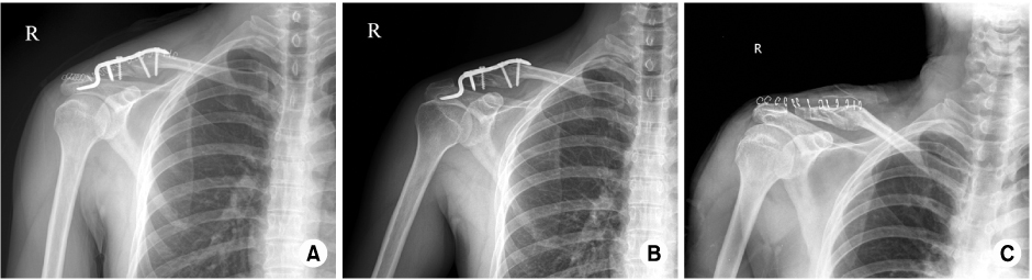
Fig. 2The hook of Hook plate can be bent to fit the clavicle anatomy. Bending is required to fit the curve of the acromoclavicular joint, that maximized the area of contact surface with hook of the Hook plate.
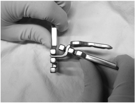
Fig. 3
Radiographs of 45-year-old female.
(A) Preoperative radiograph shows left distal clavicle fracture.
(B) Postoperative radiograph. The fracture was reduced and fixed with Hook plate. But the locking screw was being fracture site.
(C) At. POD 7 months follow-up, the radiographs shows proximal screw loosening & plate migration, but seen no bony union.
(D) At POD 10 months Hook plate was removed, internal fixation and bone graft was done.
(E) At POD 14 month follow-up, the union of fracture was deteched, eventually.
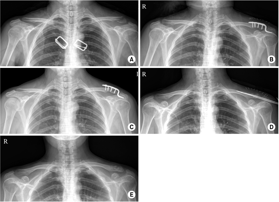
Fig. 4
Radiographs of 62-year-old female.
(A) Preoperative radiograph of the left shoulder shows neer type II distal clavicle fracture.
(B) Postoperative radiograph. Satisfactory reduction and Hook plate fixation were seen.
(C) At POD 4 weeks follow-up, postoperative radiograph shows clavicle shaft fracture.
(D) At 6 months follow-up, bony union was achieved without any complications.

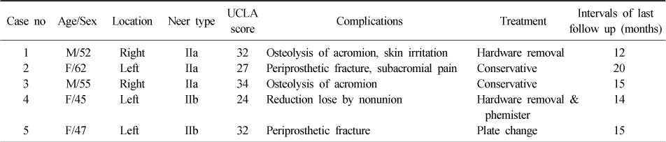
Figure & Data
REFERENCES
Citations
Citations to this article as recorded by 
- Clinical outcomes of bending versus non-bending of the plate hook in acromioclavicular joint dislocation
Min Su Joo, Hoi Young Kwon, Jeong Woo Kim
Clinics in Shoulder and Elbow.2021; 24(4): 202. CrossRef - Surgical Treatment of Unstable Distal Clavicle Fractures: Comparison of Transacromial Pin Fixation and Hook Plate Fixation
Young Sung Kim, Ho Min Lee, Han Gil Jang
The Journal of the Korean Shoulder and Elbow Society.2013; 16(2): 123. CrossRef
 Cite
CiteComplications of Hook-Plate Fixation for Distal Clavicle Fractures


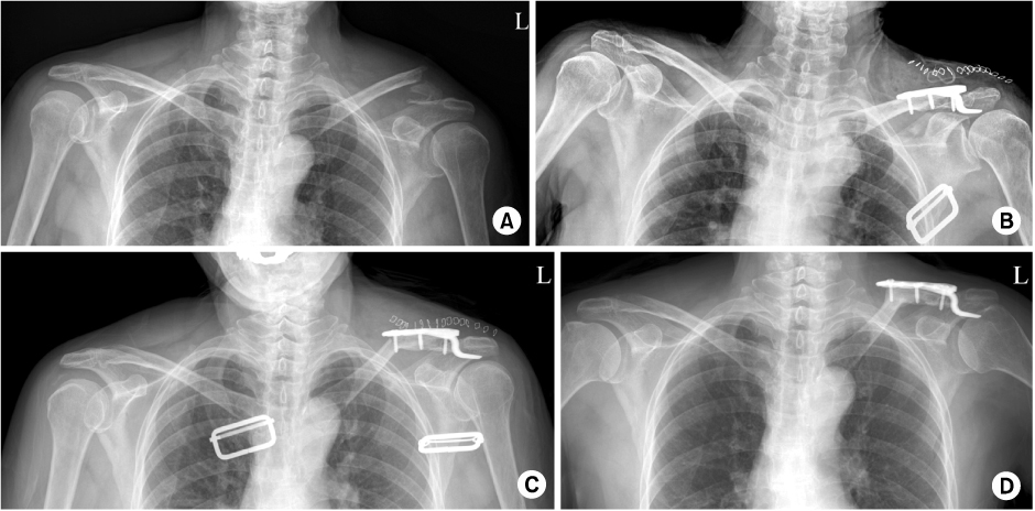
Fig. 1
Radiograhs of 52-year-old male.
(A) Postoperative radiograph of the right shoulder shows Neer type II distal clavicle fracture.
(B) At 2 months follow-up, the radiograph shows osteo-lysis of acromion by the Hook plate.
(C) At 4 month follow-up, Hook plate was removed.
Fig. 2
The hook of Hook plate can be bent to fit the clavicle anatomy. Bending is required to fit the curve of the acromoclavicular joint, that maximized the area of contact surface with hook of the Hook plate.
Fig. 3
Radiographs of 45-year-old female.
(A) Preoperative radiograph shows left distal clavicle fracture.
(B) Postoperative radiograph. The fracture was reduced and fixed with Hook plate. But the locking screw was being fracture site.
(C) At. POD 7 months follow-up, the radiographs shows proximal screw loosening & plate migration, but seen no bony union.
(D) At POD 10 months Hook plate was removed, internal fixation and bone graft was done.
(E) At POD 14 month follow-up, the union of fracture was deteched, eventually.
Fig. 4
Radiographs of 62-year-old female.
(A) Preoperative radiograph of the left shoulder shows neer type II distal clavicle fracture.
(B) Postoperative radiograph. Satisfactory reduction and Hook plate fixation were seen.
(C) At POD 4 weeks follow-up, postoperative radiograph shows clavicle shaft fracture.
(D) At 6 months follow-up, bony union was achieved without any complications.
Fig. 1
Fig. 2
Fig. 3
Fig. 4
Complications of Hook-Plate Fixation for Distal Clavicle Fractures
Summary of complications
Table 1
Summary of complications

