 E-submission
 TOTA
TOTA TOTS
TOTS
Articles
- Page Path
- HOME > J Musculoskelet Trauma > Volume 25(1); 2012 > Article
-
Original Article
- The Fate of Butterfly Fragments in Extremity Shaft Comminuted Fractures Treated with Closed Interlocking Intramedullary Nailing
- Ki-Chan An, M.D., Yoon-Jun Kim, M.D., Jang-Suk Choi, M.D., Seung Suk Seo, M.D., Hi-Chul Gwak, M.D., Dae-Won Jung, M.D., Dong-Woo Jeong, M.D.
-
Journal of the Korean Fracture Society 2012;25(1):46-51.
DOI: https://doi.org/10.12671/jkfs.2012.25.1.46
Published online: January 31, 2012
Department of Orthopedic, Busan Paik Hospital, Inje University College of Medicine, Busan, Korea.
- Address reprint requests to: Yoon-Jun Kim, M.D. Department of Orthopedic, Busan Paik Hospital, Inje University College of Medicine, 633-165, Gaegeum-dong, Busanjin-gu, Busan 614-735, Korea. Tel: 82-51-890-6129, Fax: 82-51-892-6619, fracture78@yahoo.co.kr
• Received: July 23, 2011 • Revised: October 18, 2011 • Accepted: November 7, 2011
Copyright © 2012 The Korean Fracture Society
- 2,425 Views
- 12 Download
- 7 Crossref
Abstract
-
Purpose
- For conservative treatment of shaft fractures, the butterfly fragments that were somewhat larger in the closed intra-medullary (IM) nailing. The results of treatment were monitored using radiography separately for the weight-bearing femur and non-weight-bearing humerus.
-
Materials and Methods
- 27 from Group I and 31 from Group II. In the two groups, the displacement and angulation changes in the fragments, and the degree of improvement of these two factors, were compared using follow-up radiography.
-
Results
- The mean angulation of fragments in Groups I and II were 9.2° and 9.6°, and the mean degree of displacement of the fragments in Groups I and II were 16.7 mm and 21.2 mm, respectively. Follow-up radiography showed that the above factors improved in both groups. The degree of displacement was significantly lower in the normal cases than in the complicated cases (p=0.001).
-
Conclusion
- Displacement and angulation gradually improved in both groups. It was found that the degree of displacement after the initial reduction is more important than the influence of anatomical position or weight bearing. This indicates that care should be taken when inserting IM nails to prevent displacement or angulation.
- 1. Böstman O, Varjonen L, Vainionpöö S, Majola A, Rokkanen P. Incidence of local complications after intramedullary nailing and after plate fixation of femoral shaft fractures. J Trauma, 1989;29:639-645.Article
- 2. Bucholz RW, Heckman JD, Court-Brown C. Rockwood and Green's fractures in adults, 2006;6th ed. Philadelphia, Lippincott-Williams & Wilkins Pb. 1845-1914.
- 3. Choo SK, Kim BJ, Ko HS, et al. Studies on unreduced fragments in closed interlocking nailing of comminuted femoral fracture. J Korean Orthop Assoc, 1999;34:579-586.ArticlePDF
- 4. Christie J, Court-Brown C, Kinninmonth AW, Howie CR. Intramedullary locking nails in the management of femoral shaft fractures. J Bone Joint Surg Br, 1988;70:206-210.ArticlePDF
- 5. Claes LE, Heigele CA, Neidlinger-Wilke C, et al. Effects of mechanical factors on the fracture healing process. Clin Orthop Relat Res, 1998;355 Suppl. S132-S147.Article
- 6. Farragos AF, Schemitsch EH, McKee MD. Complications of intramedullary nailing for fractures of the humeral shaft: a review. J Orthop Trauma, 1999;13:258-267.Article
- 7. Globus RK, Bikle DD, Morey-Holton E. Effects of simulated weightlessness on bone mineral metabolism. Endocrinology, 1984;114:2264-2270.Article
- 8. Habernek H, Orthner E. A locking nail for fractures of the humerus. J Bone Joint Surg Br, 1991;73:651-653.ArticlePDF
- 9. Holstein A, Lewis GM. Fractures of the humerus with radial-nerve paralysis. J Bone Joint Surg Am, 1963;45:1382-1388.Article
- 10. Jeong HJ, Kim DY, Shin JH, Chu ET, Lim SR. A comparison of using interlocking IM nail versus plate fixation in humeral shaft fracture. J Korean Orthop Assoc, 1995;30:709-716.ArticlePDF
- 11. Johnson KD, Johnston DW, Parker B. Comminuted femoral-shaft fractures: treatment by roller traction, cerclage wires and an intramedullary nail, or an interlocking intramedullary nail. J Bone Joint Surg Am, 1984;66:1222-1235.
- 12. Nam TS, Choi JW, Kim JH, Kim SY, Kim JJ, Chun JM. Nonunion of the humeral shaft. J Korean Fract Soc, 2005;18:294-298.Article
- 13. Rothwell AG, Fitzpatrick CB. Closed Küntscher nailing of femoral shaft fractures. A series of 100 consecutive patients. J Bone Joint Surg Br, 1978;60:504-509.
- 14. Sharma JC, Gupta SP, Mathur NC, et al. Comminuted femoral shaft fractures treated by closed intramedullary nailing and functional cast bracing. J Trauma, 1993;34:786-791.Article
- 15. Shin DE, Lee DH, Ahn CS, Nam KS. The shortening and rotational deformity after closed intramedullary nailing of femur shaft fracture: according to Winquist-Hansen classification. J Korean Fract Soc, 2007;20:297-301.Article
- 16. Winquist RA, Hansen ST Jr. Comminuted fractures of the femoral shaft treated by intramedullary nailing. Orthop Clin North Am, 1980;11:633-648.Article
REFERENCES
Fig. 1The degree of displacement of butterfly fragments were measured by longer pependicualr distance between shaft cortex parallel line and mid-point of fragment cortex parallel line.

Fig. 2The degree of angulation of butterfly fragments were measured by angle between parallel line of shaft cortex and main fragment cortex on AP or Lateral radiograph.
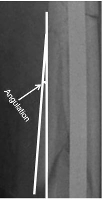
Fig. 3The fracture gap of main fragments were measured by distance between proximal and distal main fragment on AP or Lateral radiograph.
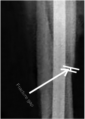
Fig. 4
(A) Complicated case Lateral radiograph showing initial post-op finding with large amount of displacement.
(B) Complicated case Lateral radiograph showing no union evidence with remnanat displacement.
(C) Immediate post-operative Lateral radiograph showing bone graft status using mini-open procedure.
(D) Three months later follow-up Lateral raidograph showing radiologic union.

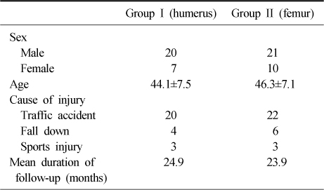
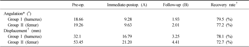
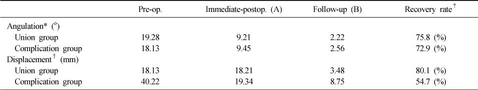
Figure & Data
REFERENCES
Citations
Citations to this article as recorded by 
- Risk Factors for Failure of Nonsurgical Management of Ulnar Shaft Fractures
Carew C. Giberson-Chen, Cassandra M. Chruscielski, Dafang Zhang, Philip E. Blazar, Brandon Earp
The Journal of Hand Surgery.2025; 50(4): 497.e1. CrossRef - The impact of the third fragment features on the healing of femoral shaft fractures managed with intramedullary nailing: a radiological study
Giovanni Vicenti, Massimiliano Carrozzo, Vincenzo Caiaffa, Antonella Abate, Giuseppe Solarino, Davide Bizzoca, Roberto Maddalena, Giulia Colasuonno, Vittorio Nappi, Francesco Rifino, Biagio Moretti
International Orthopaedics.2019; 43(1): 193. CrossRef - Reply to “Letter to the Editor on: The impact of the third fragment features on the healing of femoral shaft fractures managed with intramedullary nailing: a radiological study”
Giovanni Vicenti, Massimiliano Carrozzo, Davide Bizzoca, Biagio Moretti
International Orthopaedics.2019; 43(6): 1545. CrossRef - Letter to the Editor on “The impact of the third fragment features on the healing of femoral shaft fractures managed with intramedullary nailing: a radiological study”
Shih-Jie Lin, Kevin Liaw, Tsan-Wen Huang
International Orthopaedics.2019; 43(6): 1543. CrossRef - The impact of the third fragment features on the healing of femoral shaft fractures managed with intramedullary nailing: a radiological study
Giovanni Vicenti, Massimiliano Carrozzo, Vincenzo Caiaffa, Antonella Abate, Giuseppe Solarino, Davide Bizzoca, Roberto Maddalena, Giulia Colasuonno, Vittorio Nappi, Francesco Rifino, Biagio Moretti
International Orthopaedics.2018;[Epub] CrossRef - Comparison of the Result of the Intramedullary Nail Fixation and Plate Fixation in Humeral Shaft Fracture with Butterfly Fragments
Duk-Hwan Kho, Hyeung-June Kim, Byoung-Min Kim, Hyun-Ryong Hwang
The Korean Journal of Sports Medicine.2016; 34(2): 120. CrossRef - Clinical and Radiographical Follow-up for Residual Displacement of Fracture Fragments after Interlocking Intramedullary Nailing in Humeral Shaft Fractures
Jae-Kwang Yum, Dong-Ju Lim, Eui-Yub Jung, Su-Een Sohn
The Journal of the Korean Shoulder and Elbow Society.2013; 16(2): 107. CrossRef
 Cite
CiteThe Fate of Butterfly Fragments in Extremity Shaft Comminuted Fractures Treated with Closed Interlocking Intramedullary Nailing
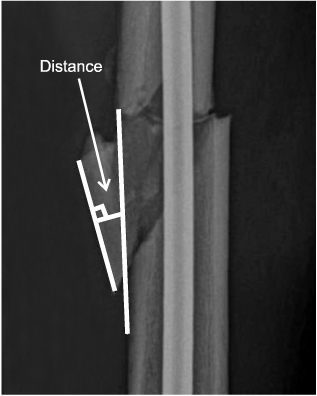


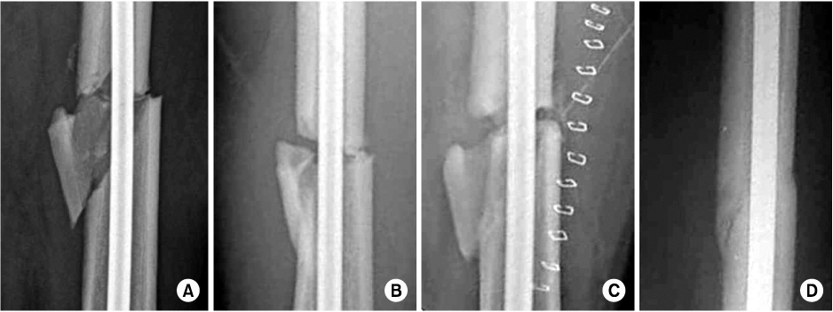
Fig. 1
The degree of displacement of butterfly fragments were measured by longer pependicualr distance between shaft cortex parallel line and mid-point of fragment cortex parallel line.
Fig. 2
The degree of angulation of butterfly fragments were measured by angle between parallel line of shaft cortex and main fragment cortex on AP or Lateral radiograph.
Fig. 3
The fracture gap of main fragments were measured by distance between proximal and distal main fragment on AP or Lateral radiograph.
Fig. 4
(A) Complicated case Lateral radiograph showing initial post-op finding with large amount of displacement.
(B) Complicated case Lateral radiograph showing no union evidence with remnanat displacement.
(C) Immediate post-operative Lateral radiograph showing bone graft status using mini-open procedure.
(D) Three months later follow-up Lateral raidograph showing radiologic union.
Fig. 1
Fig. 2
Fig. 3
Fig. 4
The Fate of Butterfly Fragments in Extremity Shaft Comminuted Fractures Treated with Closed Interlocking Intramedullary Nailing
The patient demographics
The recovery rate of angulation and displacement
*Statically no significant difference (p=0.134) beween group I and group II. †Statically no significant difference (p=0.388) beween group I and group II. ‡(A-B)/A×100.
The recovery rate of normal-union group and complication group
*Statically no significant difference (p=0.560) beween two groups. †Statically significant difference (p=0.001) beween two groups. ‡(A-B)/A×100.
Table 1
The patient demographics
Table 2
The recovery rate of angulation and displacement
*Statically no significant difference (p=0.134) beween group I and group II. †Statically no significant difference (p=0.388) beween group I and group II. ‡(A-B)/A×100.
Table 3
The recovery rate of normal-union group and complication group
*Statically no significant difference (p=0.560) beween two groups. †Statically significant difference (p=0.001) beween two groups. ‡(A-B)/A×100.

