 E-submission
 TOTA
TOTA TOTS
TOTS
Articles
- Page Path
- HOME > J Musculoskelet Trauma > Volume 27(1); 2014 > Article
-
Original Article
- Results of Intramedullary Nailing of Femoral Shaft Fracture: Trochanteric Entry Portal (Sirus Nail) versus Piriformis Entry Portal (M/DN Nail)
- Sang Ho Ha, M.D., Woong-Hee Kim, M.D., Gwang Chul Lee, M.D.
-
Journal of the Korean Fracture Society 2014;27(1):50-57.
DOI: https://doi.org/10.12671/jkfs.2014.27.1.50
Published online: January 17, 2014
Department of Orthopaedic Surgery, School of Medicine, Chosun University, Gwangju, Korea.
- Address reprint requests to: Gwang Chul Lee, M.D. Department of Orthopaedic Surgery, Chosun University Hospital, 365 Pilmun-daero, Dong-gu, Gwangju 501-717, Korea. Tel: 82-62-220-3147, Fax: 82-62-226-3379, leekci@chosun.ac.kr
• Received: September 13, 2013 • Revised: October 21, 2013 • Accepted: December 16, 2013
Copyright © 2014 The Korean Fracture Society. All rights reserved.
This is an Open Access article distributed under the terms of the Creative Commons Attribution Non-Commercial License (http://creativecommons.org/licenses/by-nc/3.0/) which permits unrestricted non-commercial use, distribution, and reproduction in any medium, provided the original work is properly cited.
- 2,164 Views
- 22 Download
- 7 Crossref
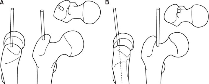
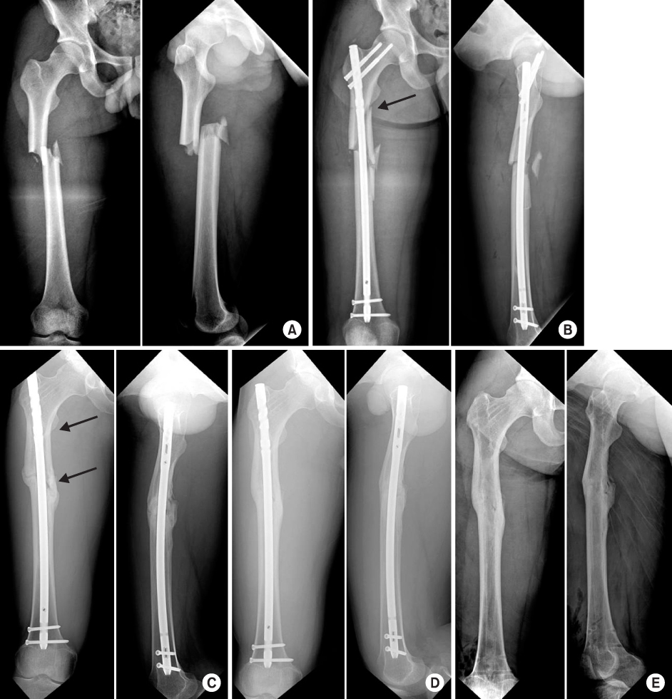
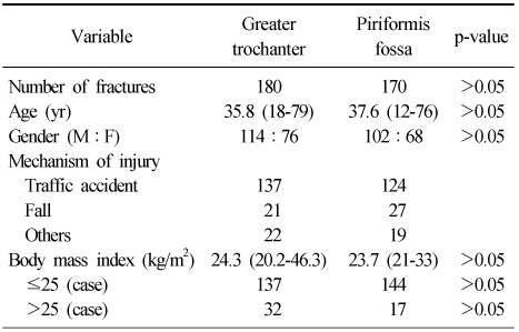
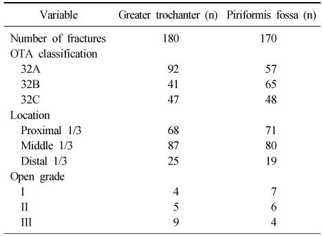
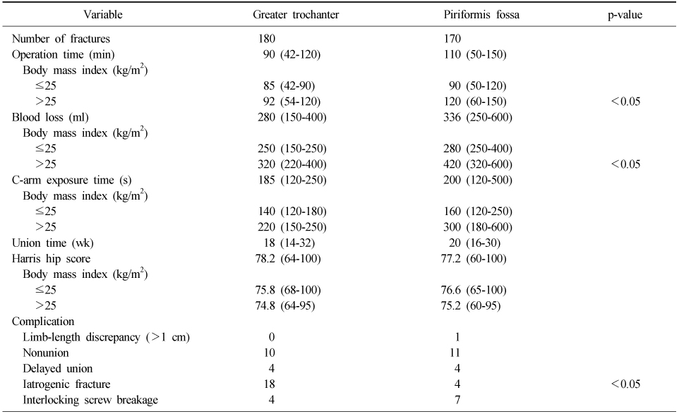
Figure & Data
REFERENCES
Citations
Citations to this article as recorded by 
- Analysis of different entry portals for femoral nail with two different nail designs-straight nail versus lateral angulated nail - Does it make a difference?
Sanjay Yadav, Saurabh Singh, Anil Kumar Rai
Journal of Clinical Orthopaedics and Trauma.2019; 10(5): 912. CrossRef - Comparing Entry Points for Antegrade Nailing of Femoral Shaft Fractures
Ujash Sheth, Chetan Gohal, Jaskarndip Chahal, Aaron Nauth, Tim Dwyer
Orthopedics.2016;[Epub] CrossRef - The Curative Effect Comparison Between Prolonged Third Generation of Gamma Nail and Prolonged Dynamic Hip Screw Internal Fixation in Treating Femoral Intertrochanteric Fracture and the Effect on Infection
Wenye He, Wei Zhang
Cell Biochemistry and Biophysics.2015; 71(2): 695. CrossRef - Treatment of Femur Subtrochanteric Fracture Using the Intramedullary Long Nail; Comparison of Closed Reduction and Minimal Open Reduction
Sang Joon Lee, Sang Hong Lee, Sang Soo Park, Hyung Seok Park
Journal of the Korean Orthopaedic Association.2015; 50(1): 18. CrossRef - Failure to Remove a Trochanteric Entry Femoral Nail and Its Cause in Adolescent Patients: Two Cases Report
Ji-Hwan Kim, Seung-Oh Nam, Young-Soo Byun, Han-Sang Kim
Journal of the Korean Fracture Society.2015; 28(1): 71. CrossRef - Treatment of the Femoral Fracture Using Sirus® Nail: A Comparison of Complication according to the Entry Potal
Young-Yool Chung, Dong-Hyuk Choi, Dae-Hyun Yoon, Jung-Ho Lee, Ji-Hun Park
Journal of the Korean Fracture Society.2015; 28(2): 103. CrossRef - Comparison of Greater Trochanter Versus Piriformis Entry Nail for Treatment of Femur Shaft Fracture
Jong-Hee Lee, Jong-Hoon Park, Si-Yeong Park, Seong-Cheol Park, Seung-Beom Han
Journal of the Korean Fracture Society.2014; 27(4): 287. CrossRef
 Cite
CiteResults of Intramedullary Nailing of Femoral Shaft Fracture: Trochanteric Entry Portal (Sirus Nail) versus Piriformis Entry Portal (M/DN Nail)


Fig. 1
(A) Trochanteric entry portal: the correct location for the trochanteric entry point immediately next to the tip of the greater trochanter on the anteroposterior view and exactly centered on the axial view. (B) Piriformis entry point: the correct position for the piriformis fossa entry point is immediately medial to the tip of the greater trochanter.
Fig. 2
(A) Anteroposterior and lateral radiograph of a 21-year-old male who received a femur midshaft fracture in a traffic accident. (B) Postoperative radiograph: iatrogenic fracture (arrow) during closed nailing of the femoral shaft fracture via the trochanteric tip. (C) Anteroposterior and lateral radiographs of femoral diaphyseal nonunion after 4 months. Nail dynamization was performed, femoral diaphyseal nonunion (arrows). (D) Bone union observed on radiographs taken 2 years after operation. (E) After implant removal.
Fig. 1
Fig. 2
Results of Intramedullary Nailing of Femoral Shaft Fracture: Trochanteric Entry Portal (Sirus Nail) versus Piriformis Entry Portal (M/DN Nail)
Patients Characteristics
Values are presented as number only or median (range).
Fracture Characteristics
OTA classification 32A: simple fractures, 32B: wedge fractures, 32C: complex fractures. Open grade: Gustilo classification.
Comparison of Results
Values are presented as number only or median (range).
Table 1
Patients Characteristics
Values are presented as number only or median (range).
Table 2
Fracture Characteristics
OTA classification 32A: simple fractures, 32B: wedge fractures, 32C: complex fractures. Open grade: Gustilo classification.
Table 3
Comparison of Results
Values are presented as number only or median (range).

