 E-submission
 TOTA
TOTA TOTS
TOTS
Articles
- Page Path
- HOME > J Musculoskelet Trauma > Volume 22(1); 2009 > Article
-
Original Article
- A Comparison of Extensile Lateral Approach and Sinus Tarsi Approach for the Sanders Type II Calcaneal Fracture
- Jeong-Seok Moon, M.D., Woo-Chun Lee, M.D.
-
Journal of the Korean Fracture Society 2009;22(1):13-18.
DOI: https://doi.org/10.12671/jkfs.2009.22.1.13
Published online: January 31, 2009
Department of Orthopedic Surgery, Seoul Paik Hospical, College of Medicine, Inje University, Seoul, Korea.
- Address reprint requests to: Jeong-Seok Moon, M.D. Department of Orthopedic Surgery, Seoul Paik Hospical, College of Medicine, Inje University, 85, Jeo-dong 2-ga, Jung-gu, Seoul 100-032, Korea. Tel: 82-2-2270-0028, Fax: 82-2-2270-0023, moonbak502@hanmail.net
• Received: September 30, 2008 • Accepted: November 20, 2008
Copyright © 2009 The Korean Fracture Society. All rights reserved.
This is an Open Access article distributed under the terms of the Creative Commons Attribution Non-Commercial License (http://creativecommons.org/licenses/by-nc/3.0/) which permits unrestricted non-commercial use, distribution, and reproduction in any medium, provided the original work is properly cited.
- 1,417 Views
- 9 Download
- 9 Crossref
Abstract
-
Purpose
- To compare the clinical results between the extensile lateral approach and sinus tarsi approach in the open reduction of the Sanders type II calcaneal fracture.
-
Materials and Methods
- From July 2002 to Februry 2007, thirty two patients having thirty three calcaneal fractures of Sanders type II were managed with open reduction and internal fixation using the extensile lateral approach or sinus tarsi approach. The mean age of 19 patients using extensile lateral approach was 43.3 years. The mean age of 13 patients using sinus tarsi approach was 46.3 years. Clinical outcome, radiographic parameters, and postoperative complications were compared between both groups.
-
Results
- There was no difference between two groups associated with patients demographs. The mean AOFAS score and VAS between both groups were not different (p=0.716, p=0.774). The mean Bohler's angle and Gissane's angle between both groups were not different (p=0.343, p=0.357). Two cases of sural nerve injury, one malunion, and one deep infection were occurred in the group of extensile lateral approach. However, patients using sinus tarsi approach had no postoperative complications.
-
Conclusion
- The clinical results of sinus tarsi approach may be comparable with those of extensile lateral approach, with the advantages of reduced risk of postoperative complications.
- 1. Bèzes H, Massart P, Delvaux D, Fourquet JP, Tazi F. The operative treatment of intraarticular calcaneal fractures. Indications, technique, and results in 257 cases. Clin Orthop Relat Res, 1993;(290):55-59.
- 2. Carr JB. Surgical treatment of intra-articular calcaneal fractures: a review of small incision approaches. J Orthop Trauma, 2005;19:109-117.
- 3. Eastwood DM, Gregg PJ, Atkins RM. Intra-articular fractures of the calcaneum. Part I: Pathological anatomy and classification. J Bone Joint Surg Br, 1993;75:183-188.ArticlePDF
- 4. Ebraheim NA, Elgafy H, Sabry FF, Freih M, Abou-Chakra IS. Sinus tarsi approach with trans-articular fixation for displaced intra-articular fractures of the calcaneus. Foot Ankle Int, 2000;21:105-113.ArticlePDF
- 5. Freeman BJ, Duff S, Allen PE, Nicholson HD, Atkins RM. The extended lateral approach to the hindfoot. Anatomical basis and surgical implications. J Bone Joint Surg Br, 1998;80:139-142.
- 6. Gavlik JM, Rammelt S, Zwipp H. Percutaneous, arthroscopically-assisted osteosynthesis of calcaneus fractures. Arch Orthop Trauma Surg, 2002;122:424-428.ArticlePDF
- 7. Gupta A, Ghalambor N, Nihal A, Trepman E. The modified Palmer lateral approach for calcaneal fractures: wound healing and postoperative computed tomographic evaluation of fracture reduction. Foot Ankle Int, 2003;24:744-753.ArticlePDF
- 8. Hwang DS, Lee JK, Oh HR, Lee SJ. Percutaneous & minimal internal fixation of displaced intraarticular calcaneal fractures. J Korean Soc Fract, 1997;10:233-241.Article
- 9. Letournel E. Open treatment of acute calcaneal fractures. Clin Orthop Relat Res, 1993;290:60-67.Article
- 10. Magnan B, Bortolazzi R, Marangon A, Marino M, Dall'Oca C, Bartolozzi P. External fixation for displaced intra-articular fractures of the calcaneum. J Bone Joint Surg Br, 2006;88:1474-1479.ArticlePDF
- 11. Maskill JD, Bohay DR, Anderson JG. Calcaneus fractures: a review article. Foot Ankle Clin, 2005;10:463-489.Article
- 12. McGarvey WC, Burris MW, Clanton TO, Melissinos EG. Calcaneal fractures: indirect reduction and external fixation. Foot Ankle Int, 2006;27:494-499.ArticlePDF
- 13. Park IH, Song KW, Shin SI, Lee JY, Kim TG, Park RS. Displaced intra-articular calcaneal fracture treated surgically with limited posterior incision. Foot Ankle Int, 2000;21:195-205.ArticlePDF
- 14. Rammelt S, Gavlik JM, Barthel S, Zwipp H. The value of subtalar arthroscopy in the management of intra-articular calcaneus fractures. Foot Ankle Int, 2002;23:906-916.ArticlePDF
- 15. Seong BY, Park DS, Park SJ, Kim SW. Treatment of intraarticular calcaneal fractures using ilizarov external fixation. J Korean Soc Fract, 1998;11:591-596.Article
- 16. Stephenson JR. Surgical treatment of displaced intra-articular fractures of the calcaneus. A combined lateral and medial approach. Clin Orthop Relat Res, 1993;290:68-75.
- 17. Thermann H, Krettek C, Hüfner T, Schratt HE, Albrecht K, Tscherne H. Management of calcaneal fractures in adults. Conservative versus operative treatment. Clin Orthop Relat Res, 1998;353:107-124.
- 18. Zwipp H, Tscherne H, Thermann H, Weber T. Osteosynthesis of displaced intraarticular fractures of the calcaneus. Results in 123 cases. Clin Orthop Relat Res, 1993;290:76-86.
REFERENCES
Fig. 1
(A) Extensile lateral approach demonstrates exellent exposure of the subtalar joint and posterior tuberosity through the wide subperiosteal dissection.
(B) Sinus tarsi approach through the small incision over the sinus tarsi can not see the posterior tuberosity.
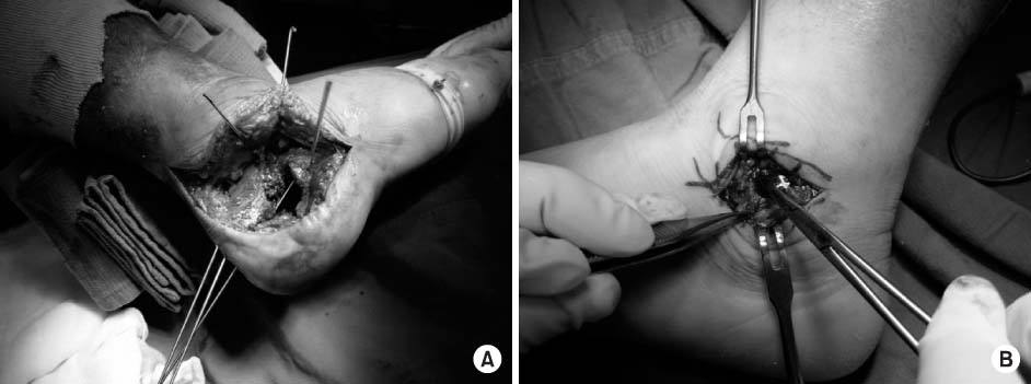
Fig. 2
(A) Lateral radiograph of the calcaneus shows measurements of the Bohler angle and the critical angle of Gissane.
(B) Broden's radiograph of the calcaneus shows measurement of the displacement between fracture fragments.
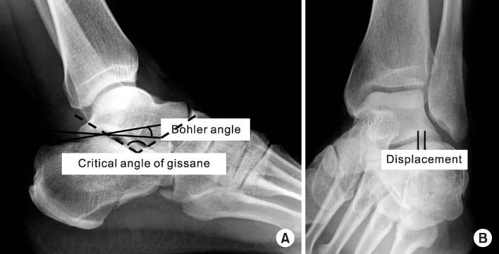
Table 1Patients demographs with comparison between two groups who underwent surgery using the extensile lateral approach or the sinus tarsi approach
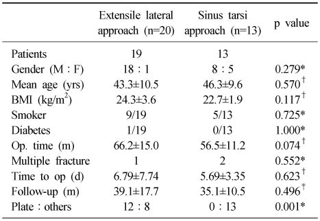
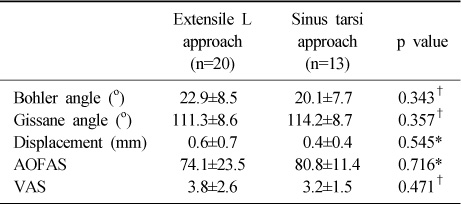
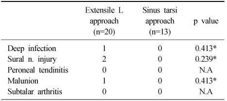
Figure & Data
REFERENCES
Citations
Citations to this article as recorded by 
- The Extensile Lateral Approach to the Calcaneus
Rohan Bhimani, Kush C. Shah, Rishin J. Kadakia
Techniques in Foot & Ankle Surgery.2025;[Epub] CrossRef - Extensile lateral versus sinus tarsi approach for calcaneal fractures
Chuangang Peng, Baoming Yuan, Wenlai Guo, Na Li, Heng Tian
Medicine.2021; 100(31): e26717. CrossRef - Lateral Extensile Approach Versus Minimal Incision Approach for Open Reduction and Internal Fixation of Displaced Intra-articular Calcaneal Fractures: A Meta-analysis
Andrea Seat, Christopher Seat
The Journal of Foot and Ankle Surgery.2020; 59(2): 356. CrossRef - Surgical Treatment of Calcaneal Fractures of Sanders Type II and III by A Minimally Invasive Technique with 6.5 mm Cancellous Screw
Yong Seung Oh, Kyung Ho Lee, Jung Ho Kim, Myoung Jin Lee
Journal of Korean Foot and Ankle Society.2019; 23(3): 116. CrossRef - A systematic review and meta-analysis of the sinus tarsi and extended lateral approach in the operative treatment of displaced intra-articular calcaneal fractures
Tomasz L. Nosewicz, Siem A. Dingemans, Manouk Backes, Jan S.K. Luitse, J. Carel Goslings, Tim Schepers
Foot and Ankle Surgery.2019; 25(5): 580. CrossRef - Meta‐analysis of two surgical approaches for calcaneal fractures: sinus tarsi versus extensile lateral approach
Fei Zhang, Hongtao Tian, Shilun Li, Bo Liu, Tianhua Dong, Yanbin Zhu, Yingze Zhang
ANZ Journal of Surgery.2017; 87(3): 126. CrossRef - Usefulness of Treatment with 6.5 mm Cancellous Screw and Steinmann Pin Fixation for Calcaneal Joint Depression Fracture
Gi-Soo Lee, Chan Kang, Deuk-Soo Hwang, Chang-Kyun Noh, Gi-Young Lee
Journal of Korean Foot and Ankle Society.2015; 19(1): 11. CrossRef - Open reduction and internal fixation with conventional plate via L-shaped lateral approach versus internal fixation with percutaneous plate via a sinus tarsi approach for calcaneal fractures – A randomized controlled trial
Shengli Xia, Yaogang Lu, Huizhong Wang, Zuming Wu, Ziping Wang
International Journal of Surgery.2014; 12(5): 475. CrossRef - Intra-articular Calcaneal Fractures Treated with Open Reduction and Internal Fixation -A Comparative Study between Groups with and without Bone Graft-
Hong Moon Sohn, Sang Ho Ha, Jun Young Lee, Sung Hwan Jo, Hoon Yang
Journal of the Korean Fracture Society.2010; 23(2): 180. CrossRef
 Cite
CiteA Comparison of Extensile Lateral Approach and Sinus Tarsi Approach for the Sanders Type II Calcaneal Fracture


Fig. 1
(A) Extensile lateral approach demonstrates exellent exposure of the subtalar joint and posterior tuberosity through the wide subperiosteal dissection.
(B) Sinus tarsi approach through the small incision over the sinus tarsi can not see the posterior tuberosity.
Fig. 2
(A) Lateral radiograph of the calcaneus shows measurements of the Bohler angle and the critical angle of Gissane.
(B) Broden's radiograph of the calcaneus shows measurement of the displacement between fracture fragments.
Fig. 1
Fig. 2
A Comparison of Extensile Lateral Approach and Sinus Tarsi Approach for the Sanders Type II Calcaneal Fracture
Patients demographs with comparison between two groups who underwent surgery using the extensile lateral approach or the sinus tarsi approach
*chi-square test, †Mann-Whitney U-test, ‡student t-test.
Comparison of the radiographic and clinical results between two groups after surgical treatment through the extensile lateral approach or through the sinus tarsi approach
*Mann-Whitney U-test, †student t-test.
Complications after surgical treatment through the extensile lateral approach or through the sinus tarsi approach
*chi-square test, N.A: not assessable.
Table 1
Patients demographs with comparison between two groups who underwent surgery using the extensile lateral approach or the sinus tarsi approach
*chi-square test, †Mann-Whitney U-test, ‡student t-test.
Table 2
Comparison of the radiographic and clinical results between two groups after surgical treatment through the extensile lateral approach or through the sinus tarsi approach
*Mann-Whitney U-test, †student t-test.
Table 3
Complications after surgical treatment through the extensile lateral approach or through the sinus tarsi approach
*chi-square test, N.A: not assessable.

