 E-submission
 TOTA
TOTA TOTS
TOTS
Articles
- Page Path
- HOME > J Musculoskelet Trauma > Volume 34(2); 2021 > Article
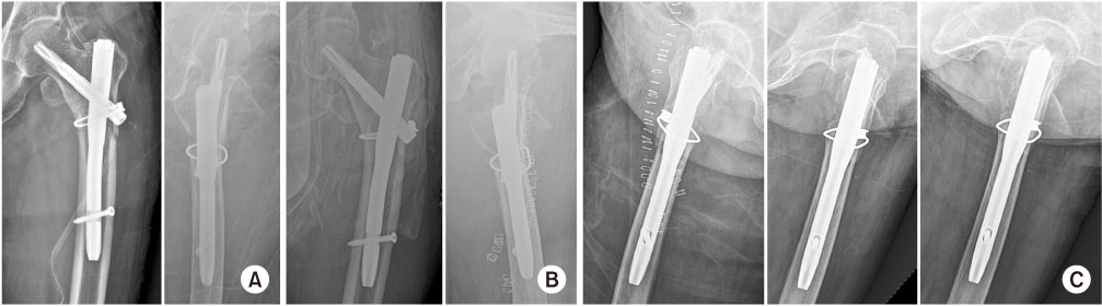
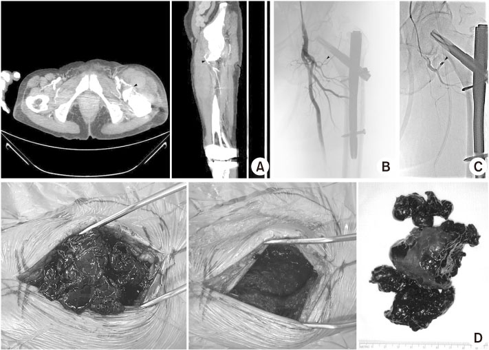
- Case Report Injury of the Ascending Branch of the Lateral Femoral Circumflex Artery Caused by a Spike of the Displaced Lesser Trochanter in an Intertrochanteric Femoral Fracture - A Case Report -
- Soon Ho Huh, Hong-Man Cho, Jiyeon Park
-
Journal of Musculoskeletal Trauma 2021;34(2):71-75.
DOI: https://doi.org/10.12671/jkfs.2021.34.2.71
Published online: April 30, 2021
1Department of Orthopedic Surgery, St. Carollo Hospital, Suncheon, Korea
2Department of Orthopedic Surgery, Gwangju Veterans Hospital, Gwangju, Korea
2Department of Orthopedic Surgery, Gwangju Veterans Hospital, Gwangju, Korea
- 1,156 Views
- 7 Download
- 2 Crossref
- 0 Scopus

Citations
Citations to this article as recorded by 
- Delayed Deep Femoral Artery Injury Secondary to Migrated Lesser Trochanter Fragment After Intertrochanteric Fracture Fixation: A Case Report and Updated Literature Review
Slavko Čičak, Josip Kocur, Vedran Farkaš, Petra Čičak, Stjepan Ištvanić, Marko Lovrić, Marko Perić, Nenad Koruga, Tomislav Ištvanić
Geriatric Orthopaedic Surgery & Rehabilitation.2025;[Epub] CrossRef - Vascular Complications Following Trans-Trochanteric Fracture: Case Report and Literature Review
Robert Bot, Adrian Tirla, Simona Daniela Cavalu
Reports.2025; 8(4): 191. CrossRef
 Cite
Cite
