 E-submission
 TOTA
TOTA TOTS
TOTS
Articles
- Page Path
- HOME > J Musculoskelet Trauma > Volume 25(1); 2012 > Article
-
Case Report
- Combined Ipsilateral Fracture and Dislocation of Hip, Knee and Foot Joints: A Case Report
- Hyoung-Soo Kim, M.D., Ph.D., Ju-Hak Kim, M.D., Ph.D., Sang-Joon Park, M.D., Jae-Won Hyung, M.D.
-
Journal of the Korean Fracture Society 2012;25(1):73-76.
DOI: https://doi.org/10.12671/jkfs.2012.25.1.73
Published online: January 31, 2012
Department of Orthopedic Surgery, Myongji Hospital, Kwandong University College of Medicine, Goyang, Korea.
- Address reprint requests to: Ju-Hak Kim, M.D., Ph.D. Department of Orthopedic Surgery, Myongji Hospital, Kwandong University College of Medicine, 697-24, Hwajeong-dong, Deogyang-gu, Goyang 412-270, Korea. Tel: 82-31-810-6530, Fax: 82-31-964-6649, hand0123@kwandong.ac.kr
• Received: November 15, 2011 • Accepted: December 19, 2011
Copyright © 2012 The Korean Fracture Society
- 887 Views
- 3 Download
Abstract
- Although clinical cases of ipsilateral knee and hip joint dislocation have been reported, there are no reports of simultaneous ipsilateral hip, knee, and foot dislocations. We report here a case of a patient who had ipsilateral hip, knee, and foot joint dislocations, and review the relevant literature.
- 1. Kim SK, Park JB, Lee JH, Chang H. Isolated plantar midtarsal dislocation: a case report. J Korean Soc Fract, 1998;11:226-229.Article
- 2. Kotter A, Wieberneit J, Braun W, Rüter A. The Chopart dislocation. A frequently underestimated injury and its sequelae. A clinical study. Unfallchirurg, 1997;100:737-741.
- 3. Levy BA, Dajani KA, Whelan DB, et al. Decision making in the multiligament-injured knee: an evidence-based systematic review. Arthroscopy, 2009;25:430-438.ArticlePDF
- 4. Park HG. Patients accompanied with simultaneous anterior dislocation of hip and anterior dislocation of knee: a case report. J Korean Fract Soc, 2009;22:185-188.Article
- 5. Roeder LF Jr, DeLee JC. Femoral head fractures associated with posterior hip dislocation. Clin Orthop Relat Res, 1980;(147):121-130.
- 6. Shelbourne KD, Porter DA, Clingman JA, McCarroll JR, Rettig AC. Low-velocity knee dislocation. Orthop Rev, 1991;20:995-1004.
REFERENCES

Fig. 2Plain knee radiograph shows posterolateral dislocation of the knee with undisplaced fracture of inferior pole of the patella.
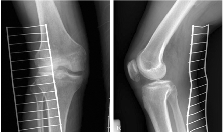


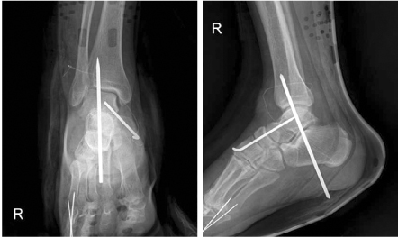

Fig. 824 months after surgery, plain pelvis radiograph shows neither avascular necrosis nor traumatic arthritic changes.


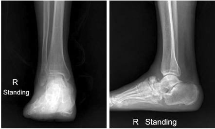
Figure & Data
REFERENCES
Citations
Citations to this article as recorded by 
 Cite
CiteCombined Ipsilateral Fracture and Dislocation of Hip, Knee and Foot Joints: A Case Report
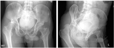

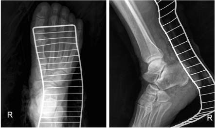
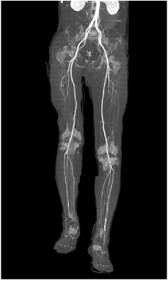
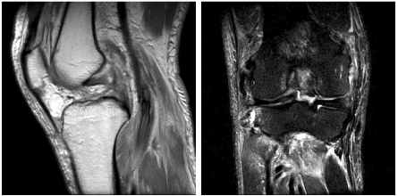

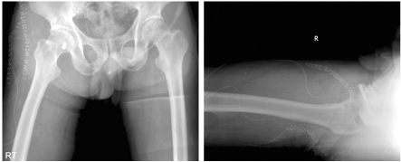
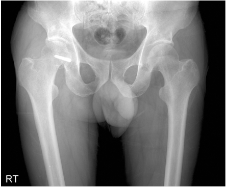
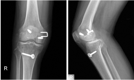

Fig. 1
Plain pelvis radiograph shows posterior dislocation of hip and fracture of femoral head.
Fig. 2
Plain knee radiograph shows posterolateral dislocation of the knee with undisplaced fracture of inferior pole of the patella.
Fig. 3
Preoperative ankle radiograph shows midtarsal plantar dislocation.
Fig. 4
Vascular CT shows no occlusion of blood flow.
Fig. 5
MRI shows ruptures of the ACL, PCL, MCL, LCL & popliteus ligaments.
Fig. 6
After surgery, reduction status of ankle is shown.
Fig. 7
Postoperative radiograph shows reduction status of hip.
Fig. 8
24 months after surgery, plain pelvis radiograph shows neither avascular necrosis nor traumatic arthritic changes.
Fig. 9
24 months after surgery, plain knee radiograph shows stable knee with all corner reconstruction status.
Fig. 10
Plain radiograph of 24 months after surgery shows residual subluxation of midtarsal joint.
Fig. 1
Fig. 2
Fig. 3
Fig. 4
Fig. 5
Fig. 6
Fig. 7
Fig. 8
Fig. 9
Fig. 10
Combined Ipsilateral Fracture and Dislocation of Hip, Knee and Foot Joints: A Case Report

