 E-submission
 TOTA
TOTA TOTS
TOTS
Search
- Page Path
- HOME > Search
Original Articles
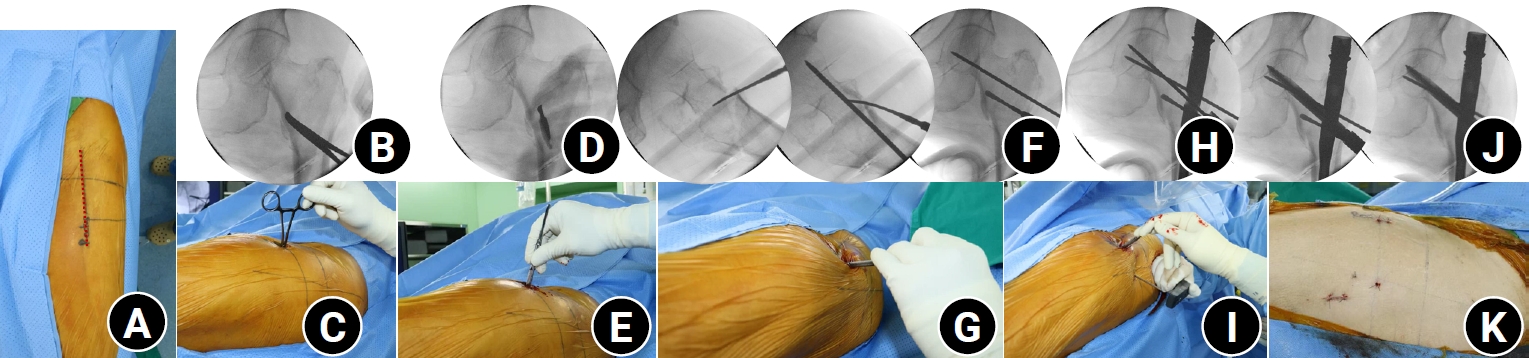
- Percutaneous anterior leverage technique for anteromedial cortical support in intertrochanteric femur fractures: a computed tomography-based validation study
- Whee Sung Son, Bum Jin Shim, Oog-jin Shon
- J Musculoskelet Trauma 2026;39(2):117-129. Published online March 27, 2026
- DOI: https://doi.org/10.12671/jmt.2025.00311
-
 Abstract
 PDF
PDF - Background
Anteromedial cortical support (AMCS) enhances stability in intertrochanteric femur fractures. However, reproducible, validated methods of achieving AMCS have not previously been reported. This study introduces a percutaneous anterior leverage technique and validates its AMCS effects using computed tomography (CT).
Methods
We retrospectively reviewed patients treated by a single surgeon between March 2022 and December 2024. The inclusion criteria were an AO/OTA classification of A1–A3, application of the percutaneous anterior leverage technique, available pre- and postoperative CT, and ≥6 months follow-up. Outcomes included CT-based AMCS (anterior on axial and medial on coronal images, classified as positive, neutral, or negative), time to union, union rate, changes in neck-shaft angle, and treatment failure (varus collapse, blade cut-through, or nonunion without the former two). The risk factors for failure were analyzed.
Results
Of 273 patients reviewed, 53 met the inclusion criteria. Follow-up was at least 6 months in all cases. Positive anterior support was achieved in 37 patients (69.8%) and positive medial support in 42 (79.25%). No patient demonstrated negative anterior support; one (1.9%) had negative medial support. Cortical support improved significantly after surgery. CT images demonstrated significant postoperative improvements (anterior P=0.026; medial P<0.001). Bone union was achieved in 50 patients (94.34%) at a mean of 3.93±1.48 months. The mean change in the neck-shaft angle at last follow-up was 1.75°±2.34° varus. Three patients (5.66%) experienced treatment failure. Anteromedial cortical breakage during follow-up differed between failure and nonfailure groups (P=0.002), but regression identified no independent predictors. No technique-related complications were observed.
Conclusions
Our percutaneous anterior leverage technique produced favorable CT-confirmed AMCS and high union with low failure, supporting its safety and effectiveness in intertrochanteric femur fractures. Level of evidence: IV.
- 1,204 View
- 41 Download

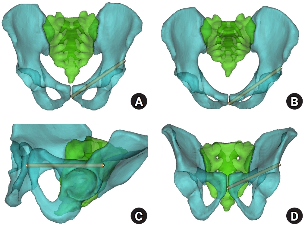
- Sex-specific bottlenecks and risk zones in the retrograde superior pubic ramus screw corridor: a 3D CT-based morphometric cadaver study
- Ji Won Jeong, Jung Tae Ahn, Gu Hee Jung, Kun Tae Kim
- J Musculoskelet Trauma 2026;39(2):103-116. Published online March 26, 2026
- DOI: https://doi.org/10.12671/jmt.2026.00066
-
Abstract
PDF
 Supplementary Material
 - Background
Superior ramus screw fixation is commonly used to stabilize anterior pelvic ring injuries but is constrained by a narrow, irregular, and curved intraosseous corridor. Trajectory-based morphometric analysis may assist in screw diameter selection and enable identification of reproducible anatomic constriction zones.
Methods
We conducted a cross-sectional computed tomography (CT)-based morphometric study of 82 cadaveric pelvises (42 males, 40 females). Bottleneck diameter was defined as the diameter of the largest fully contained virtual cylinder along the planned trajectory, and cylinder length was recorded. Orthogonal cross-sections at 9.5-mm intervals (up to 12 segments) were generated to measure segment-wise effective diameter (defined as twice the minimum centerline-to-cortex distance) and cortical clearance, which was used as a diameter-based safety margin. Segments were realigned to the acetabular start segment to define relative segment positions (Δ seg). Feasibility was assessed for prespecified screw diameters ranging from 3.5 to 7.3 mm.
Results
Mean bottleneck diameter was larger in males than in females (7.34±1.10 vs. 5.93±0.98 mm), whereas trajectory length was similar between sexes (127.85±8.54 vs. 128.85±8.20 mm). Δ seg realignment localized corridor constriction to two discrete zones: a preacetabular zone (Δ seg −6 to −4) and a periacetabular zone (Δ seg 1 to 2), where effective diameter and cortical clearance were most limited. Feasibility rates were 100% at 3.5–4.5 mm, 95.2% vs. 82.5% at 5.0 mm, 81.0% vs. 27.5% at 6.5 mm, and 59.5% vs. 10.0% at 7.3 mm in males and females, respectively.
Conclusions
Female models demonstrated smaller trajectory-wide bottleneck diameters and segment-wise effective diameters than male models. Acetabular-referenced Δ seg realignment identified two reproducible anatomic risk zones: a preacetabular zone adjacent to the obturator neurovascular bundle and a periacetabular zone near the external iliac vessels. At diameters ≥6.5 mm, cortical proximity increased more prominently in females than in males. Level of evidence: III.
- 1,066 View
- 28 Download
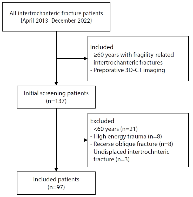
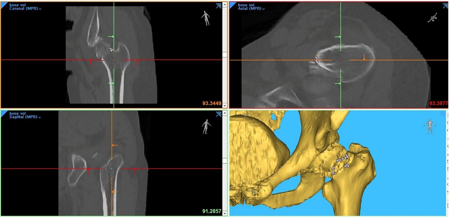
- Three-dimensional computed tomography-based differentiation of engaged versus displaced intertrochanteric fractures using the anterior fracture line: a cross-sectional study from Korea
- Jae-Suk Chang, Jin Yeob Park, Sang-Ok Chun, Chul-Ho Kim
- J Musculoskelet Trauma 2026;39(1):30-37. Published online January 25, 2026
- DOI: https://doi.org/10.12671/jmt.2025.00318
-
Abstract
PDF
- Background
With the advent of an aging society, osteoporotic fractures—particularly hip fractures—are increasing, with a 1-year mortality rate of 17%. Achieving stable fixation that enables early ambulation is essential but remains challenging because complex intertrochanteric (IT) fracture patterns are often underestimated on plain radiographs. Using three-dimensional computed tomography (3D-CT), this study analyzed whether the anterior fracture line lies medial or lateral to the IT line and examined its relationship with displacement or distal medullary canal engagement, highlighting the potential influence of the joint capsule and capsular ligaments on fracture morphology and fixation stability.
Methods
A retrospective review was conducted on 96 osteoporotic IT fractures in patients aged ≥60 years treated between April 2013 and December 2022 at National Police Hospital and Asan Medical Center, Seoul, Korea. Fractures were classified as engaged, completely displaced, and partially displaced based on 3D-CT findings. The anterior fracture-line position (medial or lateral to the IT line) and the status of the lesser trochanter (LT) were evaluated. The chi-square or Fisher exact test was used for statistical comparisons.
Results
In total, 96 patients were analyzed. Of these, 49 cases (51.0%) were classified as engaged type, 27 cases (28.1%) as completely displaced type, and 20 cases (20.8%) as partially displaced type. When comparing fracture pattern with anterior fracture-line position, the completely displaced type showed a significantly higher proportion of lateral anterior fracture lines than the other two types (P<0.001). However, no significant association was identified between fracture pattern and LT displacement. When the anterior fracture-line position and LT displacement were evaluated together, only the engaged type demonstrated a possible association between a lateral anterior fracture line and LT displacement, though the statistical significance was weak (P=0.047).
Conclusions
Fracture lines lateral to the IT line were strongly associated with displacement in IT fractures; however, their relationship with LT involvement, reflecting iliopsoas tendon traction, was not clearly demonstrated. Although the factors contributing to the engaged-type fracture remain uncertain, the statistical association between fracture pattern and anterior fracture-line position suggests that capsular structures may play a stabilizing role in select fracture configurations. Further studies are needed to clarify these anatomical interactions. Level of evidence:
- 1,409 View
- 19 Download
- Computed tomography plane reformatting to reduce projection error in measuring Pauwels angle of femoral neck fractures: a cross-sectional study
- Gyu Min Kong, Jae-Young Lim, Se-Lin Jeong, Gu-Hee Jung
- J Musculoskelet Trauma 2026;39(1):38-47. Published online January 25, 2026
- DOI: https://doi.org/10.12671/jmt.2025.00038
-
Abstract
PDF
- Objectives
This study aimed to assess fracture verticality in both coronal and axial planes after eliminating projection error in femoral neck fractures among non-older adults, and to demonstrate its clinical utility using computed tomography (CT)-based modeling at actual size.
Methods
This retrospective observational study enrolled 57 patients (30 males and 27 females), aged 20–65 years, with displaced femoral neck fractures. Based on CT images, an actual-size fracture model was constructed. The CT scanning plane was reformatted with the neck-shaft fragment realigned vertically to the ground and parallel to the femoral neck axis. Three consecutive images were used to generate coronal reformats at the centerline and posterior border to measure central and posterior coronal plane verticality as Pauwels’ angle (PA). The central image of the reformatted axial plane was used to assess axial plane verticality. Differences in verticality were analyzed using analysis of variance.
Results
Three coronal morphology types were identified: linear (n=30), concave (n=25), and convex (n=2). Two axial morphology types were observed: cephalad (n=35) and trochanteric (n=22). The mean central PA, posterior PA, and axial verticality were 55.43°±13.79°, 51.44°±11.13°, and 85.74°±18.41°, respectively. Only the central PA showed a significant difference (P<0.001). The PA was significantly higher in the linear coronal type between images (P<0.05) and in the trochanteric axial type (P<0.05).
Conclusions
After reformatting the scanning plane, the central PA showed significant variation between images. Femoral neck fractures of the linear type in the coronal plane and the trochanteric type in the axial plane demonstrated greater verticality than other morphological types. Level of evidence:
- 790 View
- 15 Download
- Relationship of lateral malleolar fracture patterns to posterior malleolar fracture morphology in supination-external rotation ankle fractures in Korea: a retrospective cohort study
- Jong-Eun Kim, Chan-Jin Park, Jun-Young Lee, Keun-Bae Lee, Gun-Woo Lee
- J Musculoskelet Trauma 2025;38(4):212-220. Published online October 24, 2025
- DOI: https://doi.org/10.12671/jmt.2025.00234
-
Abstract
PDF
- Background
Posterior malleolar fractures frequently accompany rotational ankle fractures. However, the morphological relationship between lateral and posterior malleolar fractures in supination-external rotation (SER) ankle fractures remains unclear. This study aimed to classify lateral malleolar fracture patterns in SER type 3 and 4 ankle fractures and investigated their associations with posterior malleolar fracture morphology.
Methods
We retrospectively reviewed 132 patients with SER type 3 or 4 ankle fractures and concurrent posterior malleolar fractures between January 2016 and December 2021. Lateral malleolar fractures were categorized as fibular fractures extending <4.5 cm proximal to the ankle joint (102 ankles) or fibular fractures extending ≥4.5 cm proximal to the ankle joint (30 ankles) based on posterior cortex height measured using three-dimensional computed tomography (3D-CT). Posterior malleolar fracture morphology was assessed using the Haraguchi and Bartonicek classifications. Quantitative parameters—including fracture height, angle, and articular involvement—were analyzed using 3D-CT imaging.
Results
Fibular fractures extending ≥4.5 cm proximal to the ankle joint were associated with a significantly higher frequency of Haraguchi type II and Bartonicek types 3 and 4 posterior malleolar fractures. This group also exhibited greater articular involvement (19.2% vs. 12.0%) and posterior cortical height (55.4 mm vs. 24.8 mm) compared to the <4.5 cm group (all P<0.001).
Conclusions
In SER type 3 and 4 ankle fractures, a fibular fracture extending ≥4.5 cm proximal to the ankle joint may be associated with posterior malleolar fractures exhibiting greater articular involvement and medial extension. Preoperative evaluation of the lateral malleolar fracture pattern may provide useful insights into posterior malleolar morphology and assist in surgical planning. However, these findings should be interpreted with caution due to inherent study limitations. Level of evidence: IV
- 1,945 View
- 40 Download
- Prediction of Syndesmotic Instability according to the Lateral Malleolus Fracture Pattern in Supination-External Rotation Type Ankle Fractures: Short Oblique versus Long Oblique Fracture
- Chan-Jin Park, Min-Su Lee, Keun-Bae Lee
- J Korean Fract Soc 2024;37(1):39-45. Published online January 31, 2024
- DOI: https://doi.org/10.12671/jkfs.2024.37.1.39
-
Abstract
PDF
- Purpose
This study examined whether preoperative radiological evaluations can predict syndesmotic instability according to the lateral malleolus fracture pattern in supination-external rotation-type ankle fractures.
Materials and Methods
This study enrolled 132 patients (132 ankles) with supination-external rotation stage 3 and 4 ankle fractures. Three-dimensional computed tomography was used for the morphological classification of the lateral malleolus fractures. A long oblique fracture was defined when the posterior cortical bone height of the fracture was 4.5 cm or more from the plafond of the distal tibial articular surface. A short oblique fracture was defined when the height was less than 4.5 cm. The demographic characteristics and syndesmotic instability of the two groups were evaluated.
Results
Short oblique fractures were confirmed in 102 cases, and long oblique fractures were confirmed in 30 cases. Long oblique fractures occurred at a statistically significantly higher incidence in younger ages and among males compared to short oblique fractures. Syndesmotic instability was more common in long oblique fractures.
Conclusion
In supination-external rotation-type ankle fractures, syndesmotic instability was observed in approximately 13%. Specifically, when the fracture pattern of the lateral malleolus is long oblique, the incidence of syndesmotic instability is approximately three times higher than in short oblique fractures. Therefore, meticulous evaluations of the lateral malleolus fracture pattern and establishing an appropriate treatment plan before surgery are crucial. -
Citations
Citations to this article as recorded by
- Relationship of lateral malleolar fracture patterns to posterior malleolar fracture morphology in supination-external rotation ankle fractures in Korea: a retrospective cohort stduy
Jong-Eun Kim, Chan-Jin Park, Jun-Young Lee, Keun-Bae Lee, Gun-Woo Lee
Journal of Musculoskeletal Trauma.2025; 38(4): 212. CrossRef
- Relationship of lateral malleolar fracture patterns to posterior malleolar fracture morphology in supination-external rotation ankle fractures in Korea: a retrospective cohort stduy
- 1,188 View
- 14 Download
- 1 Crossref
- Comparison of the Size of the Posterior Malleolar Fragment in Trimalleolar Ankle Fractures Measured Using Lateral Plain Radiography and Three-Dimensional Computed Tomography
- Gun-Woo Lee, Dong-Min Jung, Woo Kyoung Kwak, Keun-Bae Lee
- J Korean Fract Soc 2022;35(3):91-96. Published online July 31, 2022
- DOI: https://doi.org/10.12671/jkfs.2022.35.3.91
-
Abstract
PDF
- Purpose
This study aimed to evaluate and compare the accuracy of the size of the posterior malleolar fragment measured using lateral plain radiography and three-dimensional computed tomography (3DCT) in patients with ankle trimalleolar fractures.
Materials and Methods
This study enrolled 80 patients (80 ankles) with ankle trimalleolar fractures and analyzed the size of the posterior malleolar fragments using plain radiography and 3D-CT. The articular involvement of the posterior malleolar fragments was measured as a percentage of the articular surface in the sagittal length of the tibial plafond using lateral plain radiography, and the articular surface area was directly measured using 3D-CT. In addition, we classified the patients into three groups based on the morphology of the posterior malleolar fracture, according to the Haraguchi classification method, and evaluated and compared the accuracy of the size of the posterior malleolar fragments.
Results
The mean articular involvement of the posterior malleolar fragments on plain radiography was 27.6% (range, 6.0%-53.1%), which was significantly higher than the mean of 21.9% (range, 4.7%-47.1%) measured using 3D-CT (p=0.004). In the analysis, according to the fracture morphology, the mean difference between the two methods was the largest for type I fractures at 9.1% (range, 1.8%-19.5%) and the smallest for type II fractures at 1.1% (range, –7.7% to 8.8%).
Conclusion
The articular involvement of posterior malleolar fragments measured using plain radiography showed low accuracy and significantly higher values than the actual articular involvement. Therefore, careful evaluation using 3D-CT is crucial for accurate analysis and optimal treatment in patients with ankle trimalleolar fractures. -
Citations
Citations to this article as recorded by- Relationship of lateral malleolar fracture patterns to posterior malleolar fracture morphology in supination-external rotation ankle fractures in Korea: a retrospective cohort stduy
Jong-Eun Kim, Chan-Jin Park, Jun-Young Lee, Keun-Bae Lee, Gun-Woo Lee
Journal of Musculoskeletal Trauma.2025; 38(4): 212. CrossRef
- Relationship of lateral malleolar fracture patterns to posterior malleolar fracture morphology in supination-external rotation ankle fractures in Korea: a retrospective cohort stduy
- 928 View
- 10 Download
- 1 Crossref
- Computed Tomography Image Analysis of the Fusion Site of Subtalar Arthrodesis for Traumatic Arthritis after a Displaced Intraarticular Calcaneal Fracture
- Hong Gi Park, Jae Ang Sim, Han Soul Kim, Byung Hoon Lee
- J Korean Fract Soc 2019;32(3):121-127. Published online July 31, 2019
- DOI: https://doi.org/10.12671/jkfs.2019.32.3.121
-
Abstract
PDF
- PURPOSE
The study examined the fusion site and characteristics of the subtalar arthrodesis after intraarticular calcaneal fractures using computed tomography.
MATERIALS AND METHODS
The clinical results and computed tomographic analysis of the fusion site were reviewed in 18 patients who were followed-up for a minimum of six months after undergoing subtalar arthrodesis due to traumatic arthritis caused by an intra-articular calcaneal fracture from December 2012 to April 2017.
RESULTS
An evaluation of clinical results after subtalar arthrodesis revealed statistically significant improvements. In all cases, arthritis was found in the injured articular surface, which was displaced superolaterally from the initial primary fracture line of the calcaneus. Six months after arthrodesis, the subtalar fusion rate was 80.0% (16/20). Of these, 14 cases had a cannulated screw inserted in the uninjured site that is medial to the primary fracture line. Joint fusion was observed on the uninjured articular surface in 17 cases (85.0%).
CONCLUSION
Joint fusion was initially achieved at the uninjured posterior facet after subtalar arthrodesis due to traumatic arthritis caused by a displaced intra-articular calcaneal fracture. This suggests that meticulous surgical techniques and cannulated screw positioning at the uninjured site will promote joint fusion.
- 1,616 View
- 8 Download
- Prediction of Concomitant Lateral Meniscus Injury with a Tibia Plateau Fracture Based on Computed Tomography Assessment
- Wonchul Choi, Yunseong Choi, Go Tak Kim
- J Korean Fract Soc 2018;31(4):132-138. Published online October 31, 2018
- DOI: https://doi.org/10.12671/jkfs.2018.31.4.132
-
Abstract
PDF
- PURPOSE
This study examined whether any fracture pattern shown in computed tomography (CT) scan is associated with the presence of lateral meniscus (LM) injury in a tibia plateau fracture.
MATERIALS AND METHODS
Fifty-three tibia plateau fractures with both preoperative CT and magnetic resonance imagings (MRI) available were reviewed. The patient demographics, including age, sex, body mass index, and energy level of injury were recorded. The fracture type according to the Schatzker classification, patterns including the lateral plateau depression (LPD), lateral plateau widening (LPW), fracture fragment location, and the number of columns involved were assessed from the CT scans. The presence of a LM injury was determined from the MRI. The differences in the factors between the patients with (Group 1) and without (Group 2) LM injuries were compared and the correlation between the factors and the presence of LM injury was analyzed.
RESULTS
The LM was injured in 23 cases (Group 1, 43.4%) and intact in 30 cases (Group 2, 56.6%). The LPD in Group 1 (average, 8.2 mm; range, 3.0–20.0 mm) and Group 2 (average, 3.8 mm; range, 1.4–12.1 mm) was significantly different (p < 0.001). The difference in LPW of Group 1 (average, 6.9 mm; range, 1.2–15.3 mm) and Group 2 (average, 4.8 mm; range, 1.4–9.4 mm) was not significant (p=0.097). The other fracture patterns or demographics were similar between in the two groups. Regression analysis revealed that an increased LPD (p=0.003, odds ratio [OR]=2.12) and LPW (p=0.048, OR=1.23) were significantly related to the presence of a LM tear.
CONCLUSION
LPD and LPW measured from the CT scans were associated with an increased risk of concomitant LM injury in tibia plateau fractures. If such fracture patterns exist, concomitant LM injury should be considered and an MRI may be beneficial for an accurate diagnosis and effective treatment. -
Citations
Citations to this article as recorded by- From fracture to joint injury: association between CT-based fracture characteristics and surgically treated non-bony injuries in tibial plateau fractures
Claas Neidlein, Sebastian Colcuc, Falk Ullerich, Hendrik Schöllmann, Eva S. Steinhausen, Patric Kröpil, Nikolaus Brinkmann, Marcel Dudda, Christian Schoepp
European Journal of Trauma and Emergency Surgery.2026;[Epub] CrossRef - Current concepts in the management of high-energy tibial plateau fractures: a narrative review
Nicolas Franulic, Marco Koch, José Tomás Muñoz, Rodrigo Olivieri, Nicolas Gaggero, Rodrigo Pesantez
EFORT Open Reviews.2026; 11(6): 676. CrossRef - Tibial plateau depression and widening as predictors of meniscal and ligamentous injuries: a systematic review and meta-analysis
Sina Javidmehr, Muhammad Parsa Pashazadeh, Mohammad Reza Ramezanpour, Armin Akbarzadeh, Behnaz Behzadi, Robert F. LaPrade
Knee Surgery & Related Research.2026;[Epub] CrossRef - The value of magnetic resonance imaging in the preoperative diagnosis of tibial plateau fractures: a systematic literature review
Gregoire Thürig, Alexander Korthaus, Karl-Heinz Frosch, Matthias Krause
European Journal of Trauma and Emergency Surgery.2023; 49(2): 661. CrossRef
- From fracture to joint injury: association between CT-based fracture characteristics and surgically treated non-bony injuries in tibial plateau fractures
- 993 View
- 1 Download
- 4 Crossref
- Usefulness of Computed Tomography on Distal Tibia Intra-Articular Fracture Associated with Spiral Tibia Shaft Fracture
- Seong Eun Byun, Sang June Lee, Uk Kim, Young Rak Choi, Soo Hong Han, Byong Guk Kim
- J Korean Fract Soc 2016;29(2):114-120. Published online April 30, 2016
- DOI: https://doi.org/10.12671/jkfs.2016.29.2.114
-
Abstract
PDF
- PURPOSE
The purpose of this study is to evaluate the usefulness of computed tomography (CT) for spiral tibia shaft fracture by analyzing associated distal tibia intra-articular fractures diagnosed by CT only which met the indication of surgical fixation and were fixed.
MATERIALS AND METHODS
Ninety-five spiral tibia shaft fractures with preoperative ankle plain radiographs and CT were analyzed retrospectively. The incidence and type of associated distal tibia articular fractures were evaluated by reviewing ankle plain radiography and CT. The number of fractures diagnosed by CT that correspond with the indication of fixation and that were actually fixed were analyzed.
RESULTS
Among 95 spiral tibia shaft fractures, 62 cases (65.3%) were associated with distal tibia intra-articular fracture. There were 37 cases of posterior malleolar fracture, 5 cases of avulsion fracture of the distal anterior tibiofibular ligament, 5 cases of medial malleolar fracture, and 15 cases of complex fracture. Among 52 posterior malleolar fractures including complex fracture, 20 cases were diagnosed by ankle plain radiograph. Of these 20 cases, 16 posterior malleolar fractures (80.0%) met the indication of surgical fixation, and 14 cases were actually fixed with a screw. Among 32 posterior malleolar fractures diagnosed by CT only, 26 cases (81.3%) met the indication of surgical fixation and 18 cases (56.3%) were fixed by screw.
CONCLUSION
Approximately 50% of associated fractures were diagnosed by CT only and more than 80% of associated posterior malleolar fractures met the indication of surgical fixation and among these fractures, 18 cases (56.3%) were actually fixed by screw. This result suggests that CT is useful in diagnosis and treatment of distal tibia intra-articular fracture associated with spiral tibia shaft fracture. -
Citations
Citations to this article as recorded by- Treatment of Distal Tibial Spiral Fractures Combined with Posterior Malleolar Fractures
Young Sung Kim, Ho Min Lee, Jong Pil Kim, Phil Hyun Chung, Soon Young Park
Journal of the Korean Orthopaedic Association.2021; 56(4): 317. CrossRef
- Treatment of Distal Tibial Spiral Fractures Combined with Posterior Malleolar Fractures
- 1,274 View
- 8 Download
- 1 Crossref
- Classification and Treatment of Unstable Intertrochanteric Fracture according to the Existence of Posterior Fragment : Preliminary Report
- Lih Wang, Sung Keun Shon, Kyu Yeol Lee, Chul Hong Kim, Myung Jin Lee, Chul Won Lee, Sung Soo Kim
- J Korean Fract Soc 2008;21(2):110-116. Published online April 30, 2008
- DOI: https://doi.org/10.12671/jkfs.2008.21.2.110
-
Abstract
PDF
- PURPOSE
To predict the feature and stability of intertrochanteric fractures with posterior fragment using preoperative 3D computed tomography and to investigate the importance of the posterior fragment in treatment of unstable intertrochanteric fracture.
MATERIALS AND METHODS
15 cases of unstable fractures with posterior fragment which were treated with nail only between October 2006 to August 2007 were classified into 2 groups: study group (5 cases with cannulated screw fixation of posterior fragment) and control group (10 cases without cannulated screw fixation). The average difference of neck-shaft angle, neck screw sliding distance and the complications in the two groups were compared retrospectively after a follow up of at least 3 months.
RESULTS
The average difference of neck-shaft angle in study and control group was 3.8 and 7.5 degree (p>0.05), respectively. The average difference of neck screw sliding distance was 1.6 and 6.6 mm (p<0.05), respectively. Complication which required reoperation was not noted in study group and complications of 3 cases about neck screw lateral protrusion, proximal migration and Z-effect phenomenon were noted in control group.
CONCLUSION
The recognition and fixation of the posterior wall was found to be an important predictive factor in unstable intertrochanteric fracture treatment. -
Citations
Citations to this article as recorded by- Outcomes of dynamic hip screw augmented with trochanteric wiring for treatment of unstable type A2 intertrochanteric femur fractures
Chetan Puram, Chetan Pradhan, Atul Patil, Vivek Sodhai, Parag Sancheti, Ashok Shyam
Injury.2017; 48: S72. CrossRef - Additional Fixations for Sliding Hip Screws in Treating Unstable Pertrochanteric Femoral Fractures (AO Type 31-A2): Short-Term Clinical Results
Su Hyun Cho, Soo Ho Lee, Hyung Lae Cho, Jung Hoei Ku, Jae Hyuk Choi, Alex J Lee
Clinics in Orthopedic Surgery.2011; 3(2): 107. CrossRef
- Outcomes of dynamic hip screw augmented with trochanteric wiring for treatment of unstable type A2 intertrochanteric femur fractures
- 2,509 View
- 13 Download
- 2 Crossref
- Comparison between X-ray and Three Dimensional Computed Tomography in Trimalleolar Ankle Fractures
- Sang Jun Song, Hyung Ku Yoon, Dong Eun Shin, Soo Hong Han, Jae Hwa Kim, Hyung Kun Park, Yong Sub Han
- J Korean Fract Soc 2005;18(2):160-164. Published online April 30, 2005
- DOI: https://doi.org/10.12671/jkfs.2005.18.2.160
-
Abstract
PDF
- PURPOSE
To evaluate the accuracy of X-ray evaluation in classification, displacement and size of posterior malleolar fragment, comparing with three dimensional computed tomography (3D CT) in trimallelar ankle fractures.
MATERIALS AND METHODS
20 cases of trimalleolar ankle fractures evaluated with preoperative 3D CT, and followed up periods were at least 2 years. All cases were classified according to the Danis-Weber and Lauge-Hansen classification. Displacement and size of posterior malleolar fragment were measured using PACS. The reliability between simple X-ray and 3D CT was evaluated in the Danis-Weber and Lauge-Hansen classification (kappa analysis). The correlation between simple X-ray and 3D CT was evaluated in displacement and size of posterior malleolar fragment (correlation analysis).
RESULTS
Degree of agreement of Danis-Weber classification in simple X-ray and 3D CT was 0.700 kappa value, and that of Lauge-Hansen was 0.605 kappa value. Measurement of simple X-ray and 3D CT about displaced status of posterior malleolar fragment showed statistically significant positive linear correlation (p= 0.000), but correlation of measurement of size in simple X-ray and CT was not statistically significant (p=0.102).
CONCLUSION
CT or operative field will be more accurate than simple X-ray to select the method of treatment and operation, especially when the displacement and size of posterior malleolar fragment are important to decide. -
Citations
Citations to this article as recorded by- Comparison of the Size of the Posterior Malleolar Fragment in Trimalleolar Ankle Fractures Measured Using Lateral Plain Radiography and Three-Dimensional Computed Tomography
Gun-Woo Lee, Dong-Min Jung, Woo Kyoung Kwak, Keun-Bae Lee
Journal of the Korean Fracture Society.2022; 35(3): 91. CrossRef
- Comparison of the Size of the Posterior Malleolar Fragment in Trimalleolar Ankle Fractures Measured Using Lateral Plain Radiography and Three-Dimensional Computed Tomography
- 1,148 View
- 6 Download
- 1 Crossref
- Postoperative Evaluation of Displaced Intra-articular Calcaneal Fractures by Computed Tomography
- Woo Sik Kim, Kwang Kyoon Kim, Whan Yong Chung, Woo Suk Lee, Yong Chan Kim, Taek Soo Jeon, Dae Hwan Kim, Seong Jin Cho, Chul Mok Hwang
- J Korean Fract Soc 2004;17(3):249-256. Published online July 31, 2004
- DOI: https://doi.org/10.12671/jkfs.2004.17.3.249
-
Abstract
PDF
- PURPOSE
The purpose of the present study was to define the factors that affect the treatment and clinical result of displaced calcaneal fracture with use of the pre- operative and final follow-up computed tomography scanning.
MATERIALS AND METHODS
Present study included the 17 patients(18 feet) whom we performed surgery for displaced intra-articular calcaneal fracture at our institution between March 2000 and March 2002 and had a minimum follow-up of 12 months. For all patients, the Bohler's angle and posterior facet incongruity were measured with computed tomography pre- and post-operatively. The Creighton-Nebraska Health Foundation Assessment Scale for Fractures of the Calcaneus (CN scale) was used to evaluate the clinical results.
RESULTS
Of all eighteen fractures, the clinical results were excellent in three (16.6%), good in six (33.3%), fair in six (33.3%), and poor in three (16.6%). The Bohler's angle averaged 21degrees, 15degrees, 27degrees, 25degrees at final follow-up in each above clinical result group. The step-off averaged 1.0, 1.6, 3.9 and 6.0 mm and the average range of motion of the subtalar joint at final follow-up were 85, 76, 60 and 45% of normal. CT evaluation showed intra-articular screws in the posterior subtalar joint in three (16.6%) of the eighteen fractures but their average clinical result was good (80.3 points).
CONCLUSION
The restoration of the congruity and range of motion of posterior subtalar joint are considered important factor that affect clinical result. -
Citations
Citations to this article as recorded by- Correlation Analysis of Reduction for Intra-Articular Calcaneal Fracture and Clinical Outcomes Using Postoperative Computed Tomography
Joon-Sang Eom, Young-Deuk Joo, Seong-Jun Kim, Min-Ho Shin, Dong-Oh Lee, Hong-Geun Jung
Journal of Korean Foot and Ankle Society.2014; 18(4): 165. CrossRef
- Correlation Analysis of Reduction for Intra-Articular Calcaneal Fracture and Clinical Outcomes Using Postoperative Computed Tomography
- 1,090 View
- 6 Download
- 1 Crossref
- Prediction of Peroneal Tenosynovitis in the Intraarticular Calcaneal Fractures Using Computed Tomography
- Jeung Tak Suh, Ju Young Jung, Hui Tak Kim, Chong Il Yoo, Seong Ho Hwang, Heung Tae Chung
- J Korean Soc Fract 2000;13(3):537-543. Published online July 31, 2000
- DOI: https://doi.org/10.12671/jksf.2000.13.3.537
-
Abstract
PDF
- PURPOSE
The purpose of the current study is that CT can predict peroneal tenosynovitis in the intraarticular calcaneal fracture.
MATERIALS AND METHOD
Sixty five calcaneal fractures in 55 patients were evaluated with CT scan. The follow-up period after operation was averaged 19 months (ranging from 4 to 79 months). A classification for peroneal tendon injury was developed, based on CT scan.
RESULTS
Of the 65 intraarticular calcaneal fractures, the incidence of peroneal tenosynovitis were 14 cases(26%)[open reduction and internal fixation group 7/43(16%), Essex-Lopresti group 7/22(32%)]. According to the author's classification, the incidence of peroneal tenosynovitis among open reduction and internal fixation subgroup was followed ; type I was none(0/4), type II 11%(2/19), type III 20%(3/15) and type IV 40%(2/5) respectively(p=0.074). The incidence of peroneal tenosynovitis among Essex-Lopresti subgroup was followed ; type I was none(0/4), type II 16%(1/6), type III 33%(3/9) and type IV 100%(3/3) respectively(p=0.009).
CONCLUSION
CT can be used to evaluate the status of the peroneal tendon as well as to predict the development of peroneal tenosynovitis. The open reduction and internal fixation in type III and IV is preferable to achieve a alignment of peroneal tendon and a accurate reduction of subtalar joint.
- 635 View
- 1 Download
- Three-Dimensional Computed Tomography of Acetabular Fractures
- Poong Taek Kim, Joo Chul Ihn, Chang Wug Oh, Seung Hoon Oh
- J Korean Soc Fract 2000;13(1):46-51. Published online January 31, 2000
- DOI: https://doi.org/10.12671/jksf.2000.13.1.46
-
Abstract
PDF
- PURPOSE
In the evaluation of acetebular fractures, conventional radiography is limited by distortion, magnification, and overlap of fracture fragments. Computed tomography(CT) has already been shown to be superior in this field. The purpose of this paper was to use 3D reformations for classification of acetabular fractures and planning of operation.
MATERIALS AND METHODS
From July 1994 to December 1998, we reviewed 40 acetabular fractures. We evaluated fractures as plain X-ray(inlet & outlet view, AP view, obturator foramen & illiac wing view), axial CT with 3 mm slices, and 3D reformations. We classified fractures by classification of Letournel.
RESULTS
32 cases of 40 cases were displaced fractures, We recognized fracture easily in 3D reformations. 12 cases were posteior wall fracture. 9 cases were both column frctures. We interpretated both column fractures difficultly in plain X-ray, but we had many informations about rotation & displacement of fracture fragment by 3D reformations. Undisplaced fracture was 8 cases. We interpretated undisplaced fracture difficultly in 3D reformations and distinguished difficultly from normall 3D reformations.
CONCLUSION
3D reformations were useful for analysis of complex displaced fracture but not useful for analysis of undisplaced fracture. Acetabular internal oblique view was useful for analysis of quadrilateral space & posterior wall fractures. Acetabular external view was useful for decision of surgical approach.
- 747 View
- 0 Download
- Treatments of Intraarticular Calcaneal Fracture: Based on CT Classification and Comparison of Treatments
- Seung Rim Park, Hyoung Soo Kim, Joon Soon Kang, Woo Hyeong Lee, Ju Sik Park
- J Korean Soc Fract 1999;12(1):103-112. Published online January 31, 1999
- DOI: https://doi.org/10.12671/jksf.1999.12.1.103
-
Abstract
PDF
- The treatment modality of the displaced intraarticular calcaneal fractures is still controversial. The objectives of this study are to classify intraarticular fractures based on computed tomography and to compare the treatment results according to the classification and to consider the influence of Bohler angle to the prognosis of this injuries. From October 1989 to March 1997, 62 fractures(58 patients) who had been treated after calcaneal CT(computed tomography) were selected. The interval between the trauma and the last follw-up was mean 3.3 years(1.1-5.2 years). They had been treated with one of the three methods, that is, open reduction and internal fixation(OR/IF), Essex Lopresti or simple cast immobilization. The fracture was classified as type I(non-displced), type I(two part or split), type III(three part or split depression), type IV (four part or highly comminuted) based on CT according to Sanders et. al. The calcaneal scoring system proposed by Kerr et. al. was applied to the assessment of the treatment results, which may be more appropriate for non-parametric statistical test. The type I fractures had been treated only with cast immoobilization with or without manual reduction and all of the 4 cases(100%) have shown favorable(excellent or good) results. The OR/IF group(favorable results for 15 of 18 cases(83.3%) in type II and for 11 of 13(84.6%) in type III) have shown better results than those of other two group(P<0.05). The results between other two groups, that is, Essex-Lopresti operation group(favorable results for 4 of 8 cases(50%) in type II and for 1 of 3(33.3%) in type III)and cast immobization(2 of 5 cases(40%) in type II and for 0 of 2(O%) in type III) have made no significant differences(p>0.5). In type IV, there were no significant differences among the results of the three methods(p>0.1) and worse results than type II, III (p <0.05) probably due to difficulty in reduction of highly comminuted articular facets. The Bohler angle has given no significant influence to the final results(p>0.1). In conclusion, OR/IF has shown better results than closed modalities in the treatment of displaced intraarticular calcaneal fractures and may be the primary choice of treatment for these fractures. We have used Kruskal-Wallis H test and its approximation to chi-square distribution for comparison of three groups and Mann-Whitney U test and its approximation to normal distribution for two groups and have been aided by the computer program, SPSS in statistical calculations. The p-value was 0.05.
- 658 View
- 1 Download
- CT Classification and Surgical Treatment of Intra-Articular Fracture of the Calcaneus
- Gi Sik Hong, Eu Seup Chung, Seong Ku Chee
- J Korean Soc Fract 1997;10(1):91-98. Published online January 31, 1997
- DOI: https://doi.org/10.12671/jksf.1997.10.1.91
-
Abstract
PDF
- Computed tomography was used in the evaluation of intra-articular fractures of the calcaneus to develop and reasonable treatment program and predict prognosis. Seventeen fractures of the calcaneus in the sixteen patients were shown to involve the posterior facet and classified by the images of coronal CT scan; Type I(non-displaced), Type II(displaced) and Type III(comminuted). There were one Type I, ten Type II, and six type III fractures. All of which were treated with open reduction and internal fixation, with or without bone graft. The length of follow-up period ranged from thirteen to fourty-five months(mean : 24 months). The results were graded by a predetermined point system. The one type I had an excellent result. Of the ten type 2 fractures ; three had excellent result, four good result, two fair result, and one poor result. Ofthe six type 3 fracture, one had good result, three fair result, two poor result. On the basis a our study, we believed that open reduction and internal fixation was a good method of treatment for the displaced or mildly comminuted intraarticular fracture of the calcaneus
- 705 View
- 0 Download
 First
First Prev
Prev

