 E-submission
 TOTA
TOTA TOTS
TOTS
Search
- Page Path
- HOME > Search
Original Article
- Reverse V step-cut osteotomy for the correction of cubitus varus in adults: a retrospective study
- Jinyoung Bang, Hyung Jun Koo
- J Musculoskelet Trauma 2025;38(2):102-108. Published online April 25, 2025
- DOI: https://doi.org/10.12671/jmt.2025.00045
-
 Abstract
 PDF
PDF - Background
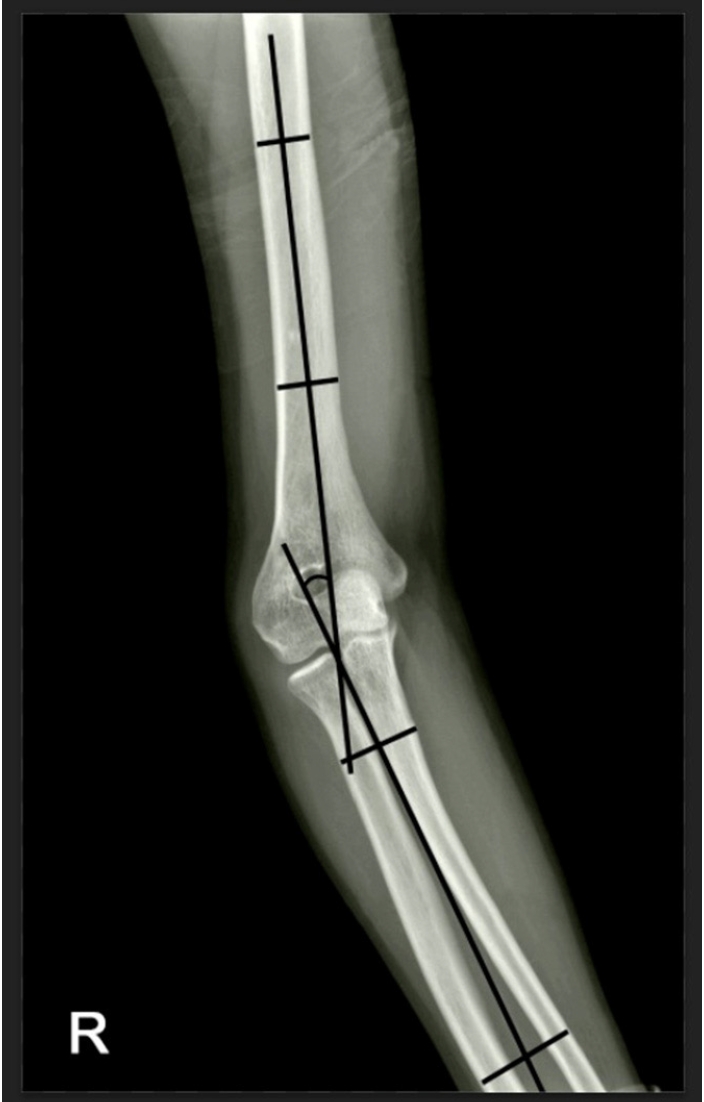
Cubitus varus deformity in adults most commonly occurs as a late complication resulting from malunion of distal humeral fractures sustained during childhood. This deformity can cause cosmetic problems and anatomical deformities that hinder normal sports activities and potentially lead to long-term complications. Although various surgical techniques exist for correcting cubitus varus, this study investigated the clinical and functional outcomes of reverse V step-cut osteotomy.
Methods
In total, 15 patients underwent surgical treatment with reverse V step-cut osteotomy between 2012 and 2023. The mean age of the patients at the time of surgery was 46.3 years (range, 20–65 years). The preoperative carrying angle was ‒11.09° of varus, which was corrected to +12.81° of valgus postoperatively. The mean preoperative lateral prominence index (LPI) was ‒10.03, and the mean postoperative LPI improved to ‒4.48. A comparison to the unaffected side showed a P-value of 0.978, indicating similarity.
Results
Preoperatively, eight patients exhibited signs of posterolateral rotatory instability, and among them, three underwent concomitant lateral ulnar collateral ligament reconstruction. Seven patients reported ulnar nerve symptoms, and all underwent concurrent ulnar nerve release. Postoperatively, improvements in elbow pain, instability, and ulnar nerve symptoms were observed. One patient required reoperation due to malunion and insufficient correction, but no other complications were noted.
Conclusions
These outcomes demonstrate that reverse V step-cut osteotomy can be an effective treatment method for cubitus varus deformity in adults. Level of evidence: IV.
- 3,898 View
- 93 Download

Review Article
- Distal Humerus Fracture: How to Choose the Approach, Implant, Fixation and Rehabilitation
- Min Ho Lee, Young Ho Lee
- J Korean Fract Soc 2019;32(1):72-81. Published online January 31, 2019
- DOI: https://doi.org/10.12671/jkfs.2019.32.1.72
-
Abstract
PDF
- Distal humerus fractures require stable fixation and early joint motion, similar to other intra-articular fractures, but are difficult to treat adequately because of the anatomical complexity, severe comminution, and accompanying osteoporosis. In most cases, surgical treatment is performed using two supporting plates. Plate fixation can be divided into right angle plate fixation and parallel plate fixation. In addition, depending on the type of fracture, surgical procedures can be performed differently, and autologous bone grafting can be required in the case of severe bone loss. The elbow joint is vulnerable to stiffness, so it is important to start joint movement early after surgery. Postoperative complications, such as nonunion, ulnar nerve compression, and heterotopic ossification, can occur. Therefore, accurate and rigid fixation and meticulous manipulation of soft tissues are required during surgery.
- 4,450 View
- 248 Download
Original Articles
- Clinical Outcome of Surgical Treatment of Distal Humerus Intercondylar Fractures Through the Transolecranon Approach Combined with Anterior Transposition of the Ulnar Nerve
- Kwang Hyun Lee, Seong Pil Lee, Kyu Tae Hwang, Joo Hak Kim
- J Korean Fract Soc 2004;17(2):70-75. Published online April 30, 2004
- DOI: https://doi.org/10.12671/jkfs.2004.17.2.70
-
Abstract
PDF
- PURPOSE
To analyze the clinical outcomes of surgical treatment of distal humerus intercondylar fractures through the transolecranon approach combined with anterior transposition of the ulnar nerve.
MATERIALS AND METHODS
Eight patients who had distal humerus intercondylar fractures were included in this study and underwent operative treatment through the transolecranon approach for sufficient operative field with anterior transposition of the ulnar nerve and fixed with reconstruction plate.
RESULTS
The results were evaluated using Riseborough and Radin rating criteria. Seven cases of eight cases were achieved good results with flexion contracture less than 30 degrees and forward flexion more than 115 degrees. However, one case was acheived poor result with 40 degrees of flexion contractue and 70 degrees of forward flexion. There were no the compressive ulnar neuropathy.
CONCLUSION
We found the transolecranon approach and anterior transposition of the ulnar nerve a viable option for surgical treatment of the distal humerus intercondylar fractures
- 669 View
- 0 Download
- Ulnar nerve palsy After Percutaneous Pinning in Childrens Supracondylar fracture
- Tai Seung Kim, Jay Rim Choi, Kuhn Sung Whang
- J Korean Soc Fract 1999;12(3):674-678. Published online July 31, 1999
- DOI: https://doi.org/10.12671/jksf.1999.12.3.674
-
Abstract
PDF
- Many authors have described percutaneous pinning techniques as the treatment of choice for most supracondylar fractures. But little information is available concerning ulnar nerve injury resulting from pinning techniques. When the surgeon is faced with a postoperative ulnar nerve palsy, it can be the result of unrecognized preoperative palsy, manipulation during surgery, or damage to the nerve by one of the medial pin placements. The options for management include exploration, medial pin removal, or observation. We reviewed our hospital records on the 132 supracondylar elbow fractures that we treated in children from 1991 to 1998 There were 16 palsies found with normal preoperative and abnormal postoperative ulnar nerve function. Normal nerve function returned without exploration and early medial pin removal in all cases. We recommand that observation is the appropriate way to manage these postoperative ulnar nerve palsies in most cases.
- 573 View
- 0 Download
- Tardy Ulnar Nerve Palsy Caused by Post-Traumatic Elbow deformities
- Seung koo Rhee, seok Whan Song, Hwa Sung Lee, Ho Tae Kim
- J Korean Soc Fract 1998;11(2):420-426. Published online April 30, 1998
- DOI: https://doi.org/10.12671/jksf.1998.11.2.420
-
Abstract
PDF
- Thirty-five patients with tardy ulnar nerve palsy caused by cubitus valgus (33 cases0 and varus (2 cases) deformities were retrospectively studied. All patients had a history of old fracture on the distal humerus during childhood. The mean interval between the previous fractures and the onset of ulnar neuropathy was 19 years. The severity of nerve palsy was classified as McGowan's grade I in 24 patients, grade II in 8 patients, and grade III in 3 patients. The mean carrying angle was average 29 degrees in 33 cases with cubitus valgus and it was decreased to average 11 degrees postoperatively, but the angle was average -23 degrees preoperatively in 2 cases with cubitus varus and it was corrected to average 9 degrees postoperatively. the cause of palsy was analysed by mechanical stetching in 11 cases, compression by a fibrous band between the two heads of flexor carpi ulnaris in 8 cases, and diffuse fibrous adhesion around the ulnar tunnel in 5 cases. All patients was treated with supracondylar closing wedge osteotomy accompanied with anterior ulnar nerve transposition in 13 patients, corrective osteotomy only in 12 patients, and anterior ulnar nerve transposition only in 10 patients. Their end results were analysed as good in 24 cases, fair in 8 cases, and poor in 3 cases within average 6 months after the operations (4 to 13 months). The poor results was obtained in 3 cases out of 9 cases with corrective osteotomy group (33.3%). Conclusively, a tardy ulnar nerve palsy caused by post-traumatic elbow deformities should be corredcted with anterior ulnar nerve transposition with or without corrective closing wedge osteotomy but not by corrective osteotomy only, because of compressive neuropathy by diffuse fibrous adhesion or bands of two heads of FCU around the ulnar tunnel in elbow.
- 1,080 View
- 8 Download
- Fracture of the Distal Radius with Ulnar Nerve Palsy
- Chil Soo Kwon, Jong Kuk Ahn, Jin Hyok Kim, Yerl Bo Sung, Jin Ho Cho
- J Korean Soc Fract 1997;10(1):171-174. Published online January 31, 1997
- DOI: https://doi.org/10.12671/jksf.1997.10.1.171
-
Abstract
PDF
- There are several complications of distal radiug fracture such as median nerve injury, malunion nonunion, rupture of EPL, and ischemic contracture. Lesion of ulnar nerve as a complication of fracture of the distal radius are very rare. The authors report 1 case of the distal radius fracture with ulnar nerve palsy. The electromyography & nerve-conduction studies showed incomplete axonotmesis of ulnar nerve on 1 month following injury. A second electromyography & nerve-conduction study two months after injury showed complete recovery of nerve function.
-
Citations
Citations to this article as recorded by
- Ulnar Nerve Palsy Following Closed Fracture of the Distal Radius: A Report of 2 Cases
Chul-Hyun Cho, Chul-Hyung Kang, Jae-Hoon Jung
Clinics in Orthopedic Surgery.2010; 2(1): 55. CrossRef
- Ulnar Nerve Palsy Following Closed Fracture of the Distal Radius: A Report of 2 Cases
- 963 View
- 2 Download
- 1 Crossref
- Treatment of Fractures of Lateral Condyle of Humerus with Compliations
- Moon Sang Chung, Sung Churl Lee
- J Korean Soc Fract 1995;8(3):659-666. Published online July 31, 1995
- DOI: https://doi.org/10.12671/jksf.1995.8.3.659
-
Abstract
PDF
- Fractures of the lateral condyle of the humerus are notorious for complications, most commonly nonunion with subsequent proximal migration of the ununited fragment, an increase in the carrying angle, and the tardy ulnar nerve palsy. In the past, the reconstructive surgery for complicated old fractures of the lateral condyle of the humerus had been hardly considered, but recently, attempts have been made to reconstruct the anatomy and function of the elbow joint. The authors have reviewed 21 cases of old fractures of the lateral condyle of the humerus, which had been treated at Seoul National University Hospital from April,1982 until March, 1990. For established nonunions of the lateral condyle fragment, better results were obtained from the procedure that includes osteosynthesis of the lateral condyle, attempting to restore the normal anatomy of the elbow joint. For tardy ulnar nerve palsies, better results were obtained from the procedure that includes medial epicondylectorny. Fractures of the lateral humeral condyle have many late problems in spite of treatment at the time of injury so early aggressive treatment is necessary. Even in cases with late problems, aggressive treatment should be done, too, as soon as possible.
-
Citations
Citations to this article as recorded by- In SituLate Metaphyseal Osteosynthesis for the Fractures of the Lateral Humeral Condyle in Children
Kun Bo Park, Seung Whan Lee, Hyun Woo Kim, Hui Wan Park, Ki Seok Lee
Journal of the Korean Fracture Society.2008; 21(2): 151. CrossRef
- In SituLate Metaphyseal Osteosynthesis for the Fractures of the Lateral Humeral Condyle in Children
- 1,098 View
- 1 Download
- 1 Crossref
 First
First Prev
Prev

