 E-submission
 TOTA
TOTA TOTS
TOTS
Search
- Page Path
- HOME > Search
Original Articles
- Open reduction and internal fixation for distal humerus fractures in older adults: a retrospective comparative study by osteoporosis status
- Tong Joo Lee, Hee Beom Shin, Yongseok Lee
- J Musculoskelet Trauma 2026;39(3):227-235. Published online July 15, 2026
- DOI: https://doi.org/10.12671/jmt.2026.00101
-
 Abstract
 PDF
PDF - Background
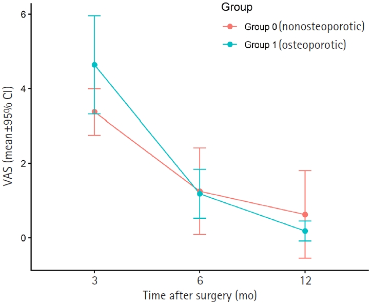
Distal humerus fractures in older patients, particularly those with osteoporosis, pose substantial treatment challenges because of increased fracture complexity and compromised bone stock. Open reduction and internal fixation (ORIF) is the preferred treatment but may be complicated by fixation failure. This study investigated the outcomes of ORIF in older osteoporotic and nonosteoporotic patients with complete articular distal humerus fractures.
Methods
This retrospective study included 19 patients with Arbeitsgemeinschaft für Osteosynthesefragen/ Orthopaedic Trauma Association (AO/OTA) 13C distal humerus fractures who underwent surgical treatment between 2012 and 2024. The mean patient age was 79.1 years. Patients were divided into osteoporotic (n=11) and nonosteoporotic (n=8) groups based on the lowest recorded dual-energy X-ray absorptiometry T-score at the femoral neck or lumbar spine. Osteoporosis was defined as a T-score of −2.5 or lower, and the nonosteoporotic group included patients with osteopenia. All fractures were treated with ORIF using bicolumnar plating. All included patients completed 12 months of clinical follow-up for visual analog scale (VAS) and Mayo Elbow Performance Score (MEPS) assessments. Radiographic follow-up was recorded separately and continued until union was confirmed; imaging follow-up extended to 12 months or longer in 14 patients and was limited to 6 months in five asymptomatic patients after confirmed union.
Results
Both groups showed significant within-group improvement in pain, as assessed using the VAS, and function, as assessed using the MEPS, over time. Between-group comparisons at each follow-up time point showed no statistically significant differences in VAS or MEPS. No radiographic nonunion was observed during the available imaging follow- up, and complications were limited to one case of screw pullout/loosening and one case of postoperative stiffness.
Conclusions
ORIF provides reliable outcomes for older patients with complex distal humerus fractures, regardless of osteoporosis status, when stable reconstruction is achievable. These findings suggest that ORIF remains a viable treatment option, with satisfactory functional recovery and low complication rates in this population. Level of evidence: III.
- 164 View
- 7 Download

- Short-term Treatment Comparison of Teriparatide and Percutaneous Vertebroplasty in Patients with Acute Osteoporotic Vertebral Compression Fractures
- Joonoh Seo, Ki Youn Kwon, Bumseok Lee, Hoon-Sang Sohn
- J Korean Fract Soc 2024;37(1):15-21. Published online January 31, 2024
- DOI: https://doi.org/10.12671/jkfs.2024.37.1.15
-
Abstract
PDF
- Purpose
This study compared the 3-month treatment effects of teriparatide and percutaneous vertebroplasty for acute osteoporotic vertebral compression fractures.
Materials and Methods
A retrospective study was conducted on 76 patients diagnosed with acute osteoporotic vertebral compression fractures from January 1, 2020 to December 31, 2022. The patients were divided into the teriparatide group and the percutaneous vertebroplasty+alendronate group. The visual analog scale (VAS), Oswestry disability index (ODI), and height of the vertebrae anterior wall were measured before treatment and at 1 and 3 months after treatment.
Results
Of the 76 patients, 42 were treated with teriparatide, and 34 were treated with percutaneous vertebroplasty. The symptoms improved in both groups, with a decrease in the VAS and ODI scores at 1 and 3 months after treatment, respectively. On the other hand, there was no significant difference in the VAS, ODI score, and anterior vertebral body height between the two groups before treatment and at 1 and 3 months after treatment.
Conclusion
In the treatment of acute osteoporotic vertebral compression fractures, conservative treatment using teriparatide showed similar short-term (3 months) treatment results to percutaneous vertebroplasty in terms of improvement in back pain and function and degree of reduction in anterior vertebral body height.
- 1,606 View
- 42 Download
Case Report
- Progressive Brachial Plexus Palsy after Fixation of Clavicle Shaft Nonunion: A Case Report
- Hong Ki Jin, Ki Bong Park, Hyung Lae Cho, Jung Il Kang, Wan Seok Lee
- J Korean Fract Soc 2019;32(2):97-101. Published online April 30, 2019
- DOI: https://doi.org/10.12671/jkfs.2019.32.2.97
-
Abstract
PDF
- The brachial plexus palsy is a rare complication of a clavicle fracture, occurring in 0.5% to 9.0% of cases. This condition is caused by excessive callus formation, which can be recovered by a spur resection and surgical fixation. In contrast, only seven cases have been reported after surgical reduction and fixation. A case of progressive brachial plexus palsy was observed after fixation of the displaced nonunion of a clavicle fracture. The symptom were improved after removing the implant.
-
Citations
Citations to this article as recorded by
- Late Presentation of Brachial Plexus Traction Injury After Fixation of Chronic Clavicle Nonunion Mimicking Brachial Plexitis: A Case Report and Literature Review
John G Skedros, John T Cronin, Kevin B Curtis, Jessie A Montgomery, Brett W Richards, Mark A Mahan
Cureus.2026;[Epub] CrossRef - Arcuate osteoplasty for brachial plexus paralysis after plate fixation of mid-clavicle fracture: a case report and literature review
Dongju Shin, Jae Hwi Han
Clinics in Shoulder and Elbow.2025; 28(3): 394. CrossRef
- Late Presentation of Brachial Plexus Traction Injury After Fixation of Chronic Clavicle Nonunion Mimicking Brachial Plexitis: A Case Report and Literature Review
- 1,577 View
- 11 Download
- 2 Crossref
Review Article
- Atypical Femoral Fractures: What Do We Know about Them?
- Beom Seok Lee, Young Kyun Lee, Heejae Won, Hyungkook Kim, Kyung Hoi Koo
- J Korean Fract Soc 2018;31(4):159-164. Published online October 31, 2018
- DOI: https://doi.org/10.12671/jkfs.2018.31.4.159
-
Abstract
PDF
- Recently, atypical femoral fractures (AFFs) have been found in patients who were prescribed bisphosphonate to prevent osteoporotic fractures. Although the occurrence of AFF is rare, there are some concerns, such as a higher risk of delayed or non-union of AFF. This paper reviews the treatment of AFF and suggests some considerations during surgery.
-
Citations
Citations to this article as recorded by- How to Improve Fracture Healing in Atypical Femoral Fractures
Sang-Jin Jeong, Chan-Woo Park, Seung-Jae Lim
Journal of the Korean Orthopaedic Association.2024; 59(1): 9. CrossRef - Atypical Femoral Fracture Occurring at a Proximal Screw Insertion Site after Plate Removal in a Distal Femoral Fracture
Jin Woo Jin, Sung Jin Shin, Jong Min Jeon
Journal of the Korean Orthopaedic Association.2024; 59(4): 314. CrossRef - Position Statement: Atypical Femoral Fracture from the Korean Society for Bone and Mineral Research in 2023
Jae-Hwi Nho, Byung-Woong Jang, Dong Woo Lee, Jae-Hyun Kim, Tae Kang Lim, Soo Min Cha, Dong-Kyo Seo, Yong-Geun Park, Dong-Geun Kang, Young-Kyun Lee, Yong-Chan Ha
Journal of Bone Metabolism.2023; 30(3): 209. CrossRef
- How to Improve Fracture Healing in Atypical Femoral Fractures
- 1,035 View
- 8 Download
- 3 Crossref
Case Report
- A Case of Surgically Treated by Transperitoneal Approach in Delayed Neurological Deficit after Sacral Fracture: A Case Report
- Young Soo Jang, Jong Seok Lee, Jae Hyuk Choi, Sung Ju Bae, Chan Il Bae
- J Korean Fract Soc 2013;26(1):69-72. Published online January 31, 2013
- DOI: https://doi.org/10.12671/jkfs.2013.26.1.69
-
Abstract
PDF
- This study reviews a case of sacral fracture with delayed onset neurological deficit that showed good results after decompressive surgery. The delayed neurological deficit appeared at 4 weeks after injury and it was treated with anterior decompression through transperitoneal approach. A 23-year-old woman was injured in a car accident and had bilateral pubic rami fractures and fractures of the sacral ala on the right side. She was treated with external fixation devices for approximately four weeks, but complained of pain and numbness. The dorsiflexion and plantalflexion of the right ankle was weakened and graded as grade 2. Preoperative pelvic and sacral radiographs, computed tomography, magnetic resonance imaging and electromyelography, and nerve conduction study were performed to identify the region of neurological deficit, and we decided to implement neurological decompression. By transperitoneal approach, we performed bone curratage and decompression around the region of sacral alar slope and S1 foramen. The pain and numbness of the right foot cleared up. Dorsiflexion and plantalflexion of the right ankle improved to grade 5. Anterior decompression by transperitoneal approach proved to bring satisfactory results in a patient, who presented delayed neurological deficit after sacral fracture.
- 665 View
- 0 Download
Original Articles
- In Situ Late Metaphyseal Osteosynthesis for the Fractures of the Lateral Humeral Condyle in Children
- Kun Bo Park, Seung Whan Lee, Hyun Woo Kim, Hui Wan Park, Ki Seok Lee
- J Korean Fract Soc 2008;21(2):151-156. Published online April 30, 2008
- DOI: https://doi.org/10.12671/jkfs.2008.21.2.151
-
Abstract
PDF
- PURPOSE
To evaluate the efficacy of the in situ late osteosynthesis for slightly displaced fractures of the lateral humeral condyle.
MATERIALS AND METHODS
From 2000 to 2004, 12 patients (8 boys and 4 girls) were managed with in situ late osteosynthesis for fractures of the lateral humeral condyle. The average age at the time of operation was 6 years 1 month (1 year 7 months~9 years 1 month), and the mean amount of fragment displacement was 3.3 mm (2.0~4.5 mm). The operative procedure included curettage and in situ fixation of the fragment RESULTS: Bony union was achieved in all cases after avg. 48 months (33~73 months) follow-up assessment. According to the score system of Dhillon et al, 7 patients had excellent, 3 had good, 2 had fair results. None of the patients developed avascular necrosis or premature closure of the epiphysis.
CONCLUSION
We suggest that in situ fixation is an effective method for the late treatment of slightly displaced fracture of the lateral humeral condyle.
- 770 View
- 3 Download
- Wedge Tibial Shaft Fractures Treated with Interlocking IM Nailing
- Sang Jun Song, Hyung Ku Yoon, Soo Hong Han, Hyung Kun Park, In Seok Lee
- J Korean Fract Soc 2006;19(3):322-328. Published online July 31, 2006
- DOI: https://doi.org/10.12671/jkfs.2006.19.3.322
-
Abstract
- PURPOSE
To investigate the bone union time of patients treated with interlocking intramedullary nailing in wedged tibial shaft fracture and to evaluate the factors that influence this result.
MATERIALS AND METHODS
32 patients treated with interlocking intramedullary nailing for wedge tibial shaft fracture were reviewed with a follow-up period of more than 1 year. Radiographic results were assessed with diameter (%) and length (mm) of wedge fragment, pre and postoperative displacement (mm) of wedge fragment. We also checked the bone union time of the main fragment and the wedge fragment (paired t-test). We investigated the bone union time acocording to the diameter, length of wedge fragment, pre and postopertvie displacement (correlation analysis).
RESULTS
Bone union time of the main fragments averaged 15.3 weeks (6~53 weeks) and that of wedge fragment averaged 24.2 weeks (8~64 weeks) (p=0.005). There was no correspondence between wedge fragment diameter and bone union time (p=0.681), but the bone union time of wedge fragment increased in proportion to its diameter (r2=0.747, p=0.031). There was no correspondence between preoperative displacement of wedge fragment and bone union time (p=0.574), but the bone union time increased in proportion to postoperative displacement of wedge fragment (r2=0.730, p=0.001).
CONCLUSION
Wedge fragments need longer time for bone union than main fragments in interlocking intramedullary nailing for wedge tibial shaft fractures. We need to pay attention to the displacemet of fragments in treating tibial shaft fractures with large wedge fragment. -
Citations
Citations to this article as recorded by- RNA modifications: molecular orchestrators of wound healing
Xiuying Guo, Lele Liu, Junqi Yang, Yuhe Dai, Qianbo Zhang, Rifang Gu, Min Tan, Ming Tang, Xuqiang Nie
Burns & Trauma.2026;[Epub] CrossRef
- RNA modifications: molecular orchestrators of wound healing
- 740 View
- 0 Download
- 1 Crossref
Review Article
- Bone Graft Substitute
- Tae Joon Cho, Ki Seok Lee
- J Korean Fract Soc 2006;19(1):109-116. Published online January 31, 2006
- DOI: https://doi.org/10.12671/jkfs.2006.19.1.109
-
Abstract
- No abstract available.
-
Citations
Citations to this article as recorded by- The Effect of Gyejibokryunghwan on Fracture Union in Tibia-fractured Rats
Jeong-Won Yoon, K.M.D., Soon-Joong Kim, K.M.D.
Journal of Korean Medicine Rehabilitation.2019; 29(4): 1. CrossRef - Evaluation of equine cortical bone transplantation in a canine fracture model
S.H. Heo, C.S. Na, N.S. Kim
Veterinární medicína.2011; 56(3): 110. CrossRef
- The Effect of Gyejibokryunghwan on Fracture Union in Tibia-fractured Rats
- 895 View
- 0 Download
- 2 Crossref
Original Articles
- The Effect of Fibular Malreduction on Ankle Joint after Tibial Interlocking IM Nailing of Tibial and Fibular Fractures
- Dong Eun Shin, Duck Yun Cho, Hyung Ku Yoon, Jin Soo Lee, Yoon Seok Lee, Hyoung Jun Kim
- J Korean Fract Soc 2005;18(1):29-35. Published online January 31, 2005
- DOI: https://doi.org/10.12671/jkfs.2005.18.1.29
-
Abstract
PDF
- PURPOSE
To investigate the effect of fibular malreduction on ankle joint after tibia interlocking IM nailing of tibial and fibular fractures according to type of fibular fractures at preoperation.
MATERIALS AND METHODS
Thirty-nine patients who had ipsilateral tibiofibular fracture were analyzed clinically and radiographically. The talocrural angle and the distance from joint line to the tip of fibular were measured on both ankle standing AP view. The difference of angle and distance of both ankle were analyzed by paired t-test and correlation between defference and AOFAS score by Spearman correlation coefficients.
RESULTS
The difference of The talocrural angle and the distance from joint line to the tip of fibular of both ankle was statistically significant (p<0.05). The correlation between this difference and AOFAS score was statistically insignificant (p>0.05).
CONCLUSION
In tibia interlocking IM nailing of tibia and fibula fracture, malreduction of fibula could cause the change of ankle joint. -
Citations
Citations to this article as recorded by- The Risk Factors Associated with Nonunion after Surgical Treatment for Distal Fibular Fractures
Jun Young Lee, Kwi Youn Choi, Sinwook Kang, Kang Yeol Ko
Journal of Korean Foot and Ankle Society.2018; 22(3): 95. CrossRef
- The Risk Factors Associated with Nonunion after Surgical Treatment for Distal Fibular Fractures
- 1,013 View
- 2 Download
- 1 Crossref
- Early Postoperative Complications of Calcaneal Fractures Following Operative Treatment by a Lateral Extensile Approach
- Young Soo Byun, Young Ho Cho, Jun Woo Park, Jin Seok Lee, Ji Hwan Kim
- J Korean Fract Soc 2004;17(4):323-327. Published online October 31, 2004
- DOI: https://doi.org/10.12671/jkfs.2004.17.4.323
-
Abstract
PDF
- PURPOSE
To analyze early postoperative complications of calcaneal fractures operated by a lateral extensile approach and to identify risk factors for wound complications.
MATERIALS AND METHODS
From July 1990 to February 2003, 116 calcaneal fractures in 104 patients were treated by open reduction and internal fixation through a lateral extensile approach. The patient's records were reviewed for early postoperative complications. Statistical analysis was performed to determine significant relationships between predicted variables and the development of wound complications.
RESULTS
Fourteen fractures (12.0%) developed infection. Ten of them were superficial infection and four were deep infection that required surgical treatment. Eight fractures (6.9%) developed skin necrosis. Six of them were marginal skin necrosis and two were flap necrosis that required surgical treatment. Seven fractures (6.0%) developed sural nerve injury, but their symptoms were improved without additional treatment. Open fracture (p=0.003) and prolonged operating time (p=0.049) increased significantly the rate of wound complications.
CONCLUSION
The rate of early postoperative complications of calcaneal fractures operated by a lateral extensile approach is high. These complications can be reduced by meticulous treatment of an open wound, reduced operating time within 90 minutes through preoperative planning and skillful technique, and correct incision to avoid damage of the sural nerve. -
Citations
Citations to this article as recorded by- Results in Operative Treatment of Open Calcaneal Fracture
Ba Rom Kim, Jun Young Lee, Donghyuk Cha
Journal of Korean Foot and Ankle Society.2021; 25(3): 133. CrossRef - Bilateral Open Transcalcaneal Fracture with Talonavicular Dislocation - A Case Report -
Hun Park, Sung Jin Shin, Sang Rim Kim, Kwang Woo Nam, Sung Wook Choi, Kyu Bum Seo, Jun Young Seo
Journal of the Korean Fracture Society.2011; 24(1): 87. CrossRef - Treatment of Intra-articular Calcaneal Fractures Using Minimally Invasive Sinus Tarsi Approach in Diabetic Patients
Hong-Moon Sohn, Sang-Ho Ha, Sang-Hong Lee, Jun-Young Lee, Jeong-Ho Kim, Sang-Jun Lee
Journal of the Korean Fracture Society.2008; 21(3): 195. CrossRef
- Results in Operative Treatment of Open Calcaneal Fracture
- 993 View
- 1 Download
- 3 Crossref
- Operative Treatment of Proximal Tibial Plateau Fractures through Lateral Submeniscal Approach
- Hyug Su An, Se Ang Chang, Jun Woo Park, Jin Seok Lee, Hun Ho Bang
- J Korean Fract Soc 2004;17(3):237-242. Published online July 31, 2004
- DOI: https://doi.org/10.12671/jkfs.2004.17.3.237
-
Abstract
PDF
- PURPOSE
The purpose of this study was conducted to evaluate the clinical results of proximal tibial plateau fractures treated with open reduction and internal fixation through the lateral submeniscal approach and allowed early motion of the knee and to evaluate the effectiveness of the approach.
MATERIALS AND METHODS
From January 1998 to December 2002, fifty four patients who underwent open reduction through the lateral submeniscal approach for proximal tibia plateau fracture and had a follow-up more than one year were included in this study. Clinical results were evaluated by postoperative radiographs taken at the last follow-up and Porter's assessment method.
RESULTS
Anatomical reduction was achieved under direct vision through the submeniscal approach in most of the cases in this study. The postoperative radiographs showed anatomical reduction in 32 cases (59%) and adequate reduction with displacement within 2 mm in 20 cases (37%). The clinical evaluation by Porter's assessment method revealed that 49 cases (91%) were acceptable results of excellent or good at the final follow-up CONCLUSION: This study indicates that open reduction and internal fixation through the lateral submeniscal approach can be a good option for proximal tibia plateau fractures because it allows accurate reduction of the articular fractures, which is confirmed directly during operation, identification and repair of associated soft tissue injuries are facilitated, sufficient bone graft and stable fixation of the articular fragments under direct vision allow early motion of the knee.
- 799 View
- 9 Download
- Development of a Computer-assisted Surgery System for Screw Fixation of the Sacro-iliac Joint
- Jin Sup Yeom, Won Sik Choy, Ha Yong Kim, Whoan Jeang Kim, Jong Won Kang, Yeongho Kim, Hyungmin Kim, Donghyun Seo, Seok Lee, Jae Bum Lee, Namkug Kim, Cheol Young Kim
- J Korean Soc Fract 2003;16(1):1-7. Published online January 31, 2003
- DOI: https://doi.org/10.12671/jksf.2003.16.1.1
-
Abstract
PDF
- PURPOSE
The purposes of this study were to develop a computer-assisted surgery system for percutaneous screw fixation of the sacro-iliac joint and to evaluate its accuracy.
MATERIALS AND METHODS
We have developed a navigation system composed of an optical tracking device (Polaris, Northern Digital, Canada) and a personal computer. The registration error and target localization error at hybrid registration were measured using a phantom. The errors were measured 30 times for each. Sixteen 6.5 mm cannulated screws were inserted into four plastic bone models (Sawbones, USA), and the accuracy was evaluated.
RESULTS
The registration error was 0.76 +/-0.33 mm, and the target localization error was 1.43 +/-0.42 mm. All of the 16 screws were inserted well across the sacro-iliac joint, and there was neither penetration of the cortical bones nor collision between screws or washers.
CONCLUSION
The accuracy of the developed system was similar to existing ones, and its usefulness and helpfulness was proven with screw insertion into plastic bone models.
- 848 View
- 3 Download
- Biomechanical Analysis of Korean Radiolucent Carbon/Graphite Ring Fixator
- In Ho Choi, Jun kyung Kim, Kui won Choi, Chin Youb Chung, Tae Joon Cho, Ki Seok Lee
- J Korean Soc Fract 2000;13(1):1-12. Published online January 31, 2000
- DOI: https://doi.org/10.12671/jksf.2000.13.1.1
-
Abstract
PDF
- PURPOSE
The mechanical stiffness of Korean radiolucent carbon/graphite ring fixator(KRCRF) was analyzed and compared with those of conventional stainless steel Ilizarov system and the Smith- Nephew carbon fiber circular external fixator.
MATERIALS AND METHODS
The transfixing olive pins of the circular fixator on the acryl pylon were assembled in 90degrees- 90degrees and 135degrees- 45degrees configuration, respectively. And the fixator-pylon model was loaded with Instron model No. 8500 in three testing modes: axial compression, anteroposterior(AP) bending and lateral bending.
RESULTS
As compared with stainless steel Ilizarov fixator, the KRCRF was significantly more stiff on the axial compression test regardless of the ring size(140 mm and 200 mm diameters) and transfixation configuration. But, it was less stiff on the anteroposterior(AP) and lateral bending tests. When compared with the Smith-Nephew carbon fiber circular external fixator, the KRCRF was generally more stiff on the axial compression, AP and lateral bending tests regardless of the ring size(140 mm and 180 mm diameters) and configuration, except the AP bending stiffness in 90degrees- 90degrees configuration and lateral bending stffness in 135degrees- 45degrees configuration on the 180 mm diameter frame.
CONCLUSION
Considering the radiolucency, weight and biomechanical stffness, we think that the KRCRF is an excellent substitute for the imported circular fixators made of stainless steel or carbon/graphite. -
Citations
Citations to this article as recorded by- A Study on the Development of the Off-Line Software for Regulating the 6 D.O.F. Circular Fixator
Bum-Seok PARK, In-Ho CHOI, Jin-Woo KIM, Seung-Yeol LEE, Chang-Soo HAN
JSME International Journal Series C.2006; 49(4): 1123. CrossRef
- A Study on the Development of the Off-Line Software for Regulating the 6 D.O.F. Circular Fixator
- 1,071 View
- 0 Download
- 1 Crossref
- Ipsilateral Femoral Shaft and Neck fracture
- Dong Chul Lee, Yun Seok Lee, Duk Seop Shin
- J Korean Soc Fract 1999;12(2):245-252. Published online April 30, 1999
- DOI: https://doi.org/10.12671/jksf.1999.12.2.245
-
Abstract
PDF
- The occurrence of ipsilateral hip and femoral shaft fracture is uncommon and this problematic combination occur in 2.5% to 6% of femoral fracture. This combination of fractures result from high energy trauma and occurs in young multiply injured patient. As most orthopedists attention is directed to the shaft fructure of femur, the neck fracture can be commonly missed initially. Ipsilateral femoral shaft and neck fracture has the characteristics that has multiple associated injuries and many complications, such at avascular necrosis and nonunion of the femur neck, coxa vara. We analysed the outcome of treatment to know the results of treatment and its complication in 10 patients who were treated at Orthopedic Department, Yeungnam University Hospital from May 1991 to May 1996. The results were as follows : 1. Sixty percent of femoral neck fracture was basicervical type; Ninty Percent of femoral shaft fracture occured in the middle one third and most of the fractures were comminuted. There was one open fracture of femoral shaft. 2. All of the causes of the fractures occurred in motor vehicle accident. Mean age was 40.3 years(range: 15-59 years). There were 24 associated injuries in ten patients: especially, the injuries around knee were most common(6 tibial condylar fractures, 3 patellar fractures, 1 ligament injuries). 3. The bone union was obtained at average 2.8 months after operation(2 months - 5 months) in femur neck fractures, at average 5.5 months after operation(4 months - 12 months) in femur shaft fractttres and weight hearing was permitted at average 2.3 months after operation(1.5 months - 5 months). 4. There were 3 cases of nonunion of femoral shaft fracture, but they were treated by autogenous iliac bone graft Early bone graft may be necessary in comminuted femoral shaft fracture.
- 748 View
- 6 Download
 First
First Prev
Prev

