 E-submission
 TOTA
TOTA TOTS
TOTS
Search
- Page Path
- HOME > Search
Original Articles
- Postoperative sagittal alignment and reoperation predict late outcomes after plate fixation of AO/OTA 33C distal femur fractures: a retrospective cohort study
- Hee Gon Park
- J Musculoskelet Trauma 2026;39(3):216-226. Published online July 24, 2026
- DOI: https://doi.org/10.12671/jmt.2026.00073
-
 Abstract
 PDF
PDF - Background
Intra-articular distal femoral fractures are associated with high complication rates and variable late outcomes. Although the AO Foundation/Orthopaedic Trauma Association (AO/OTA) classification system is useful for describing injury severity and anticipating complications, whether fracture subtype alone determines clinically relevant late outcomes remains unclear. This study aimed to evaluate postoperative complications and late outcomes following plate fixation of intra-articular distal femoral fractures and to identify factors independently associated with final clinical outcomes.
Methods
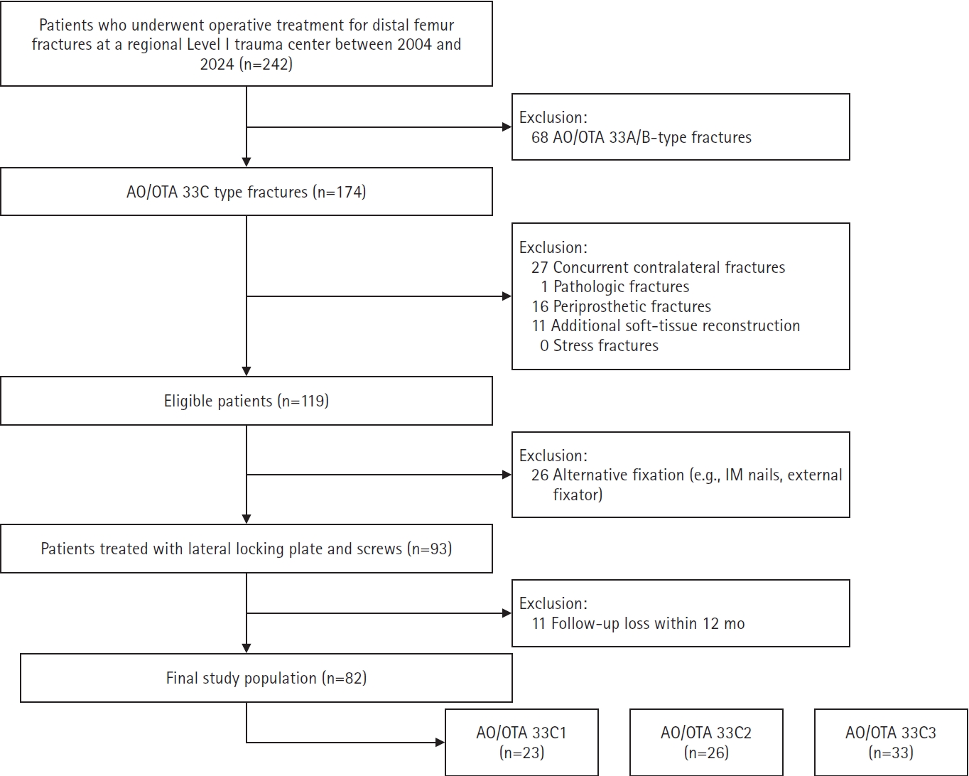
This retrospective cohort study was conducted at a regional Level I trauma center. Patients with AO/OTA 33C intra-articular distal femoral fractures who underwent plate fixation and were followed for at least 12 months were included. Radiographic parameters reflecting coronal and sagittal alignment were assessed using standardized measurement methods. Postoperative complications and reoperation were recorded as intermediate events. Clinically relevant late outcomes included final knee range of motion (ROM), leg length discrepancy, and radiographic osteoarthritis severity, assessed as the difference in Kellgren-Lawrence grade. Multivariable linear regression analyses were performed to identify factors independently associated with late outcomes.
Results
The 33C3 group (n=33) had significantly higher rates of open fracture (54.5%), nonunion (39.4%), and reoperation (45.5%) than the 33C1–2 group (n=49). At the final follow-up, 33C3 fractures were associated with lower mean ROM (P<0.001) and greater osteoarthritis progression (P<0.001). However, multivariable analysis showed that sagittal malalignment (Δ anatomical posterior distal femoral angle: β=−2.35, P=0.001) and reoperation (β=−17.5, P=0.001), rather than AO/OTA subtype itself, were independent predictors of final ROM.
Conclusions
Although fracture subtype according to the AO/OTA classification was associated with predictable complication patterns, clinically relevant late outcomes following intra-articular distal femoral fractures were more closely associated with postoperative alignment quality and the treatment course than with fracture classification alone. These findings highlight the importance of optimizing radiographic alignment and preventing complications that lead to reoperation to improve long-term outcomes after plate fixation. Level of evidence: III.
- 38 View
- 3 Download

- Comparison of the Radiological Outcomes of an Anatomical Quadrilateral Surface Plate with a Pelvic Reconstruction Plate in Acetabulum Fractures
- Sung Hyun Yoon, Hee Gon Park, Dong Uk Lee
- J Korean Fract Soc 2024;37(2):95-101. Published online April 30, 2024
- DOI: https://doi.org/10.12671/jkfs.2024.37.2.95
-
Abstract
PDF
- Purpose
This study compared the radiological outcomes of fixation using an anatomical quadrilateral surface plate with those using a traditional pelvic reconstruction plate for fractures involving the quadrilateral surface or superomedial wall of the acetabulum.
Materials and Methods
From 2015 to 2022, 47 patients who met the inclusion and exclusion criteria were analyzed retrospectively. Internal fixation of an acetabular fracture was achieved with a pelvic reconstruction plate (n=28) or an anatomical quadrilateral surface plate (n=19). The ability to achieve immediate postoperative anatomical reduction and the long-term outcomes were assessed by confirming the arthritic changes. Immediate postoperative reduction quality and long-term radiological outcomes for post-traumatic arthritis were assessed using the Matta scoring system on standard radiographs.
Results
The assessment of immediate postoperative reduction in the pelvic reconstruction plate group was satisfactory in 16 patients (57.1%) and unsatisfactory in 12 patients (42.9%). In the anatomical quadrilateral surface plate group, the results were satisfactory in 16 patients (84.2%) and unsatisfactory in 3 patients (15.8%). When evaluating over an extended follow-up period in the pelvic reconstruction plate group, 19 patients (67.9%) demonstrated satisfactory, while 9 patients (32.1%) had unsatisfactory outcomes. In the anatomical quadrilateral surface plate group, 12 patients (63.2%) achieved satisfactory, and 7 patients (36.8%) had unsatisfactory outcomes. The immediate postoperative reduction quality was superior in the anatomical quadrilateral surface plate group (p=0.03). Comparing longterm results, the anatomical quadrilateral surface plate group did not have statistically more favorable outcomes (p=0.49).
Conclusion
In this study, the anatomical quadrilateral surface plate achieved sufficiently good radiological results without significant difference from the existing pelvic reconstruction plate. It was concluded that it is a useful option that can replace the existing metal plate in the selection of surgery for acetabular fractures.
- 1,522 View
- 18 Download
- Results after Less Invasive Locking Plating in Intra-Articular Fractures of the Distal Femur
- Sung Hyun Kim, Sung Hyun Yoon, Hee Gon Park, Jae Uk Jung
- J Korean Fract Soc 2019;32(1):14-20. Published online January 31, 2019
- DOI: https://doi.org/10.12671/jkfs.2019.32.1.14
-
Abstract
PDF
- PURPOSE
The purpose of this study was to determine the clinical outcomes after a less invasive locking plating technique in intra-articular fractures of the distal femur.
MATERIALS AND METHODS
This was a retrospective 19 case series of patients with distal femoral intraarticular fractures treated with a less invasive locking plating technique in a single center (Dankook University Hospital) from June 2010 to April 2016. Nineteen patients (11 males and 8 females) with a mean age of 55.9 years were enrolled. The functional outcomes were evaluated using the visual analogue scale (VAS), range of knee joint motion (flexion & extension), and Knee Society score. The radiology outcomes were evaluated with parameters measured in a plain radiograph (deviation angle of alignment axis on coronal and sagittal plane, mechanical lateral distal femur angle).
RESULTS
The mean follow-up period was 26.4 months (range, 12–72 months) and the mean duration to union was 15.94 weeks (range, 11–28 weeks). The mean VAS was 1.36 (range, 0–8) and the range of motion of the knee joint was extension 4.73° (range, 0°–30°) and flexion 107.36° (range, 60°–135°). The mean Knee Society score was 85.47 (range, 47–100). The mean deviation angle of the coronal alignment axis was 4.07° (range, 1.3°–8.8°), the mean deviation angle of the sagittal alignment axis was 3.23° (range, 0.7°–7.0°), and the mechanical lateral femoral angle was 87.75° (range, 82.8°–95.5°). Six patients had traumatic osteoarthritis at the final follow-up.
CONCLUSION
The purpose of this study was to evaluate the clinical and radiologic outcomes of intraarticular fractures of the distal femur in patients who underwent an anatomical reduction through an open reduction, and converted to an extra-articular fracture with rigid internal fixation. The results were relatively satisfactory.
- 966 View
- 3 Download
- The Cause of Primary Reduction Failure in Hip Dislocation with or without Hip Fracture
- Hee Gon Park, Yong Eun Shin, Sung Hyun Kim
- J Korean Fract Soc 2017;30(1):9-15. Published online January 31, 2017
- DOI: https://doi.org/10.12671/jkfs.2017.30.1.9
-
Abstract
PDF
- PURPOSE
A rapid and accurate reduction is important for hip dislocated patients to avoid various potential complications, including avascular necrosis of the femoral head. We analyzed hip dislocation cases, ones that particularly failed during the primary reduction trial.
MATERIALS AND METHODS
Eighty-seven patients with hip dislocation, who visited the emergency department between January 2007 and September 2015, were retrospectively analyzed. Of them, 68 patients were successfully treated in the first closed reduction trial, and the remaining 19 patients were unsuccessful. Of the 19 unsuccessful first trial, 12 patients were successfully treated in the second closed reduction; however, in the remaining 7 patients, open reduction was performed in the operation room with general anesthesia. Every closed reduction was practiced by at least 2 orthopedic doctors, and open reduction was performed by a single senior author.
RESULTS
The rate of first reduction failure was higher, with statistical significance, in patients aged under 50 years, male gender, and those with combined around hip fractures, including femoral head and acetabular fracture (p<0.05). In particular, the presence of impacted fracture fragment in the hip joint and large size of the impacted fracture fragment was highly related to the failure of second closed reduction trial requiring open reduction. Conversely, the method of reduction, Thompson-Epstein classification, Pipkin classification were not related to the failure of closed reduction statistically (p>0.05).
CONCLUSION
To evaluate the patients with hip dislocation, realizing the type of dislocation, presence of accompanied fracture, location and size of fracture fragment, age, as well as gender of patients is important. If the fracture fragment is impacted in the hip joint and the size of the fragment is large, then the operative treatment is considered, rather than the repetitive trial of closed reduction by constraint. -
Citations
Citations to this article as recorded by
- Atypical and unclassifiable hip dislocation with capsule and labrum incarceration: a case report and review of the literature
Francis Zifa Pentèce Zengui, Moise Radam Ellah, Kevin Bienvenu Parfait Bouhelo-Pam, Arnauld Sledge Wilfrid Bilongo-Bouyou, Nevil Stève Ngona Gampio Mvili, Marius Monka
International Journal of Surgery Case Reports.2025;[Epub] CrossRef - Traumatic obturator dislocation of the hip joint
Z. F. Zengui, O. El Adaoui, M. Fargouch, O. Adnane, Y. El Andaloussi, M. Fadili
International Journal of Surgery Case Reports.2022; 93(C): 106983. CrossRef
- Atypical and unclassifiable hip dislocation with capsule and labrum incarceration: a case report and review of the literature
- 1,673 View
- 8 Download
- 2 Crossref
- Missed Fractures in Severely Injured Patients
- Hee Gon Park, Jae Sung Yoo, Hyung Suk Yi
- J Korean Fract Soc 2014;27(2):113-119. Published online April 30, 2014
- DOI: https://doi.org/10.12671/jkfs.2014.27.2.113
-
Abstract
PDF
- PURPOSE
The purpose of this study is to analyze anatomic distributions, diagnostic methods, and prognosis of missed fractures in patients with severe injury.
MATERIALS AND METHODS
A review of single-institutional medical records between January 2001 and May 2012 identified 58 patients with 62 delayed diagnoses of fractures among 4,643 severely injured patients older than 20 years with Injury Severity Scores higher than 16. We evaluated combined injuries, location of fractures, diagnostic methods, and reasons for missed diagnosis at initial exam.
RESULTS
Among 62 missed fractures, there were eight cases of spine fracture, 10 cases of peri-shoulder joint fracture, eight cases of upper extremity fracture, 10 cases of pelvis of acetabulum fracture, and 26 cases of lower extremity fracture. Head injury was the most common concomitant injury (23 cases). Initially missed fractures were most commonly discovered by official reading by radiologists. The most common reasons for misdiagnosis were the use of improper radiologic study and missed-reading of proper radiologic studies.
CONCLUSION
In order to prevent misdiagnosis of fractures in patients with severe injury, meticulous physical examination with suspicion of fractures should come first. In addition, obtaining proper radiologic study and thorough evaluation of radiologic images are important to decreasing the rates of missed fracture diagnoses. In addition, thorough surveillance for ipsilateral fractures is important in extremities with identified fractures. -
Citations
Citations to this article as recorded by- Neglected upper-extremity fractures at a severe trauma center in the Republic of Korea
Dong Hee Kim, Seung Soo Han, Sang Hyun Lee
Archives of Hand and Microsurgery.2023; 28(4): 233. CrossRef
- Neglected upper-extremity fractures at a severe trauma center in the Republic of Korea
- 1,434 View
- 13 Download
- 1 Crossref
- Treatment of Tibial Plateau Fractures Using a Locking Plate and Minimally Invasive Percutaneous Osteosynthesis Technique
- Hee Gon Park, Dae Hee Lee, Kyung Joon Lee
- J Korean Fract Soc 2012;25(2):110-116. Published online April 30, 2012
- DOI: https://doi.org/10.12671/jkfs.2012.25.2.110
-
Abstract
PDF
- PURPOSE
To acknowledge the importance of precise reduction of articular surface of tibial plateau fractures and to make a guideline of treatment by evaluating outcomes and effectiveness of using locking plate and minimally invasive percutaneous osteosynthesis technique.
MATERIALS AND METHODS
Twenty-nine patients who underwent surgery for tibial plateau fracture from November 2005 to March 2010 were enrolled with 12 months follow-up in a retrograde manner. The Shatzker classification was used to classify fractures, and we used lateral submeniscal approach to make a precise reduction of articular surface. Radiologic evaluation was determined by presence of bone union, malalignment, and reduction loss or joint depression of articular surface. Post-operative infection, time of active movement of the knee joint, time of partial weight loading, and range of motion (ROM) of knee joint were evaluated. Lysholm Knee Score was used for functional evaluation.
RESULTS
Bone union took place in all but one case that developed osteomyelitis. Angulation deformity of more than 10degrees and reduction loss or joint depression of more than 5 mm were not observed. There was one case of osteomyelitis and one case of superficial surgical site infection. There were satisfactory clinical results, with an average time of active knee joint movement and weight loading of 6 weeks. The average ROM of knee joint was 125degrees in the last follow up. As for functional evaluation using Lysholm Knee Score, cases showed an average Lysholm Knee Score of 94 which was a satisfactory result.
CONCLUSION
In cases of tibial plateau fractures, if a surgeon accurately reduces the articular surface of joint and use minimally invasive locking plate it will help in bone union biologically, reducing the incidence of soft tissue injuries, and biomechanically maintaining the articular surface of the joint, proving itself to be a useful method of treatment.
- 1,547 View
- 11 Download
- Clinical Outcome of Surgical Treatment for Fracture of the Femoral Shaft with Ipsilateral Fracture of the Proximal Femur
- Hee Gon Park, Jae Sung Yoo
- J Korean Fract Soc 2011;24(4):307-312. Published online October 31, 2011
- DOI: https://doi.org/10.12671/jkfs.2011.24.4.307
-
Abstract
PDF
- PURPOSE
To analyze diagnostic process and clinical data in cases of fracture of the femoral shaft with fracture of the proximal femur.
MATERIALS AND METHODS
We reviewed 24 cases of patient who undergone surgery for fracture of the femoral shaft with ipsilateral fracture of the proximal femur and more than 1 year of examination of follow up was available. Age, sex.location and classification of the fracture, the time of diagnosis and operation, the method of operation, the associated injuries, the time of bony union and complication were investigated, postoperative function was evaluated on Friedman and Wyman criteria.
RESULTS
Bony union showed significant difference in the displacement and comminution of fracture, postoperative function revealed significant difference according to the associated injuries. The 6 cases (25%) out of 24 cases are failed early diagnosis, 4 cases out of 6 cases was detected during operation and 2cases was found after surgery. 21 cases out of 24 cases of femoral shaft fractures showed union, 23 cases out of 24 cases of femoral neck fractures showed union. There were eleven good, eleven fair, and two poor functional result according to Friedman and Wyman criteria.
CONCLUSION
Precious clinical and radiologic examination is needed not to miss the diagnosis of proximal femur fractures in ipsilateral femoral shaft fractures with proximal femur fractures. Anatomical reduction and rigid fixation of proximal femur are important to reduce avascular necrosis of femoral head and nonunion of proximal femoral fractures.
- 703 View
- 2 Download
Case Reports
- Bilateral PCL Avulsion Fracture from Tibial Attatchment Site in a 16-years-old Male : A Case Report
- Hee Gon Park
- J Korean Fract Soc 2009;22(3):189-192. Published online July 31, 2009
- DOI: https://doi.org/10.12671/jkfs.2009.22.3.189
-
Abstract
PDF
- Posterior cruciate ligament avulsion fracture is occurred by high energy trauma, usually in motor vehicle accident or sports injury. Bilateral posterior cruciate ligament avulsion fracture is not yet reported in Korea. Authors report a case of bilateral posterior cruciate ligament avulsion fracture in 16-years-old man treated with anatomical reduction and internal fixation with a review of literature.
- 718 View
- 1 Download
- Patient Accompanied with Simultaneous Anterior Dislocation of Hip and Anterior Dislocation of Knee : A Case Report
- Hee Gon Park
- J Korean Fract Soc 2009;22(3):185-188. Published online July 31, 2009
- DOI: https://doi.org/10.12671/jkfs.2009.22.3.185
-
Abstract
PDF
- We are reporting a case that a 61-year-old patient who had simultaneous anterior dislocation of left hip and anterior dislocation of right knee after fall from a height injury was treated by closed reduction respectively.
-
Citations
Citations to this article as recorded by- Combined Ipsilateral Fracture and Dislocation of Hip, Knee and Foot Joints - A Case Report -
Hyoung-Soo Kim, Ju-Hak Kim, Sang-Joon Park, Jae-Won Hyung
Journal of the Korean Fracture Society.2012; 25(1): 73. CrossRef
- Combined Ipsilateral Fracture and Dislocation of Hip, Knee and Foot Joints - A Case Report -
- 1,081 View
- 1 Download
- 1 Crossref
Original Article
- Evaluation of the Patterns of Fractures and the Soft Tissue Injury Using MRI in Tibial Plateau Fractures
- Ji Yong Chun, Hee Gon Park, Sung Su Hwang
- J Korean Fract Soc 2007;20(4):302-308. Published online October 31, 2007
- DOI: https://doi.org/10.12671/jkfs.2007.20.4.302
-
Abstract
PDF
- PURPOSE
To compare information about fracture type in MRI with simple radiograph in tibial plateau fractures and evaluate tibial plateau fractures type and accompanying soft tissue injury, and evaluate usefulness of MRI in tibial plateau fractures.
MATERIALS AND METHODS
Compared MRI with simple radiograph about Schatzker classification, depression of articular surface and displacement of bone fragment from the 68 examples who checked MRI and we evaluated soft tissue injury around knee joint.
RESULTS
There were 7 examples of Schatzker type change after MRI check. Average depression of articular surface in simple radiograph was 2.93 mm and 4.28 mm in MRI. It increased by 1.35 mm and it was meaningful statistically (p<0.05). There was no significant difference between MRI and simple radiograph of displaced bone fragment (p=0.168). There were 58 (85.3%) cases of soft tissue injury in MRI.
CONCLUSION
MRI can find additional fracture line or articular depression that can't be found in simple radiograph and gives more information about articular depression and soft tissue that is useful in surgical plans. I think preoperative MRI is necessary to better treatment of fracture & treatment of periarticular soft tissue injury in tibial plateau fracture. -
Citations
Citations to this article as recorded by- The Use of Fresh Frozen Allogenic Bone Graft in the Impacted Tibial Plateau Fractures
Yeung Jin Kim, Soo Uk Chae, Jung Hwan Yang, Ji Wan Lee, Dae Han Wi, Duk Hwa Choi
Journal of the Korean Fracture Society.2010; 23(1): 26. CrossRef
- The Use of Fresh Frozen Allogenic Bone Graft in the Impacted Tibial Plateau Fractures
- 1,663 View
- 5 Download
- 1 Crossref
Case Report
- Fatal Hemothorax Following Percutaneous Vertebroplasty: A Case Report
- Hee Gon Park, Joo Hong Lee
- J Korean Fract Soc 2007;20(2):202-205. Published online April 30, 2007
- DOI: https://doi.org/10.12671/jkfs.2007.20.2.202
-
Abstract
PDF
- Overall, the percutaneous vertebroplasty has low complication rate. Nevertheless, severe complications can occur. The majority of these are related to cement leakage. The cement migration through perivertebral venous system can lead to fatal complication. We present a case of death by hemothorax due to cement leakage following percutaneous vertebroplasty with literature review.
- 658 View
- 4 Download
Original Articles
- A Clinical Analysis of 260 Percutaneous Vertebroplasty in the Treatment of Osteoporotic Compression Fracture
- Sang Hyuk Min, Myung Ho Kim, Hee Gon Park, Ho Dong Paik
- J Korean Fract Soc 2006;19(3):357-362. Published online July 31, 2006
- DOI: https://doi.org/10.12671/jkfs.2006.19.3.357
-
Abstract
- PURPOSE
To evaluate retrospectively the results regarding pain relief, complication after percutaneous vertebroplasty, for an osteoporotic compression fractures.
MATERIALS AND METHODS
260 patients (male 55, female 260, mean age 69.4 years old) treated by percutaneous vertebroplasty in Dankook University Hospital from July 1997 to July 2004 were reviewed. We performed percutaneous vertebroplasty and observed the degree of pain relief using pain scale pre-/postoperation. we evaluate the complication by plain radiographs and computed tomography, ABGA and chest X-ray. we evaluate pain relief and complication for 1 week by follow-up plain radiographs. we recommended BMD follow-up per 1 year and osteoporosis medication at least 2 years. A clinical result was evaluated as excellent, good, fair, poor and visual analogue scale (VAS 0~10) for 1 year. We prefaced a statistical analysis by T-test using SPSS (version 11.0) correlating 1 week and 1 years effects.
RESULTS
73 (28.3%) of the patients were evaluated as excellent: 123 (45.5%), as good: 45 (17.8%), as fair; and 23 (8.5%), as poor, show 73.8% over good in 1 week. 76 (29.3%) of the patients were evaluated as excellent; 120 (44.3%), as good; 43 (16.8%), as fair; and 25 (9.6%), as poor in 1 year, show 73.6% over good result. 1 week follow-up and 1 year follow-up show similar results. 1 patient had death (hemothorax), 4 patients had arrhythmia, 15 patients (21 vertebrae) had fracture around vertebroplasty.
CONCLUSION
Percutaneous vertebroplasty using PMMA is valuable method in the treatment of osteoporotic compression fracture, providing immediately pain relief, long term pain relief, prevention of complication originated from long term traction and bed rest, unwearing brace and early ambulation. -
Citations
Citations to this article as recorded by- Comparison of Outcomes of Conservative Treatment, Early Vertebroplasty, and Delayed Vertebroplasty in Patients with Osteoporotic Vertebral Compression Fractures
Se-Hyuk Im, Young-Joon Ahn, Bo-Kyu Yang, Seung-Rim Yi, Ye-Hyun Lee, Ji-Eun Kwon, Jong-Min Kim
Journal of Korean Society of Spine Surgery.2016; 23(3): 139. CrossRef - Comparison of Outcomes of Conservative Treatment, Early Vertebroplasty, and Delayed Vertebroplasty in Patients with Osteoporotic Vertebral Compression Fractures
Se-Hyuk Im, Young-Joon Ahn, Bo-Kyu Yang, Seung-Rim Yi, Ye-Hyun Lee, Ji-Eun Kwon, Jong-Min Kim
Journal of Korean Society of Spine Surgery.2016; 23(3): 139. CrossRef - Large Pulmonary Embolus after Percutaneous Vertebroplasty - A Case Report -
Sang Ho Moon, Soo Won Lee, Byoung Ho Suh, Sung Hwan Kim
Journal of Korean Society of Spine Surgery.2009; 16(1): 46. CrossRef - Risk Factors of New Compression Fractures in Adjacent Vertebrae after Percutaneous Vertebroplasty
Myung-Ho Kim, Sang-Hyuk Min, Suk-Ha Jeon
Journal of the Korean Fracture Society.2007; 20(3): 260. CrossRef
- Comparison of Outcomes of Conservative Treatment, Early Vertebroplasty, and Delayed Vertebroplasty in Patients with Osteoporotic Vertebral Compression Fractures
- 1,056 View
- 0 Download
- 4 Crossref
- Treatment of Diaphyseal Fractures of Forearm Both Bones: Comparison between Plate Fixation and Rush Pin Intramedullary Nailing
- Myung Ho Kim, Moon Jib Yoo, Hong Geun Jung, Hee Gon Park, Woo Sup Byun, Ji Yong Chun, Suk Ha Jeon
- J Korean Fract Soc 2006;19(2):215-220. Published online April 30, 2006
- DOI: https://doi.org/10.12671/jkfs.2006.19.2.215
-
Abstract
- PURPOSE
To compare the functional results between the plate fixation and Rush pin insertion for the treatment of diaphyseal fractures of the forearm both bones.
MATERIALS AND METHODS
We reviewed 51 patients who were treated for diaphyseal fractures of the both forearm bones from 1995 to 2003, and evaluated them with Anderson's method. Eighteen patients were treated with plate fixation of both bones (group I), 14 patients treated with of the Rush pin insertion of the radius and plate fixation of the ulna (group II), 11 patients treated with plate fixation of the radius and Rush pin insertion of the ulna (group III), and 8 patients treated with Rush pin insertion of forearm both bones (group IV).
RESULTS
Seventeen out of eighteen cases obtained favorable result (94.4%) in group I, 12 out of 14 cases (85.7%) in group II, 7 out of 11 cases (63.3%) in group III, and 4 out of 8 cases (50.0%) in group IV with statistically significant differences between the groups (p=0.04).
CONCLUSION
Plate fixation of forearm both bones yield the best result. Thus, plate fixation of both forearm bones is recommended in treating the diaphyseal fractures of both forearm bones. At least one bone is recommended to be fixed with a plate if it is not possible to fix both forearm bones with plates. -
Citations
Citations to this article as recorded by- Shaft Fractures of Both Forearm Bones: The Outcomes of Surgical Treatment with Plating Only and Combined Plating and Intramedullary Nailing
Sang Bum Kim, Youn Moo Heo, Jin Woong Yi, Jung Bum Lee, Byoung Gu Lim
Clinics in Orthopedic Surgery.2015; 7(3): 282. CrossRef - Treatment of Forearm Shaft Fracture with Modified Interlocking Intramedullary Nail
Kwang-Yul Kim, Moon-Sup Lim, Shin-Kwon Choi, Hyeong-Jo Yoon
Journal of the Korean Fracture Society.2008; 21(2): 157. CrossRef
- Shaft Fractures of Both Forearm Bones: The Outcomes of Surgical Treatment with Plating Only and Combined Plating and Intramedullary Nailing
- 1,079 View
- 0 Download
- 2 Crossref
- Treatment of the Distal Femur Fracture with Retrograde Intramedullary Nailing
- Moon Jib Yoo, Myung Ho Kim, Hee Gon Park, Woo Sup Byun, Ki Choul Kim
- J Korean Fract Soc 2005;18(3):238-243. Published online July 31, 2005
- DOI: https://doi.org/10.12671/jkfs.2005.18.3.238
-
Abstract
PDF
- PURPOSE
To evaluate the results and complications of the retrograde intramedullary nailing for the treatment of distal femur fracture.
MATERIALS AND METHODS
Thirty three patients who received retrograde IM nailing for fractures of the distal femur between October 1998 to December 2003. Average age was 53.8+/-17 (17~86) years. The average follow up period was 19.4 (12~36) months. Clinical information included age, sex distribution, associated fracture and fracture was classified by AO classification. Functional result was evaluated by Schatzker's criteria.
RESULTS
The most common cause of injury was traffic accident (60%). The type of fracture were 6 A1 cases, 5 A2 cases, 11 A3 cases, 5 C2 cases, 6 C3 cases by AO classification. Among the 33 cases, 15 cases were excellent, 9 good, 6 fair and 1 failure according to Schatzker's criteria. Average union time was 9.7+/-3.5 months.
CONCLUSION
Treatment of distal femur fracture with retrograde intramedullary nailing was useful due to its minimal invasiveness and early range of motion, more rigid fixation. -
Citations
Citations to this article as recorded by- Retrograde Intramedullary Nailing for Periprosthetic Supracondylar Fractures of the Femur after Total Knee Arthroplasty
Hyuk-Soo Han, Kyu-Won Oh, Seung-Baik Kang
Clinics in Orthopedic Surgery.2009; 1(4): 201. CrossRef - Retrograde Nailing for Supracondylar Fracture after Total Knee Replacement: The Compatibility of Femoral Implant with Supracondylar Nail
Moon-Jib Yoo, You-Jin Kim, Jin-Won Lee
Journal of the Korean Fracture Society.2008; 21(1): 19. CrossRef - Midterm Results of Treatment with a Retrograde Nail for Periprosthetic Fractures of the Femur Following Total Knee Arthroplasty
Kyung-Taek Kim, Jin-Hun Kang, Lih Wang, Jae-Sung Hwang
Journal of the Korean Fracture Society.2007; 20(4): 309. CrossRef
- Retrograde Intramedullary Nailing for Periprosthetic Supracondylar Fractures of the Femur after Total Knee Arthroplasty
- 1,134 View
- 3 Download
- 3 Crossref
- Treatment of Tibial Plateau Fractures using Ilizarov Fixation (Schatzker Type IV, V, VI)
- Hee Gon Park, Moon Jib Yoo, Myung Ho Kim, Woo Sup Byun, Ji yong Chun
- J Korean Fract Soc 2004;17(3):230-236. Published online July 31, 2004
- DOI: https://doi.org/10.12671/jkfs.2004.17.3.230
-
Abstract
PDF
- PURPOSE
To evaluate the effectiveness of Ilizarov fixation in tibial plateua fractures (Schatzker type IV, V, VI), the clinical and radiological results were analysed retrospectively.
MATERIALS AND METHODS
Of the tibial plateau fractures (Schatzker type IV, V, VI) which had been treated by using Ilizarov fixatrion method at Dankook university from June 1995 to June 2004, we clinically, radiologically analysed the 47 cases with follow-up study of a mean 38 months. Overall results which were evaluated according to Blokker's evaluation system.
RESULTS
The average start time of the range of motion excercise was 4.2 weeks, and the average start time of partial weight bearing was 4.6 weeks. Results which were evaluated according to Blokker's evaluation systems were "satisfactory" in 8 cases (80%) of the type IV fractures, in 9 cases of the type V fracures, and in 18 cases (69%) of the type VI. Overall results were "satisfactory" in 35 cases (74.4%), "unsatisfactory" in 12 cases (25.5%).
CONCLUSION
When use Ilizarov fixation in tibial plateau fracture (Schatzker type IV, V, VI), we have many advantages that the early start time of the range of motion, the early start time of weight bearing, the acceptable results of Blokker's evaluation system. Therefore, we conclude that Ilizarov fixation in tibial plateua fracure (Schatzker type IV, V, VI) is effective. -
Citations
Citations to this article as recorded by- Treatment of Shatzker Type VI Tibia Plateau Fracture Using Lateral and Posteromedial Dual Incision Approach and Dual Plating
In-Jung Chae, Sang-Won Park, Soon-Hyuck Lee, Won Noh, Ho-Joong Kim, Seung-Beom Hahn
Journal of the Korean Fracture Society.2009; 22(4): 252. CrossRef - Dual Plate Fixation Compared with Hybrid External Fixator Application for Complex Tibial Plateau Fractures
Jae-Sung Lee, Yong-Beom Park, Han-Jun Lee
Journal of the Korean Fracture Society.2008; 21(2): 124. CrossRef
- Treatment of Shatzker Type VI Tibia Plateau Fracture Using Lateral and Posteromedial Dual Incision Approach and Dual Plating
- 1,193 View
- 15 Download
- 2 Crossref
- Complications after Vertebroplasty of Treatment for Compression Fracture with Osteoporosis
- Hee Gon Park, Myung Ho Kim, Moon Jib Yoo, Sung Chul Lee, Jin Young Park, Woo Yeon Hwang, Jin Woo An
- J Korean Soc Fract 2003;16(4):534-540. Published online October 31, 2003
- DOI: https://doi.org/10.12671/jksf.2003.16.4.534
-
Abstract
PDF
- PURPOSE
To evaluate the complications of percutaneous vertebroplasty using PMMA (polymethylmethacrylate) in the treatment of osteoporotic compression fractures.
MATERIALS AND METHODS
Authors reviewed 113 patients treated by percutaneous vertebroplasty from 1998 to 2001. After treatment, Simple x-ray and computed tomography were done of methods for analysis of complication, especially bone cement leakage.
RESULTS
In each case, we injected bone cement (PMMA) in one vertebra, average amount is 5.6 cc. The complications were 39 cases (34.5%): 1 case was dead by hemothorax, 1 case was arrhythmia, 12 cases were intercostals neuralgia, 7 cases were back pain, 2 cases were mild dyspnea, 14 cases were abdominal pain and 2 case were injection site pain. In follow-up x-ray and CT, bone cement (PMMA) leakage were 45 cases (39.8%).
CONCLUSION
Bone cement (PMMA) leakage can be cause of complications in vertebroplasty. We try to avoid the complication of bone cement leakage. -
Citations
Citations to this article as recorded by- A Review of Korean Medicine Treatment for Managing the Thoracolumbar Compression Fractures: A Retrospective Observational Study
Min-Jin Cho, Jiyun Lee, Myeong-Jong Lee, Hojun Kim, Kyungsun Han
Journal of Korean Medicine Rehabilitation.2023; 33(4): 109. CrossRef - Clinical and radiological outcomes of denosumab and teriparatide treatment in elderly patients with osteoporotic spinal compression fracture without vertebroplasty
Joo Young Jung, Byoung Hun Lee, Jong Young Lee, Hong Jun Jeon, Byung Moon Cho, Su Yeon Kim, Se Hyuck Park
Journal of Korean Society of Geriatric Neurosurgery.2021; 17(2): 69. CrossRef - A Retrospective Clinical Survey of Vertebral Compression Fractures
Ji Hye Oh, Yun Kyu Lee, Jae Soo Kim, Hyun Jong Lee, Sung Chul Lim
Journal of Acupuncture Research.2018; 35(4): 219. CrossRef - Survival Analysis of Conservative Treatement in Osteoporotic Vertebral Fracture
Young Do Koh, Jong-Oh Kim, Rag Gyu Kim, Dae Youn Kim, Nam-Ki Kim, Dong Jun Kim
Journal of Korean Society of Spine Surgery.2012; 19(4): 138. CrossRef - Factor Analysis Affecting the Leakage of Bone Cement After Vertebroplasty
Jae-Hoon Kim, Kyung-Jin Song, Tai-Seung Kim, Jae-Lim Cho, Ye-Soo Park
Journal of Korean Society of Spine Surgery.2010; 17(1): 13. CrossRef
- A Review of Korean Medicine Treatment for Managing the Thoracolumbar Compression Fractures: A Retrospective Observational Study
- 1,215 View
- 3 Download
- 5 Crossref
- Surgical Treatment of Unstable Pelvic Bone Fracture Involving Sacroiliac Joint
- Myung Ho Kim, Hee Gon Park, Moon jib Yoo, Jin Woo An
- J Korean Soc Fract 2003;16(4):433-440. Published online October 31, 2003
- DOI: https://doi.org/10.12671/jksf.2003.16.4.433
-
Abstract
PDF
- PURPOSE
To evaluate the results of surgical method using plate and screws in the treatment of unstable pelvic bone fracture involving Sacroiliac Joint.
MATERIALS AND METHOD
Authors reviewed 21 patients treated by surgical method from 1998 to 2002. Mean follow-up period was 15 months (12~24 month). Male were 16 and female were 5. We used plate and screws in 18 cases, just screws in 3 cases. We classified the type of fracture by Tile's classification and evaluated the results with Moon's criteria that based on reduction state in simple x-ray and patient's subjective satisfaction.
RESULTS
We got the bony union in all cases. By Moon's criteria, 10 cases were good, 7 cases were fair and 4 cases were poor. In 17 cases (80.9%), we got the results over fair. Mean weight bearing exercise periods were 6.4 weeks. There were 2 infection and 2 sacroiliac arthritis after operation.
CONCLUSION
As a method of surgical treatment on unstable pelvic bone fracture involving sacroiliac joint, we recommend open reduction and internal fixation with plate and screws and it may has particular advantages in early ambulation and satisfactory functional outcome.
- 768 View
- 1 Download
- A Comparative Study of Reamed and Unreamed Nail for Femoral Shaft Fracture's Treatment
- Hee gon Park, Myoung ho Kim, Mun jib Yoo, Woo sup Byun
- J Korean Soc Fract 2003;16(2):169-176. Published online April 30, 2003
- DOI: https://doi.org/10.12671/jksf.2003.16.2.169
-
Abstract
PDF
- PURPOSE
The comparative analysis of clinical difference between the use of reamed nail and unreamed nail in treatment of femoral shaft fracture.
MATERIALS AND METHODS
In 105 patients with femoral shaft fracture who were treated with reamed nail or unreamed nail between June of 1997 and April of 2000, 95 patients who underwent more than a year of follow-up were selected. Winquist-Hansen criteria was applied for the classification of fracture. Based on the medical records and radiological examinations, conducted a retrospective, statistical analysis of the duration of operation, the amount of bleeding during operation, the first time of callus formation, union time, and complications.
RESULTS
The average duration of operation was 107 minutes for reamed nail group, and 94 minutes for unreamed nail group, and the difference was statistically significant (p<0.005). The amount of bleeding during the operation was 400 mL for reamed nail group and 250 mL for unreamed nail group, and the difference was statistically significant (p<0.001). There was no statistical difference in the first time of callus formation and union time between the two groups but, in general union time tend to be long in unreamed nail group.
CONCLUSION
In the treatment of femoral shaft fracture, the use of unreamed nail was shown to have an advantage over the use of reamed nail in terms of the duration of operation and the amount of bleeding. We recommend restrictive cases.
- 690 View
- 3 Download
- Interlocking Intramedullary Nailing of the Proximal Humerus Fracture in Elderly Patients over 65 Years old
- Hee Gon Park
- J Korean Soc Fract 2002;15(3):385-390. Published online July 31, 2002
- DOI: https://doi.org/10.12671/jksf.2002.15.3.385
-
Abstract
PDF
- PURPOSE
The purpose of this study is to document the result of the interlocking intramedullary nailing of the proximal humerus fracture in eldery patients over 65 years old.
MATERIALS AND METHODS
We performed a clinical and radiographic assessment after a follow up period exceeding 12months of 14cases of interlocking intramedullary nailing of proximal humerus fracture.
RESULTS
By Kronberg 's radiogrphic evaluation, 9 cases were good, 4 cases were acceptable, 1 case was poor. The average pain index was 3.2 point by Howkins guide line. All patients complained about final range of motion, especially in abduction and flexion movement.
CONCLUSION
Though the interlocking intramedullary nailing was an attractive alternative for the proximal humerus fracture stabilization for early rehabilitation in eldery patients over 65 years old, but should be consideration for postoperative shouder pain and loss of motion
- 649 View
- 1 Download
 First
First Prev
Prev

