
 E-submission
 KOTA
KOTA TOTA
TOTA TOTS
TOTS
Search
- Page Path
- HOME > Search
Case Reports
- Same-site recurrence of an incomplete atypical femoral fracture 8 years after prophylactic intramedullary nailing following bisphosphonate re-exposure in an 81-year-old woman in Korea: a case report
- Youngho Cho, Changhun Lim
- Received May 31, 2026 Accepted July 1, 2026 Published online September 21, 2026
- DOI: https://doi.org/10.12671/jmt.2026.00227 [Epub ahead of print]
-
 Abstract
 PDF
PDF - An 81-year-old woman with osteoporosis presented in April 2024 with recurrent right thigh pain, 8 years after prophylactic intramedullary nailing for an incomplete atypical femoral fracture (AFF) in February 2016. She had a history of bisphosphonate use for approximately 10 years followed by a 5-year drug holiday and three ibandronate infusions before the index fracture in 2016; after a subsequent 4-year antiresorptive-free interval, bisphosphonate therapy was resumed in 2020–2021 following a new vertebral compression fracture. Plain radiographs in 2024 showed recurrent lateral cortical thickening and a transverse radiolucent line at the same site as the previous lesion, with intact hardware. Bone scintigraphy confirmed intense uptake consistent with a recurrent stress reaction. Laboratory testing showed markedly suppressed bone turnover markers, with a serum β-isomerized C-terminal telopeptide of type I collagen (β-CTX) level of 0.048 ng/mL, below the lower limit of the reference range (0.112 ng/mL). The lesion healed during conservative management consisting of bisphosphonate discontinuation, calcium and vitamin D supplementation, retained mechanical stabilization, and a 4-month course of off-label teriparatide. The independent contribution of teriparatide cannot be determined from this single case. This case highlights the rare phenomenon of same-site incomplete AFF recurrence long after prophylactic fixation in association with bisphosphonate resumption and describes successful conservative healing with short-term teriparatide in the presence of preexisting hardware. Level of evidence: V.
- 169 View
- 1 Download

- Surgical management of a displaced proximal fibular fracture with peroneal nerve injury in Korea: a case report
- Sang-Gon Kim, Sang-Heon Oh, Ki-Bong Park
- Received May 15, 2026 Accepted July 1, 2026 Published online September 21, 2026
- DOI: https://doi.org/10.12671/jmt.2026.00213 [Epub ahead of print]
-
Abstract
PDF
- This case report describes a rare inferomedially displaced proximal fibular fracture associated with acute common peroneal nerve injury in a 73-year-old man after an industrial crush injury. The patient presented with severe knee pain and a subjective sensory deficit on the dorsum of the right foot. Given the marked inferomedial displacement of the fracture fragment and the associated neurological symptoms, surgical intervention was selected to achieve anatomical reduction and permit direct nerve assessment. After four days of soft-tissue management, open reduction and internal fixation were performed via a posterior approach with the patient in the prone position. The fragment was securely fixed with 4.5-mm cannulated screws, and nerve exploration confirmed mild nerve sheath injury without complete disruption; therefore, neurorrhaphy was not required. This case illustrates that operative intervention may be considered in selected patients with markedly inferomedially displaced proximal fibular fractures, particularly when acute neurological deficits are present. In this specific displacement pattern, a posterior approach in the prone position may provide a useful surgical option for achieving anatomical reduction while allowing direct neurovascular protection. Level of evidence: V.
- 277 View
- 4 Download
- Reconstruction of a 20-cm tibial bone defect after recurrent fracture-related infection using the induced membrane technique and rhBMP-2 augmentation with limited autogenous bone availability: a case report
- Whee Sung Son
- Received March 30, 2026 Accepted June 29, 2026 Published online August 21, 2026
- DOI: https://doi.org/10.12671/jmt.2026.00143 [Epub ahead of print]
-
Abstract
PDF
- Fracture-related infection (FRI) poses the combined challenges of infection control and reconstruction of critical-sized bone defects. These challenges are amplified in recurrent cases with severely limited autogenous bone availability. We report a 61-year-old male patient with recurrent right tibial FRI after multiple operations and a 20-cm segmental defect. Suspected residual infectious foci were mapped by comparing preoperative fluorodeoxyglucose positron emission tomography/computed tomography (PET/CT) with post-debridement CT and were evaluated using site-specific pre- and post-debridement cultures. After a second targeted debridement, all post-debridement cultures were negative, and no clinical or laboratory evidence of active infection was observed during a 2-week antibiotic-free interval. The defect was subsequently reconstructed using the induced membrane technique with 30 cm³ of autogenous cancellous bone, 60 cm³ of demineralized bone matrix, and 9 mg of recombinant human bone morphogenetic protein-2 (rhBMP-2) delivered on a 20 cm³ hydroxyapatite carrier. Radiographic union was achieved, and no clinical evidence of infection recurrence was observed at 1 year. This case suggests the feasibility of combining PET/CT- and culture-guided anatomically targeted debridement with rhBMP-2-augmented induced membrane reconstruction when autogenous graft volume is severely limited. Level of evidence: V.
- 298 View
- 9 Download
- Open distal radius fracture with ulnar neurovascular injury treated with dorsal spanning plate fixation and vein interposition grafting: a case report
- Kyeong-Eon Kim, Ho-Yong Lee, Shin-Woo Lee, Ji-Sup Kim
- J Musculoskelet Trauma 2026;39(3):262-268. Published online July 20, 2026
- DOI: https://doi.org/10.12671/jmt.2026.00164
-
Abstract
PDF
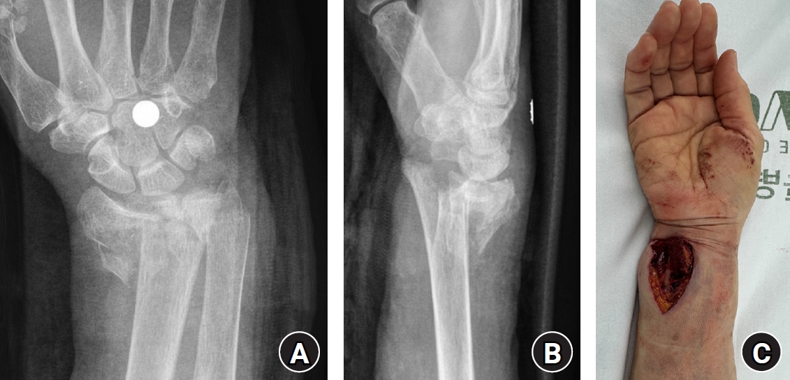
- Open comminuted intra-articular distal radius fractures with associated ulnar neurovascular injury are rare; furthermore, these injuries are challenging because skeletal stabilization, arterial reconstruction, nerve assessment, and soft-tissue coverage must be coordinated. We report the case of an 80-year-old female patient who presented after a fall from stairway with a severe open distal radius fracture and a volar-ulnar distal forearm wound. She reported numbness and tingling in the ring and small fingers, with a cold sensation and delayed capillary refill. Imaging showed a severely comminuted intra-articular distal radius fracture (AO/OTA type 23-C3). Emergency surgery was performed within 6 hours. First, a dorsal spanning plate was applied, and the small volar lunate facet fragment was fixed with a volar lunate facet plate. After debridement of the injured ulnar artery, a segmental defect remained and was reconstructed using a reversed basilic vein interposition graft harvested from the ipsilateral proximal forearm. The contused but continuous ulnar nerve was treated with neurolysis. The soft-tissue defect was managed with an acellular dermal matrix and negative-pressure wound therapy, followed by staged full-thickness skin grafting 1 week later. At 1 year postoperatively, bony union was achieved, and the functional outcomes were satisfactory. Level of evidence: V.
- 469 View
- 11 Download
- Fixation with two headless compression screws and a distal-radius bone graft for proximal scaphoid nonunion with dorsal intercalated segment instability deformity: a report of three cases
- Seonjeong Lee, Won Sun Lee, Jae Kwang Kim
- J Musculoskelet Trauma 2026;39(3):252-261. Published online July 7, 2026
- DOI: https://doi.org/10.12671/jmt.2026.00136
-
Abstract
PDF
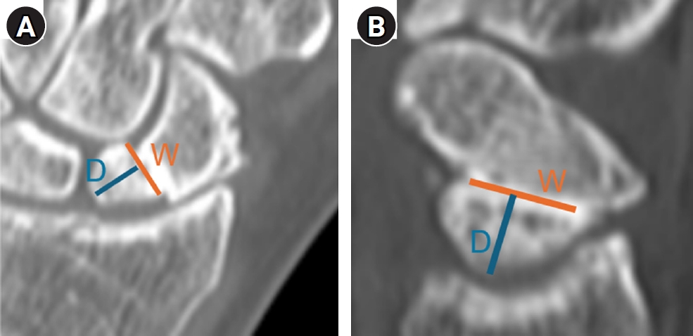
- Proximal scaphoid nonunion is difficult to treat because the proximal fragment is small and biologically compromised. Secure fixation can be especially challenging when dorsal intercalated segment instability (DISI) is present and requires correction. We report three male patients with proximal scaphoid nonunion and DISI deformity who underwent volar fixation with two 2.4-mm headless compression screws and cancellous bone graft harvested from the distal radius at the Lister tubercle. Preoperative computed tomography confirmed a proximal nonunion line and proximal fragment dimensions considered sufficient for dual-screw fixation, and magnetic resonance imaging showed no definite avascular necrosis. Early postoperative computed tomography showed maintained fixation and alignment in all three patients. Although CT-confirmed union at final follow-up was not uniformly available, radiographic union defined on plain radiographs was achieved in all cases without loss of alignment or carpal collapse. Carpal alignment improved in two patients, but DISI persisted in one. One patient underwent secondary removal of a preexisting loose body and one screw. This small case series suggests that dual-screw fixation with cancellous grafting may be technically feasible in selected proximal scaphoid nonunions with concomitant DISI. Level of evidence: V.
- 396 View
- 12 Download
- Radiocarpal subluxation after volar plating due to an insufficiently supported dorsal key fragment: a case report
- Yeongyoon Koh, Kanghun Yu, Jong Woong Park, In Cheul Choi
- J Musculoskelet Trauma 2026;39(3):269-274. Published online July 3, 2026
- DOI: https://doi.org/10.12671/jmt.2026.00185
-
Abstract
PDF
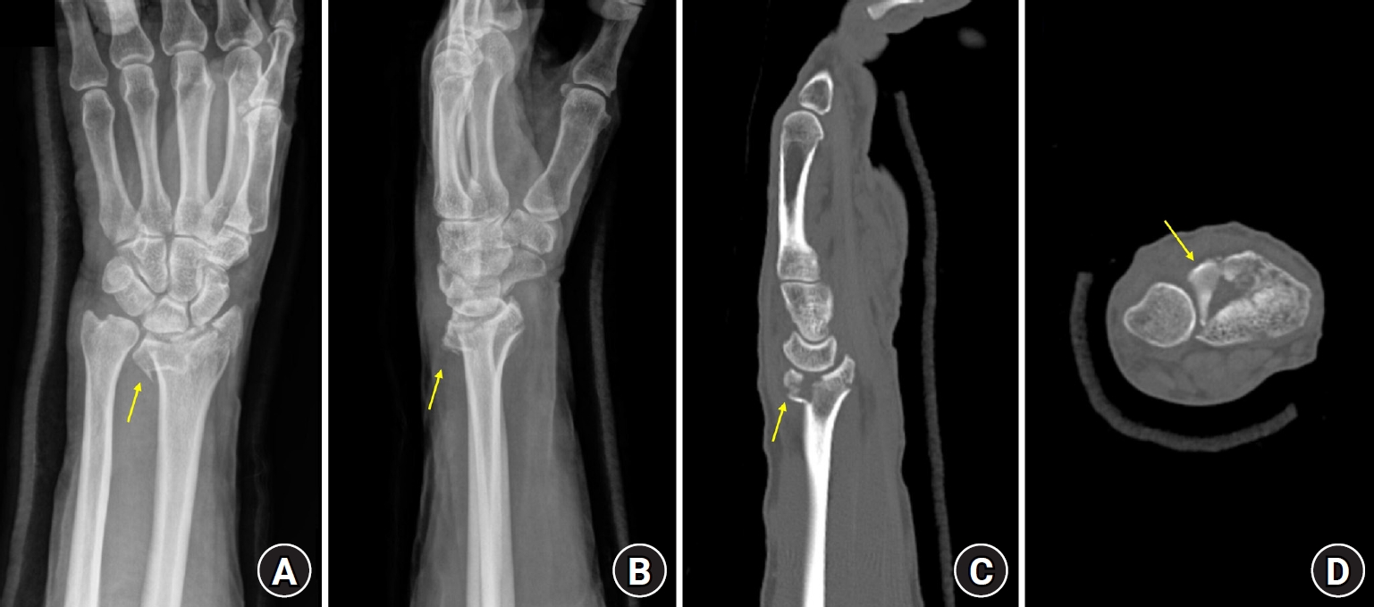
- Distal radius fractures are commonly managed using volar locking plate fixation, which provides stable fixation in most cases. However, certain fracture patterns involving dorsal key fragments may not be adequately stabilized with a volar approach alone, even when intraoperative reduction appears satisfactory. We report the case of a 58-year-old male patient with a complex intra-articular distal radius fracture involving a dorsoulnar corner (DUC) fragment. Preoperative computed tomography revealed a dorsal fragment associated with subtle dorsal radiocarpal subluxation. Despite this finding, the fragment was considered amenable to fixation through a volar approach because it was relatively large. Although satisfactory reduction was achieved intraoperatively, early postoperative imaging demonstrated progressive dorsal radiocarpal subluxation due to displacement of the DUC fragment, while overall alignment parameters remained preserved. Revision surgery with additional dorsal buttress fixation successfully restored stability. This case highlights the critical importance of recognizing dorsal key fragments and achieving adequate dorsal support during fixation, as failure to provide sufficient support may result in delayed instability despite acceptable initial reduction. Level of evidence: V.
- 437 View
- 12 Download
 First
First Prev
Prev


