
 E-submission
 KOTA
KOTA TOTA
TOTA TOTS
TOTS
Articles
- Page Path
- HOME > J Musculoskelet Trauma > Volume 39(3); 2026 > Article
-
Case Report
- Open distal radius fracture with ulnar neurovascular injury treated with dorsal spanning plate fixation and vein interposition grafting: a case report
- Kyeong-Eon Kim1, Ho-Yong Lee2, Shin-Woo Lee1, Ji-Sup Kim1
-
Journal of Musculoskeletal Trauma 2026;39(3):262-268.
DOI: https://doi.org/10.12671/jmt.2026.00164
Published online: July 20, 2026
1Department of Orthopedic Surgery, Ewha Womans University Seoul Hospital, Ewha Womans University College of Medicine, Seoul, Korea
2Department of Orthopedic Surgery, Ewha Womans University Mokdong Hospital, Ewha Womans University College of Medicine, Seoul, Korea
- Correspondence to: Ji-Sup Kim Department of Orthopedic Surgery, Ewha Womans University Seoul Hospital, Ewha Womans University College of Medicine, 260 Gonghang-daero, Gangseo-gu, Seoul 07804, Korea Tel: +82-2-6956-1656 Email: kkimjsno1@naver.com
• Received: April 7, 2026 • Revised: May 1, 2026 • Accepted: May 11, 2026
© 2026 The Korean Orthopaedic Trauma Association
This is an Open Access article distributed under the terms of the Creative Commons Attribution Non-Commercial License (http://creativecommons.org/licenses/by-nc/4.0/) which permits unrestricted noncommercial use, distribution, and reproduction in any medium, provided the original work is properly cited.
- 571 Views
- 12 Download


Abstract
-
Open comminuted intra-articular distal radius fractures with associated ulnar neurovascular injury are rare; furthermore, these injuries are challenging because skeletal stabilization, arterial reconstruction, nerve assessment, and soft-tissue coverage must be coordinated. We report the case of an 80-year-old female patient who presented after a fall from stairway with a severe open distal radius fracture and a volar-ulnar distal forearm wound. She reported numbness and tingling in the ring and small fingers, with a cold sensation and delayed capillary refill. Imaging showed a severely comminuted intra-articular distal radius fracture (AO/OTA type 23-C3). Emergency surgery was performed within 6 hours. First, a dorsal spanning plate was applied, and the small volar lunate facet fragment was fixed with a volar lunate facet plate. After debridement of the injured ulnar artery, a segmental defect remained and was reconstructed using a reversed basilic vein interposition graft harvested from the ipsilateral proximal forearm. The contused but continuous ulnar nerve was treated with neurolysis. The soft-tissue defect was managed with an acellular dermal matrix and negative-pressure wound therapy, followed by staged full-thickness skin grafting 1 week later. At 1 year postoperatively, bony union was achieved, and the functional outcomes were satisfactory.
-
Level of evidence V.
Introduction
Distal radius fractures are among the most common fractures encountered in orthopedic practice. However, vascular injury associated with distal radius fracture is uncommon, and combined injury of the ulnar artery and ulnar nerve is particularly rare [1-3]. Reported vascular complications include not only acute arterial laceration or thrombosis, but also delayed pseudoaneurysm formation after distal radius and ulna fractures [1]. Wang et al. [1] described a delayed ulnar artery pseudoaneurysm after distal radius and ulna fracture, highlighting that vascular injury may present in a delayed fashion even when the hand initially appears perfused. De Witte et al. [2] reported six cases of acute vascular injury associated with distal radius fracture and noted that these injuries were more likely to occur in open fractures, high-energy injuries, or fractures with an associated ulnar lesion. In their series, most single-artery injuries at the wrist were not associated with ischemia, but arterial reconstruction was considered necessary in the unusual patient with a dysvascular hand [2]. Sahin et al. [3] also emphasized that the ulnar neurovascular bundle may be vulnerable in severely displaced comminuted distal radius fractures.
Severe intra-articular distal radius fractures with marked metaphyseal comminution remain difficult to treat, although similar fracture patterns may also occur after a fall from standing height in elderly patients [4]. Restoration and maintenance of radial length and alignment are essential in these fractures [4,5]. Small volar lunate facet fragments may require fragment-specific fixation because inadequate stabilization of this fragment can lead to persistent instability or secondary displacement [6]. In highly comminuted intra-articular distal radius fractures, a dorsal spanning plate may provide indirect reduction through distraction and ligamentotaxis, functioning as an internal bridge construct [5]. Management becomes even more challenging when vascular compromise and soft-tissue loss are also present, because skeletal stabilization, revascularization, nerve evaluation, and soft-tissue coverage must be addressed in a coordinated manner [7]. We report a rare case of an open comminuted intra-articular distal radius fracture with combined ulnar artery and ulnar nerve injury in an elderly patient, treated with dorsal spanning plate fixation, fixation of a small volar lunate facet fragment with a volar lunate facet plate, basilic vein interposition grafting, neurolysis, and staged soft-tissue reconstruction.
Case report
This study was reviewed by the Institutional Review Board (IRB) of Ewha Womans University Seoul Hospital (IRB No. 2026-05-041-001). Written informed consent for publication of clinical details and accompanying images was obtained from the patient.
An 80-year-old female patient with a medical history of hypertension and chronic kidney disease presented to our emergency department after a fall from stairway, during which she landed with her outstretched right hand striking the ground. Physical examination revealed gross deformity of the wrist with the fragment protruding through an approximately 8 cm open wound over the volar-ulnar aspect of the distal forearm, with minimal contamination (Fig. 1). The patient reported numbness and tingling in the ring and small fingers. The hand was cool to touch, with capillary refill delayed to approximately 4 seconds. The radial pulse was palpable, whereas the ulnar pulse was nonpalpable distal to the injury site. Persistent clinical hypoperfusion prompted operative exploration and vascular reconstruction.
Radiographs and computed tomography demonstrated a severely comminuted intra-articular distal radius fracture, classified as AO/OTA type 23-C3 (Fig. 1). After operative exploration confirmed an ulnar arterial injury requiring reconstruction, the open fracture was classified as Gustilo-Anderson type IIIC. Given the open injury and signs of impaired distal perfusion, preoperative angiography was deferred because urgent exploration, skeletal stabilization, and revascularization were prioritized. Emergency surgery was initiated within 6 hours after injury, and flow through the reconstructed ulnar artery was restored approximately 8 hours after injury.
On arrival at the emergency department, intravenous cefazolin and gentamicin were administered immediately and continued for 72 hours, and a tetanus toxoid booster was given after confirmation of prior immunization history. Following thorough irrigation with 9 L of normal saline using pulsed lavage and meticulous debridement of devitalized and contaminated soft tissue, a segmental defect of the ulnar artery remained and required reconstruction (Fig. 2). Because of the marked metaphyseal and intra-articular comminution with residual instability, a combined fixation strategy was chosen. Although revascularization was a priority, skeletal stabilization was performed first to provide a stable framework for the subsequent microvascular anastomosis and to allow accurate measurement of the required vein graft length. A dorsal spanning plate was applied first to restore length and provide provisional stability to the severely comminuted fracture, with particular attention to restoration of coronal alignment. As a bridge construct, the dorsal spanning plate allowed indirect reduction through longitudinal distraction and ligamentotaxis. Distal fixation over both the second and third metacarpals was assessed intraoperatively, and fixation to the second metacarpal was ultimately selected because it provided more satisfactory coronal alignment. After overall length and alignment had been re-established, the small volar lunate facet fragment was reduced through a volar approach and fixed using a volar lunate facet plate (Fig. 3) [6].
The ulnar artery was explored through the volar-ulnar wound, and the injured segment was debrided back to healthy proximal and distal ends with intact intima. Because of the open wound, gross deformity, and clinical signs of impaired distal perfusion at presentation, a formal preoperative Allen test could not be reliably performed. After debridement to healthy intima, an approximately 2-cm segmental defect remained, precluding tension-free primary repair. Because clinical evidence of distal hypoperfusion persisted after skeletal realignment, we elected to reconstruct the ulnar artery using a reversed basilic vein interposition graft. A segment of the ipsilateral basilic vein was harvested from the proximal forearm, just distal to the elbow crease, reversed, and interposed between the arterial ends. End-to-end anastomoses were performed under ×2.5 loupe visualization using interrupted nylon sutures. After completion of the reconstruction, distal perfusion was restored. Exploration of the ulnar nerve revealed contusion without loss of continuity; the nerve was found to be entrapped by hematoma at the level of injury, and neurolysis was performed.
During the initial operation, the residual soft-tissue defect was covered with an acellular dermal matrix (AlloDerm), and negative-pressure wound therapy was applied. At re-exploration on postoperative day 7, the wound was free of infection and the acellular dermal matrix demonstrated granulation, with no remaining exposure of the arterial graft, tendon, bone, or hardware. A full-thickness skin graft harvested from the ipsilateral groin was applied over the incorporated matrix/the vascularized wound bed. Graft take was approximately 95%, and no additional coverage procedure was required.
The postoperative course was uneventful. A conservative antiplatelet regimen was selected; aspirin 100 mg once daily was administered for 4 weeks postoperatively. Cefazolin and gentamicin were administered according to the institutional protocol for type III open fractures, with gentamicin dosing adjusted for renal function. Antibiotic prophylaxis was discontinued after 3 days. Postoperative three-dimensional computed tomography angiography, performed on postoperative day 3 after reassessment of renal function, confirmed patency of the interposition graft (Fig. 2C), with no deterioration in renal function. Clinical examination demonstrated a warm, well-perfused hand with a capillary refill time of 1 second. The dorsal spanning plate was removed at 2 months postoperatively. Ulnar nerve-related symptoms persisted for up to 3 months postoperatively, but gradually improved and had resolved by 6 months. At the 1-year follow-up, bony union had been achieved, and the patient demonstrated a favorable functional outcome, with a Disabilities of the Arm, Shoulder and Hand score of 6.7 and a modified Mayo wrist score of 85. Objective sensory assessment using Semmes-Weinstein monofilament testing demonstrated 2.83 at the ring and small finger pulps. At 1 year, wrist flexion and extension were 50° and 70°, respectively, and forearm pronation and supination were 80° and 80°. Grip strength was 72% of that of the contralateral hand. Finger range of motion was full without stiffness (Figs. 4, 5). The hand remained warm and well-perfused, with a palpable radial pulse and palpable ulnar pulse and a capillary refill time of 1 second. No postoperative vascular complication, infection, or graft-related adverse event occurred. The clinical timeline from injury through final follow-up is summarized in Table 1.
Discussion
The clinical challenge in our patient was the need to coordinate skeletal stabilization, arterial reconstruction, nerve evaluation, and staged soft-tissue reconstruction within a single treatment plan in an elderly patient with significant comorbidities. Several case-specific factors guided our decision-making. First, the presence of impaired distal perfusion, including cold sensation and delayed capillary refill, indicated that the residual radial artery inflow alone was unlikely to provide adequate perfusion, and that arterial reconstruction rather than simple ligation should be considered. Second, the markedly comminuted intra-articular fracture pattern (AO/OTA 23-C3) with a small volar lunate facet fragment required a combined fixation strategy capable of restoring overall length and alignment while also addressing the articular fragment. Third, because definitive soft-tissue coverage could not be achieved primarily, a staged reconstructive approach was planned from the outset. The following discussion focuses on how each of these decisions was made and how they were integrated into the overall treatment sequence. Although single-artery injury at the wrist can often be managed with ligation when the hand remains well perfused through the remaining intact artery, this principle did not fully apply in our patient [2]. Unlike the ligation cases reported by de Witte et al. [2], our patient demonstrated evidence of distal hypoperfusion, delayed capillary refill, and a nonpalpable ulnar pulse. In addition, debridement resulted in a 2-cm segmental ulnar arterial defect that precluded tension-free primary repair. Although ligation may be acceptable for an isolated single-artery injury in a well-perfused hand, these two factors—persistent hypoperfusion and a defect too large for primary repair—supported reconstruction as a case-specific strategy to restore adequate hand perfusion. This case does not establish the superiority of reconstruction over ligation in a well-perfused hand; rather, it is consistent with the observation by de Witte et al. [2] that interpositional vein grafting may be necessary in the unusual patient with a dysvascular hand, particularly when crush injury or thrombosis involves a several-centimeter arterial segment.
The pattern of ulnar neurovascular injury in our case also deserves attention. Sahin et al. [3] reported a delayed case of combined ulnar artery and ulnar nerve injury after comminuted distal radius fracture, in which scar entrapment, thrombosis, and a segmental arterial defect were identified during secondary exploration and treated with basilic vein grafting. In contrast, our patient underwent urgent exploration at the index procedure because the fracture was open and accompanied by impaired distal perfusion. The ulnar nerve remained in continuity and was therefore treated with neurolysis alone. Ulnar nerve-related symptoms gradually improved and recovery occurred during the follow-up period.
Stable skeletal fixation and coordinated staged treatment were also essential. Severe metaphyseal comminution in elderly patients may occur even after a fall from standing height [4], and dorsal spanning plate fixation can serve as an internal bridge construct that restores length and alignment through distraction and ligamentotaxis [5]. In our case, provisional restoration of length and coronal alignment provided a stable framework for vascular reconstruction and subsequent soft-tissue management. Completing skeletal stabilization before arterial anastomosis also allowed accurate determination of the required vein graft length and minimized the risk of disrupting the microvascular repair during subsequent fracture manipulation. Because hand perfusion was reduced but not completely absent, rapid skeletal stabilization was completed first to restore alignment and protect the subsequent vascular repair. Skeletal stabilization required approximately 60 minutes, after which arterial reconstruction was performed. More broadly, this case illustrates the feasibility of coordinated management of debridement, skeletal stabilization, revascularization, and soft-tissue coverage in a patient with combined orthopedic and vascular injury [7]. Shahien et al. [7] reported high complication and amputation rates in patients with combined orthopedic and vascular injuries presenting with ischemia and emphasized the importance of an individualized salvage strategy.
Throughout management, the patient’s advanced age and chronic kidney disease were taken into account in key decisions, including antibiotic dosing, staged soft-tissue reconstruction, and selection of a conservative antiplatelet regimen. At 1 year postoperatively, bony union was achieved, postoperative computed tomography angiography confirmed graft patency, and the patient had no perfusion-related symptoms such as cold intolerance. Although this is a single case, it suggests that satisfactory results can be achieved in severe open distal radius fractures with combined ulnar neurovascular injury when arterial reconstruction is selected appropriately based on perfusion status and defect morphology, and when fixation, revascularization, and soft-tissue reconstruction are carefully coordinated.
This report has several limitations: it is a single case without a comparison group, and the 1-year follow-up may be insufficient to detect late vascular or neurologic complications. Given the rarity of this injury pattern, additional cases, multicenter registries, and longer vascular surveillance would help clarify optimal management.
-
Author contributions
Conceptualization: KEK, JSK. Data curation: KEK, HYL, SWL. Investigation: KEK, HYL, SWL, JSK. Methodology: KEK, JSK. Project administration: KEK, JSK. Resources: JSK. Supervision: JSK. Visualization: KEK, JSK. Writing-original draft: KEK. Writing-review & editing: KEK, HYL, SWL, JSK. All authors read and approved the final manuscript.
-
Conflicts of interest
No potential conflict of interest relevant to this article was reported.
-
Funding
None.
-
Data availability
Not applicable.
-
Acknowledgments
This case report was presented in the Case Discussion section of the 2026 Annual Congress of the Korean Orthopaedic Trauma Association (KOTA).
-
Supplementary material
None.
Article Information
Fig. 1.
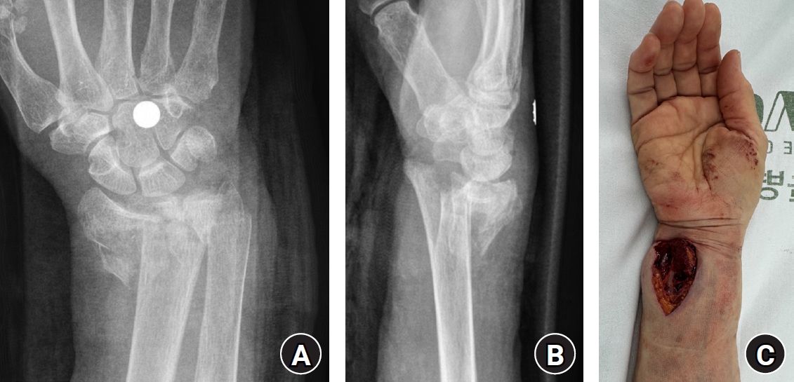
Initial images of an 80-year-old female patient with a severe open distal radius fracture after a fall from stairway. (A) Posteroanterior radiograph showing a markedly displaced comminuted intra-articular distal radius fracture. (B) Lateral radiograph showing severe dorsal displacement and metaphyseal comminution. (C) Clinical photograph demonstrating a volar-ulnar open wound at the distal forearm.
Fig. 2.
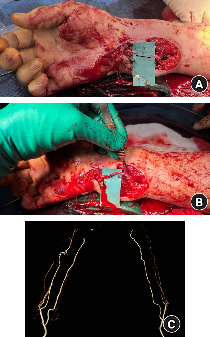
Intraoperative photographs showing ulnar artery reconstruction for a segmental defect. (A) After debridement, an approximately 2-cm segmental defect remained in the ulnar artery. (B) The defect was reconstructed with a reversed interposition vein graft harvested from the ipsilateral basilic vein. (C) Postoperative three-dimensional computed tomography angiography confirming patency of the reconstructed ulnar artery.
Fig. 3.
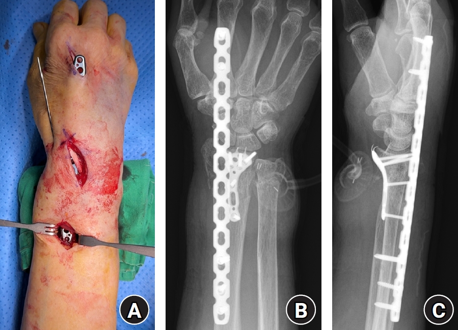
Dorsal spanning plate and volar locking plate fixation. (A) Intraoperative photograph showing the dorsal spanning plate passed beneath the extensor tendons. (B, C) Immediate postoperative posteroanterior and lateral radiographs demonstrating combined dorsal spanning plate and volar plate fixation; the dorsal spanning plate was used to restore length and alignment, and the volar plate was used to restore the articular surface.
Fig. 4.
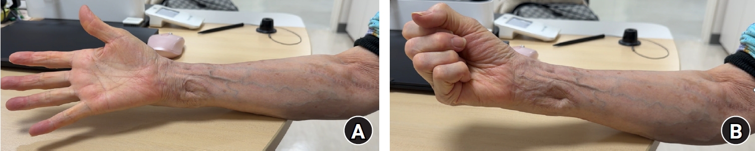
Clinical photographs obtained 1 year postoperatively. (A, B) Full finger range of motion without stiffness, no residual ulnar nerve-related symptoms, and a well-healed skin graft site are shown.
Fig. 5.
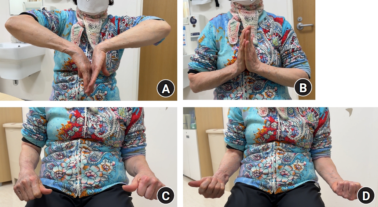
Clinical photographs obtained at final follow-up demonstrating wrist and forearm range of motion: (A) flexion, (B) extension, (C) pronation, and (D) supination. Mild limitation of wrist flexion is noted, whereas wrist extension and forearm rotation are full.
Table 1.
Clinical timeline from injury to final follow-up
- 1. Wang AA, Strauch RJ, Moore JA. Pseudoaneurysm of the ulnar artery occurring after fracture of the distal radius and ulna: a case report. J Hand Surg Am 1998;23:933-7.ArticlePubMed
- 2. de Witte PB, Lozano-Calderon S, Harness N, Watchmaker G, Green MS, Ring D. Acute vascular injury associated with fracture of the distal radius: a report of 6 cases. J Orthop Trauma 2008;22:611-4.ArticlePubMed
- 3. Sahin MS, Gokkus K, Sargin MB. Ulnar nerve and ulnar artery injury caused by comminuted distal radius fracture. J Orthop Case Rep 2020;10:25-30.Article
- 4. Cha SM. The clinical outcome of treating elderly distal radius fractures by long volar locking plate with the elimination of irreducible metaphyseal comminuted volar cortical fragments: a retrospective case series. J Musculoskelet Trauma 2025;38:13-25.ArticlePDF
- 5. Calem D, Weintraub M, Vosbikian M, Ahmed I. Radiographic outcomes of dorsal spanning plate with and without volar plate fixation for comminuted intraarticular distal radius fractures. J Clin Orthop Trauma 2025;64:102949.ArticlePubMedPMC
- 6. Park HJ, Kim JH. Hook plate versus periarticular-type volar locking plate for distal radius fractures involving the volar lunate facet in Korea: a retrospective cohort study. J Musculoskelet Trauma 2025;38:221-8.ArticlePDF
- 7. Shahien AA, Sullivan M, Firoozabadi R, et al. Combined orthopaedic and vascular injuries with ischemia: a multicenter analysis. J Orthop Trauma 2021;35:512-6.ArticlePubMed
References
Figure & Data
REFERENCES
Citations
Citations to this article as recorded by 
 ePub Link
 Cite
Cite XML Download
Open distal radius fracture with ulnar neurovascular injury treated with dorsal spanning plate fixation and vein interposition grafting: a case report
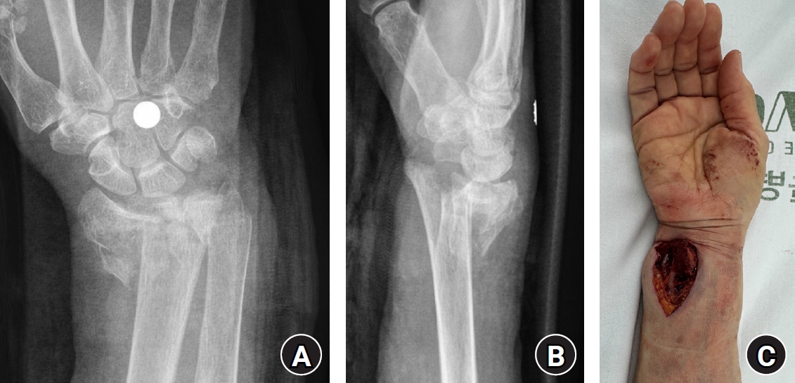
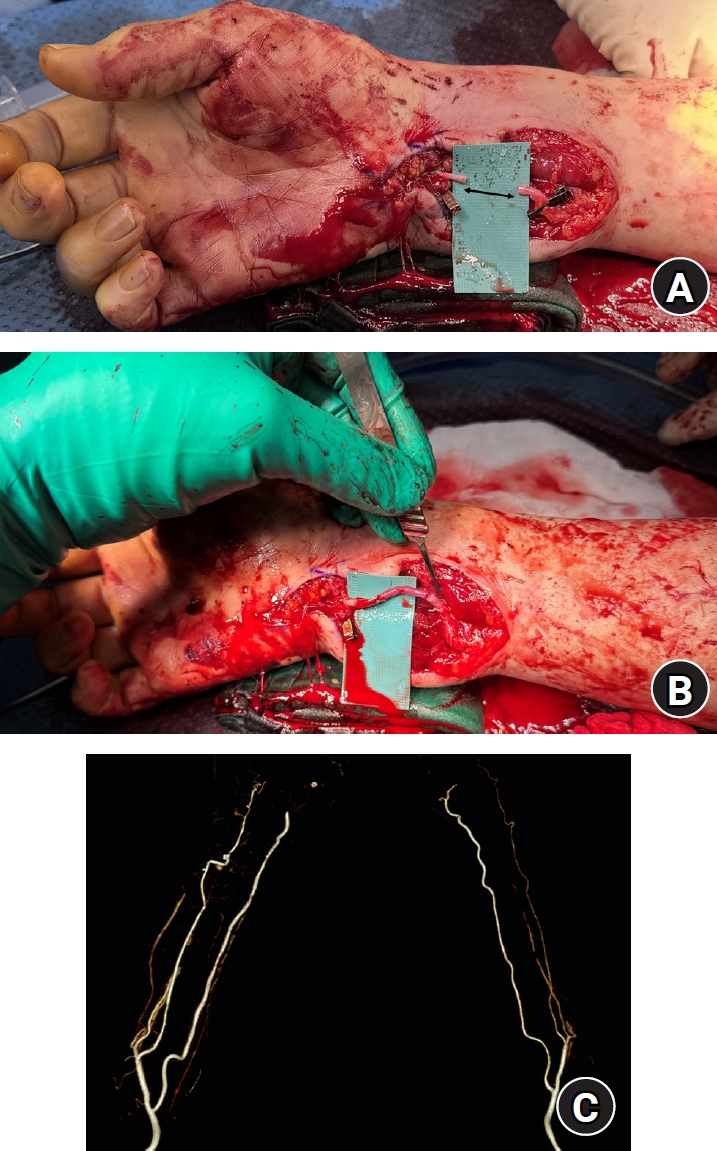
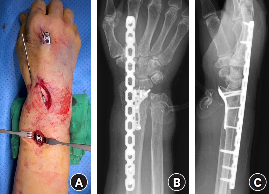
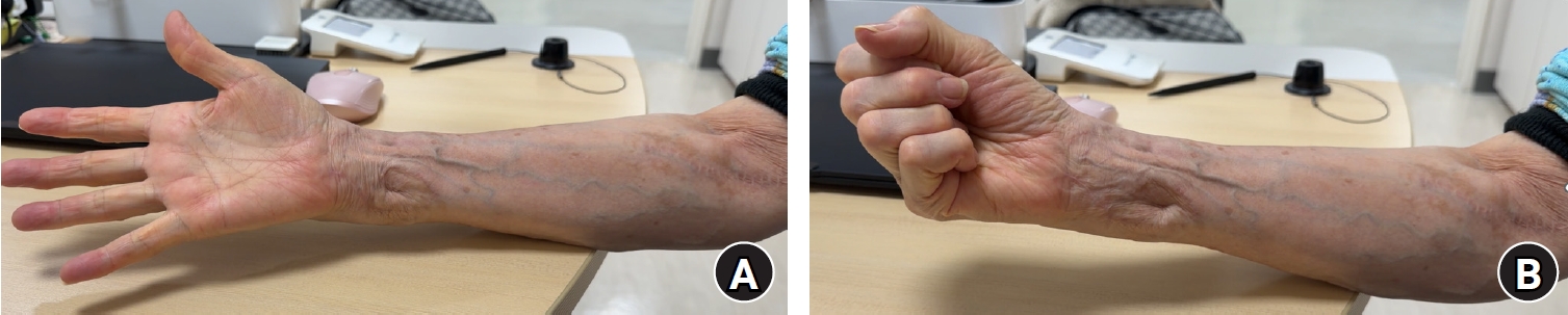
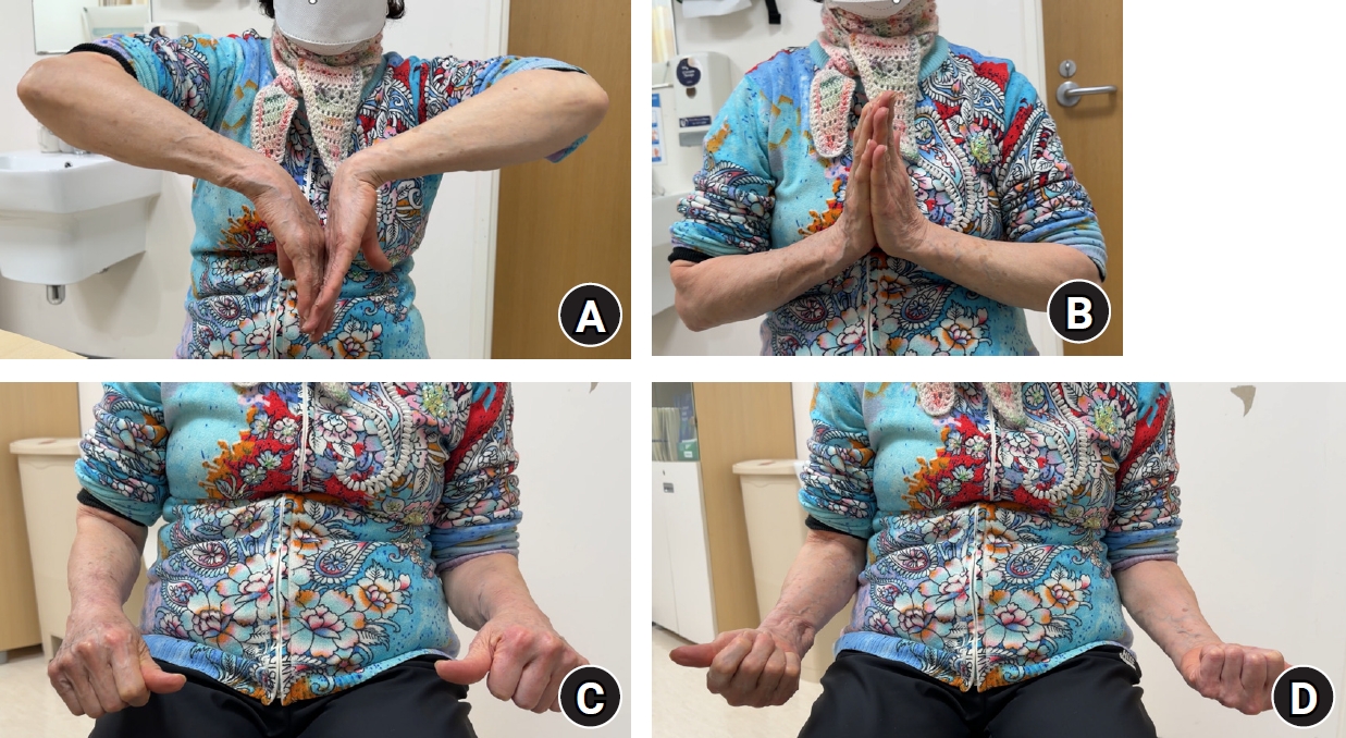
Fig. 1. Initial images of an 80-year-old female patient with a severe open distal radius fracture after a fall from stairway. (A) Posteroanterior radiograph showing a markedly displaced comminuted intra-articular distal radius fracture. (B) Lateral radiograph showing severe dorsal displacement and metaphyseal comminution. (C) Clinical photograph demonstrating a volar-ulnar open wound at the distal forearm.
Fig. 2. Intraoperative photographs showing ulnar artery reconstruction for a segmental defect. (A) After debridement, an approximately 2-cm segmental defect remained in the ulnar artery. (B) The defect was reconstructed with a reversed interposition vein graft harvested from the ipsilateral basilic vein. (C) Postoperative three-dimensional computed tomography angiography confirming patency of the reconstructed ulnar artery.
Fig. 3. Dorsal spanning plate and volar locking plate fixation. (A) Intraoperative photograph showing the dorsal spanning plate passed beneath the extensor tendons. (B, C) Immediate postoperative posteroanterior and lateral radiographs demonstrating combined dorsal spanning plate and volar plate fixation; the dorsal spanning plate was used to restore length and alignment, and the volar plate was used to restore the articular surface.
Fig. 4. Clinical photographs obtained 1 year postoperatively. (A, B) Full finger range of motion without stiffness, no residual ulnar nerve-related symptoms, and a well-healed skin graft site are shown.
Fig. 5. Clinical photographs obtained at final follow-up demonstrating wrist and forearm range of motion: (A) flexion, (B) extension, (C) pronation, and (D) supination. Mild limitation of wrist flexion is noted, whereas wrist extension and forearm rotation are full.
Fig. 1.
Fig. 2.
Fig. 3.
Fig. 4.
Fig. 5.
Open distal radius fracture with ulnar neurovascular injury treated with dorsal spanning plate fixation and vein interposition grafting: a case report
| Timepoint | Event |
|---|---|
| Injury | Fall from stairway |
| Within 6 hr | Surgery initiated |
| Approximately 7 hr | Skeletal stabilization completed (dorsal spanning plate + volar lunate facet plate) |
| Approximately 8 hr | Ulnar arterial flow restored (reversed basilic vein interposition graft) |
| Day 0 | Acellular dermal matrix + negative-pressure wound therapy applied |
| Day 3 | Postoperative computed tomography angiography confirms graft patency |
| Day 7 | Full-thickness skin graft |
| 2 Months | Dorsal spanning plate removed |
| 3 Months | Peak ulnar nerve-related symptoms |
| 6 Months | Ulnar nerve symptoms resolved |
| 1 Year | Final functional and perfusion assessment |
Table 1. Clinical timeline from injury to final follow-up


