
 E-submission
 KOTA
KOTA TOTA
TOTA TOTS
TOTS
Search
- Page Path
- HOME > Search
Review Article
- Current concepts in distal clavicle fractures: an evidence-based update
- Du-Han Kim, Hyuk-Joon Sohn, Byung-Chan Choi, Chul-Hyun Cho
- Received June 24, 2026 Accepted July 20, 2026 Published online September 4, 2026
- DOI: https://doi.org/10.12671/jmt.2026.00241 [Epub ahead of print]
-
 Abstract
 PDF
PDF - Distal clavicle fractures (DCFs) account for 10%–30% of all clavicle fractures and remain difficult to manage because fracture stability depends heavily on coracoclavicular ligament integrity, plain radiographic classification has limited reliability, and distal fragment size varies substantially, complicating fixation planning. This review provides an evidence- based update on the anatomy, classification, imaging, and management of DCFs, incorporating recent literature, including population-specific cadaveric data and three-dimensional fracture mapping. The conoid and trapezoid ligaments have distinct osseous footprints that influence fracture stability in both the Neer/Craig and Cho classification systems; however, interobserver reliability remains only fair-to-moderate for both systems, particularly when conoid ligament integrity must be inferred. Three-dimensional computed tomography mapping has shown that posterior displacement and angulation are nearly universal but often underrecognized on standard radiographs, supporting selective computed tomography use when radiographs do not adequately characterize fracture morphology, comminution, or multiplanar displacement. Magnetic resonance imaging should be reserved for cases in which ligament integrity remains diagnostically ambiguous. Stable fractures generally have excellent outcomes with conservative treatment using a simple arm sling. For unstable fractures, operative and conservative treatment yield similar 1-year functional outcomes despite different union rates (96% vs. 63%), with conservative treatment carrying a nonunion risk of approximately one-third and operative treatment carrying a technique-dependent reoperation burden, ranging from planned implant removal in most hook plate cases to less than 10% with coracoclavicular stabilization constructs. When surgery is indicated, technique selection should be guided by fracture morphology and surgeon experience: anatomic locking plate fixation with or without coracoclavicular augmentation for Neer IIA/Cho IIA fractures, dedicated coracoclavicular stabilization for Neer IIB fractures (Cho IIB/IIC), and hook plate fixation as a salvage construct for Neer V/Cho IID fractures. Management should integrate classification, imaging findings, union risk, reoperation risk, expected recovery, and patient-specific factors through shared decision- making.
- 441 View
- 7 Download

Original Article
- Comparative results of the femoral neck system versus the dynamic hip screw for stable femoral neck fractures in older adults in Korea: a retrospective cohort study
- Byung-Chan Choi, Byung-Woo Min, Kyung-Jae Lee, Jun-Sik Hong
- J Musculoskelet Trauma 2025;38(4):203-211. Published online October 24, 2025
- DOI: https://doi.org/10.12671/jmt.2025.00276
-
Abstract
PDF
- Background
This study aimed to compare the clinical and radiological outcomes of the femoral neck system (FNS) and the dynamic hip screw (DHS) for the internal fixation of stable femoral neck fractures in older adults.
Methods
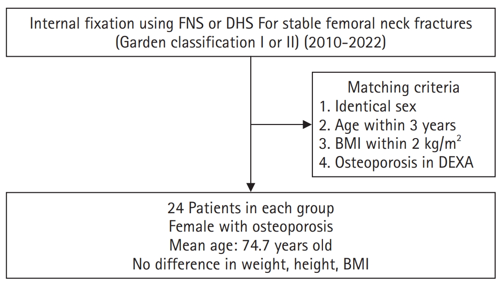
This retrospective cohort study included 48 matched older adult patients based on sex, age, BMI, and osteoporosis status, who had undergone internal fixation with either FNS or DHS for stable femoral neck fractures between January 2010 and December 2022. To minimize selection bias, a 1:1 case-control matching was performed based on sex, age, body mass index (BMI), and the presence of osteoporosis. A total of 48 patients (24 in each group) were included. We compared perioperative data (operation time, hemoglobin change, transfusion rate), functional outcomes using the Koval score, and radiological outcomes, including union rate, femoral neck shortening, and complication rates.
Results
The mean operation time was significantly shorter in the FNS group than in the DHS group (60.9 minutes vs. 70.8 minutes; P=0.007). There were no statistically significant differences between the two groups in the union rate (87.5% in FNS vs. 95.8% in DHS), femoral neck shortening, final Koval score distribution, or overall complication rates (12.5% in both groups).
Conclusions
For treating stable femoral neck fractures in older adults, the FNS demonstrated comparable clinical and radiological outcomes to the DHS, with the distinct advantage of a shorter operation time. While these findings suggest that the FNS is a promising and safe alternative that may reduce the surgical burden, definitive conclusions are precluded by the small sample size, warranting further research to corroborate these results. Level of evidence: IV. -
Citations
Citations to this article as recorded by
- Characteristics of the Extent and Onset of Osteonecrosis of the Femoral Head After Femoral Neck System (FNS) Fixation: A Minimum Two-Year Follow-Up Comparative Study with Cannulated Screws
Incheol Kook, Sihoon Choi, Soo-Young Jeong, Kyu Tae Hwang
Journal of Clinical Medicine.2026; 15(16): 6405. CrossRef
- Characteristics of the Extent and Onset of Osteonecrosis of the Femoral Head After Femoral Neck System (FNS) Fixation: A Minimum Two-Year Follow-Up Comparative Study with Cannulated Screws
- 3,543 View
- 49 Download
- 1 Crossref
Review Article
- Osteoporotic Hip Fracture: How We Make Better Results?
- Byung-Chan Choi, Kyung-Jae Lee
- J Korean Fract Soc 2024;37(1):52-59. Published online January 31, 2024
- DOI: https://doi.org/10.12671/jkfs.2024.37.1.52
-
Abstract
PDF
- The prevalence of osteoporosis and incidence of osteoporotic fractures is increasing gradually as life expectancy is prolonged and the aged population increases. Osteoporotic hip fractures (femoral neck fractures and femoral intertrochanteric fractures) have high mortality because the patients with these fractures are elderly and have several comorbidities. Thorough preparation and a multidisciplinary approach in the preoperative period are critical, and early surgery is recommended. There are also several principles to treat osteoporotic hip fractures and prevent fixation failures. Many studies have suggested various treatment methods for femoral neck fractures and femoral intertrochanteric fractures. Functional recovery treatment is essential based on the patient’s health and activity levels. Finally, aggressive management of osteoporosis and the prevention of falling is needed to treat osteoporotic hip fractures successfully.
- 1,484 View
- 43 Download
 First
First Prev
Prev


