-
Definitions and assessment of residual distal radioulnar joint instability after operative fixation of distal radius fractures: a methodological systematic review
-
Jae Ho Cho, Seungyeob Sakong, Sumin Lim
-
J Musculoskelet Trauma 2026;39(4):323-340. Published online October 19, 2026
-
DOI: https://doi.org/10.12671/jmt.2026.00290
-
-
 Abstract  PDF  Supplementary Material
- Background
Residual distal radioulnar joint (DRUJ) instability after operative fixation of distal radius fractures serves as an eligibility criterion, treatment indication, and outcome, but previous syntheses report inconsistent definitions. This methodological systematic review examined how clinical reports define, assess, and report it, and whether they distinguish it from structural triangular fibrocartilage complex (TFCC) lesions.
Methods
MEDLINE (PubMed) and Embase were searched from 2015 to 3 August 2026 for adult clinical reports in which residual DRUJ instability was assessed after fixation, or inferred from TFCC lesion characteristics, and used for a treatment decision or as an outcome. Every report entering full-text eligibility was assessed independently in duplicate; data were locked before agreement was calculated and disagreements disclosed. No riskof- bias tool was applied: neither treatment effects nor diagnostic accuracy were estimated. Findings are counts and proportions; no meta-analysis was performed.
Results
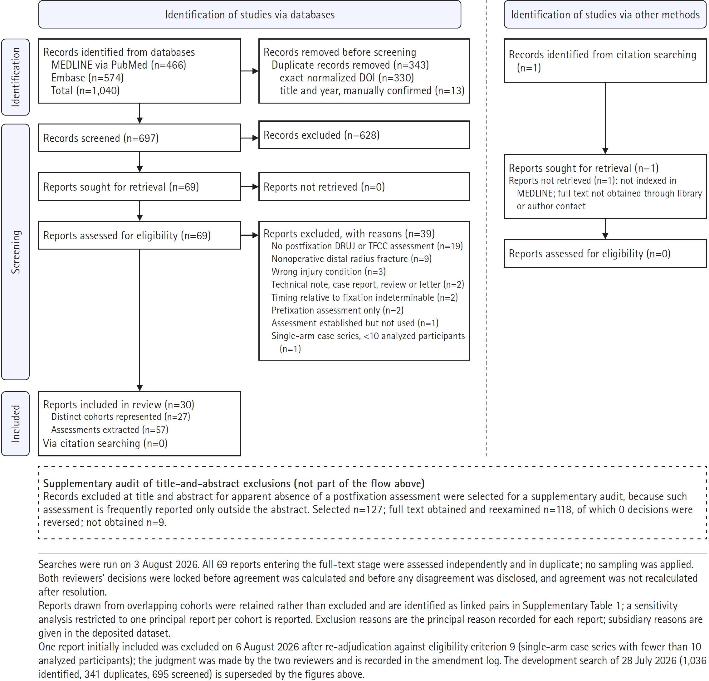
Of 1,040 records, 697 were screened and 69 assessed as full texts; 30 reports of 27 cohorts were included, contributing 57 assessments. In the treatment-decision context, 12 reports stated an operational definition (40.0% of all 30; 50.0% of the 24 containing such an assessment), four stated neither required element (16.7% of 24), and six contained no such assessment and were classified as not applicable. At the assessment level, the positive criterion was vague in 43.9% and absent in 17.5%, and explicit for 48.0% of intraoperative but 25.9% of final follow-up assessments. No report inferred instability from a TFCC lesion instead of testing the joint; one used the two constructs interchangeably, 14 (46.7%) distinguished them, and 15 (50.0%) addressed instability without structural verification. A comparable prevalence estimate was extractable from only 9 of 57 assessments (15.8%).
Conclusions
Residual DRUJ instability is therefore assessed frequently but is often incompletely defined, and until these elements are stated consistently, frequency estimates cannot be compared across studies.
Level of evidence: Not applicable.
-
Treatment of avulsion fractures around the knee
-
Jeong-Hyun Koh, Hyung Keun Song, Won-Tae Cho, Seungyeob Sakong, Sumin Lim
-
J Musculoskelet Trauma 2025;38(2):63-73. Published online March 31, 2025
-
DOI: https://doi.org/10.12671/jmt.2025.00073
-
-
Abstract
PDF
- Avulsion fractures of the knee occur when tensile forces cause a bone fragment to separate at the site of soft tissue attachment. These injuries, which frequently affect adolescent athletes, can involve the cruciate and collateral ligaments, arcuate complex, iliotibial band, and patellar and quadriceps tendons. Radiographs aid in the initial diagnosis, while computed tomography and magnetic resonance imaging facilitate a comprehensive evaluation of injury severity and concomitant damage. Specific avulsion fracture types include: anterior cruciate ligament avulsions (tibial site, Meyers and McKeever classification), posterior cruciate ligament avulsions (tibial attachment, Griffith's classification), Segond fractures (anterolateral complex injury), iliotibial band avulsions, medial collateral ligament avulsions (reverse Segond, Stieda fractures), arcuate complex avulsions ("arcuate sign"), medial patellofemoral avulsions (patellar dislocations), and patellar/quadriceps tendon avulsions. The treatment depends on the fracture location, displacement, and associated injuries. Non-displaced fractures can be managed conservatively, while displaced fractures or those with instability require surgical reduction and fixation. Prompt recognition and appropriate intervention prevent complications such as deformity, nonunion, malunion, and residual instability. This review provides an overview of the pathogenesis, diagnosis, and management of knee avulsion fractures to guide clinical decision-making.
-
Citations
Citations to this article as recorded by  - Avulsionsfrakturen am Kniegelenk – eine Übersicht
Anton Ferdinand Schmidt, Christoph Kittl, Adrian Deichsel, Larissa Eckl, Riccardo D’Ambrosi, Arasch Wafaisade, Josina Maiti Münchgesang, Jakob Ackermann, Dominic T. Mathis, Lukas Münch, Gergo Merkely, Lena Eggeling, Andreas Martin Seitz, Karl F. Schüttler
Arthroskopie.2026; 39(4): 255. CrossRef - Lateral marginal fractures of the patella and patellofemoral pain
Jae-Ang Sim, Chul-Ho Kim, Ji Wan Kim
Journal of Musculoskeletal Trauma.2025; 38(3): 152. CrossRef
-
50,105
View
-
351
Download
-
2
Crossref
-
Treatment of Avulsion Fractures around the Knee
-
Sumin Lim
-
J Korean Fract Soc 2024;37(2):117-124. Published online April 30, 2024
-
DOI: https://doi.org/10.12671/jkfs.2024.37.2.117
-
-
Abstract
PDF
- Avulsion fractures are common in athletes and result from high-impact or sudden, forceful movements involving the separation of a bone fragment at the ligament or tendon attachment site. The key focus areas include the anterior and posterior cruciate ligaments, medial collateral ligament, anterolateral complex, arcuate complex, medial patellofemoral ligament, patellar tendon, and quadriceps tendon. Diagnostic approaches combine radiography with advanced imaging techniques, such as computed tomography and magnetic resonance imaging, to elucidate the extent of injury and guide treatment decisions. Treatment ranges from conservative management for non-displaced fractures to surgical intervention for displaced fractures, with strategies customized based on the specific ligament involved and the nature of the fracture.
|

 E-submission
 KOTA
 TOTA
 TOTS