 E-submission
 TOTA
TOTA TOTS
TOTS
Articles
- Page Path
- HOME > J Musculoskelet Trauma > Volume 22(1); 2009 > Article
-
Original Article
- Interlocking Intramedullary Nailing of Forearm Shaft Fractures in Adults
- Sanglim Lee, M.D., Hee-Sung Lee, M.D., Yerl-Bo Sung, M.D., Jae-Kwang Yum, M.D.
-
Journal of the Korean Fracture Society 2009;22(1):30-38.
DOI: https://doi.org/10.12671/jkfs.2009.22.1.30
Published online: January 31, 2009
Department of Orthopedic Surgery, Sanggye Paik Hospital, College of Medicine, Inje University, Seoul, Korea.
- Address reprint requests to: Jae-Kwang Yum, M.D. Department of Orthopedic Surgery, Sanggye Paik Hospital, 761-1, Sanggye-7-dong, Nowon-gu, Seoul 139-707, Korea. Tel: 82-2-950-1032, Fax: 82-2-934-6342, shoulder@paik.ac.kr
• Received: February 15, 2008 • Revised: October 27, 2008 • Accepted: November 11, 2008
Copyright © 2009 The Korean Fracture Society. All rights reserved.
This is an Open Access article distributed under the terms of the Creative Commons Attribution Non-Commercial License (http://creativecommons.org/licenses/by-nc/3.0/) which permits unrestricted non-commercial use, distribution, and reproduction in any medium, provided the original work is properly cited.
- 1,314 Views
- 4 Download
- 3 Crossref
Abstract
-
Purpose
- To evaluate the usefulness of interlocking intramedullary nailing for operative treatment of forearm shaft fractures in adults.
-
Materials and Methods
- Thirteen forearm shaft fractures in 12 patients were fixated with 13 Acumed forearm intramedullary rods (ulna: 8, radius: 5). The average age was 36.7 years and mean follow-up period was 15.2 months. The union time was measured when there was no tenderness over the fracture site and the bridging callus was evident in at least two sides of the cortex. The range of motion of the joint and the rotation of the forearm was measured and the functional results were evaluated with Grace and Eversmann's rating system.
-
Results
- Radiologic union was observed at 11.8 weeks postoperatively in 11 cases out of 13. No limitation of motion was observed. Nine had excellent or good functional results. In one Galeazzi fracture, radial shaft became displaced after nailing and should be re-stabilized with plate. Proximal interlocking screws were improperly inserted in one ulnar nail. Implants were removed in 7 cases. Removal guide screw was broken while removing the intramedullary nail in one case of ulnar shaft fracture.
-
Conclusion
- Interlocking intramedullay nailing might be a treatment option for the middle 1/3 shaft fractures of the adult forearm bone with favorable results.
- 1. Beaupre GS, Csongradi JJ. Refracture risk after plate removal in the forearm. J Orthop Trauma, 1996;10:87-92.Article
- 2. Brumback RJ, Virkus WW. Intramedullary nailing of the femur: reamed versus nonreamed. J Am Acad Orthop Surg, 2000;8:83-90.Article
- 3. De Pedro JA, Garcia-Navarrete F, Garcia De, Otero R, Oteo A, Lopez-Duran Stern L. Internal fixation of ulnar fractures by locking nail. Clin Orthop Relat Res, 1992;283:81-85.
- 4. Finkemeier CG, Schmidt AH, Kyle RF, Templeman DC, Varecka TF. A prospective, randomized study of intramedullary nails inserted with and without reaming for the treatment of open and closed fractures of the tibia shaft. J Orthop Trauma, 2000;14:187-193.
- 5. Gao H, Luo CF, Zhang CQ, Shi HP, Fan CY, Zen BF. Internal fixation of diaphyseal fractures of the forearm by interlocking intramedullary nail: short-term results in eighteen patients. J Orthop Trauma, 2005;19:384-391.
- 6. Grace TG, Eversmann WW Jr. Forearm fracture: treatment by rigid fixation with early motion. J Bone Joint Surg Am, 1980;62:433-438.
- 7. Jones DJ, Henley MB, Schemitsch EH, Tencer AF. A biomechanical comparison of two methods of fixation of fractures of the forearm. J Orthop Trauma, 1995;9:198-206.Article
- 8. Kang CN, Kim JO, Kim DW, et al. The operative treatment of the shaft fractures of the forearm bone: operative comparison in intramedullary fixation to plate fixation on treatment of the both forearm bone fracture. J Korean Soc Fract, 1998;11:63-69.Article
- 9. Kim KY, Lim MS, Choi SK, Yoon HJ. Treatment of forearm shaft fracture with modified interlocking intramedullary nail. J Korean Fract Soc, 2008;21:157-164.Article
- 10. Labosky DA, Cermak MB, Waggy CA. Forearm fracture plates: to remove or not to remove. J Hand Surg Am, 1990;15:294-301.Article
- 11. Langkamer VG, Ackroyd CE. Removal of forearm plates. A review of the complications. J Bone Joint Surg Br, 1990;72:601-604.ArticlePDF
- 12. Lee YH, Lee SK, Chung MS, Baek GH, Gong HS, Kim KH. Interlocking contoured intramedullary nail fixation for selected diaphyseal fractures of the forearm in adults. J Bone Joint Surg Am, 2008;90:1891-1898.Article
- 13. Matthews LS, Kaufer H, Garver DF, Sonstegard DA. The effect on supination-pronation of angular malalignment of fractures of both bones of the forearm. J Bone Joint Surg, 1982;64:14-17.Article
- 14. Rosson JW, Shearer JR. Refracture after the removal of plates from the forearm. An avoidable complication. J Bone Joint Surg Br, 1991;73:415-417.ArticlePDF
- 15. Sage FP, Smith H. Medullary fixation of forearm fractures. J Bone Joint Surg Am, 1957;39:91-98.Article
- 16. Sarmiento A, Ebramzadeh E, Brys D, Tarr R. Angular deformities and forearm function. J Orthop Res, 1992;10:121-133.Article
- 17. Schemitsch EH, Jones D, Henley MB, Tencer AF. A comparison of malreduction after plate and intramedullary nail fixation of forearm fractures. J Orthop Trauma, 1995;9:8-16.Article
- 18. Shin HD, Rhee KJ, Yang JY, Yun SH, Lee MJ. Comparison of clinical results between the plate fixation and intramedullary nailing for the diaphyseal both forearm bone fractures. J Korean Soc Fract, 1999;12:135-144.Article
- 19. Stannard JP, Harris HW, McGwin G Jr, Volgas DA, Alonso JE. Intramedullary nailing of humeral shaft fractures with a locking flexible nail. J Bone Joint Surg Am, 2003;85:2103-2110.Article
- 20. Street DM. Intramedullary forearm nailing. Clin Orthop Relat Res, 1986;212:219-230.Article
- 21. Weckbach A, Blattert TR, Weisser Ch. Interlocking nailing of forearm fractures. Arch Orthop Trauma Surg, 2006;126:309-315.ArticlePDF
REFERENCES
Fig. 1
(A) Photograph shows the left radial rod (anterior view), right radial rod (anterior view), right radial rod (lateral view), ulnar rod (anterior view), and ulnar rod (lateral view) from left to right.
(B) The radial rod is assembled with the targeting guide. The drill guide for an interlocking screw is inserted through the cannula.
(C) The ulnar rod is inserted with the targeting guide.
(D) The probe is tapped on the skin to indicate the incision site for the screw.
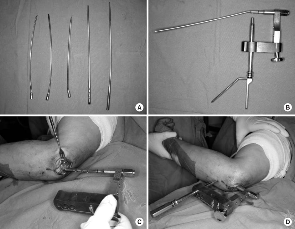
Fig. 2
(A) Preoperative radiographs of the forearm of 17-year-old male patient showed ulnar oblique and radial transverse shaft fracture after slip-down from the stairs.
(B) Ulna was fixated with plate and screws and radius was reduced closely and stabilized with the intramedullary nail.
(C) Fracture gap was not observed on the radiograph and the patient had no tenderness on the fracture site at 8 weeks postoperatively.
(D) At postoperative one year, metal was removed. No complication had been observed till one year after removal with 'excellent' functional result in the Grace and Eversmann rating system.
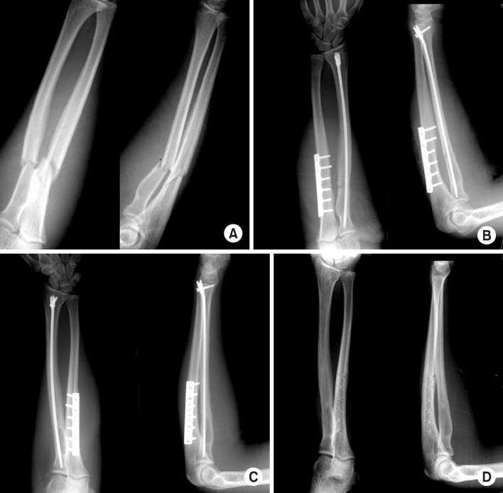
Fig. 3
(A) Preoperative X-ray showed ulnar shaft comminuted fracture.
(B) Immediate postoperative X-ray showed that ulna was fixated with intramedullary nail.
(C) Radiographs showed increased gap in the fracture site at four weeks postoperatively and patient reported pain during the motion of wrist and elbow. Then, they were immobilized with long arm splint until the pain was subsided for about two weeks.
(D) Radiographs at postoperative one year showed solid union without additional bone grafting.

Fig. 4
(A) Preoperative radiographs of the forearm of 28-year-old female patient showed ulnar transverse shaft fracture.
(B) Immediate postoperative X-ray showed that ulna was fixated with intramedullary nail.
(C) Intraoperative C-arm film showed removal of IM nail.
(D) At postoperative 17 months, metal was removed.
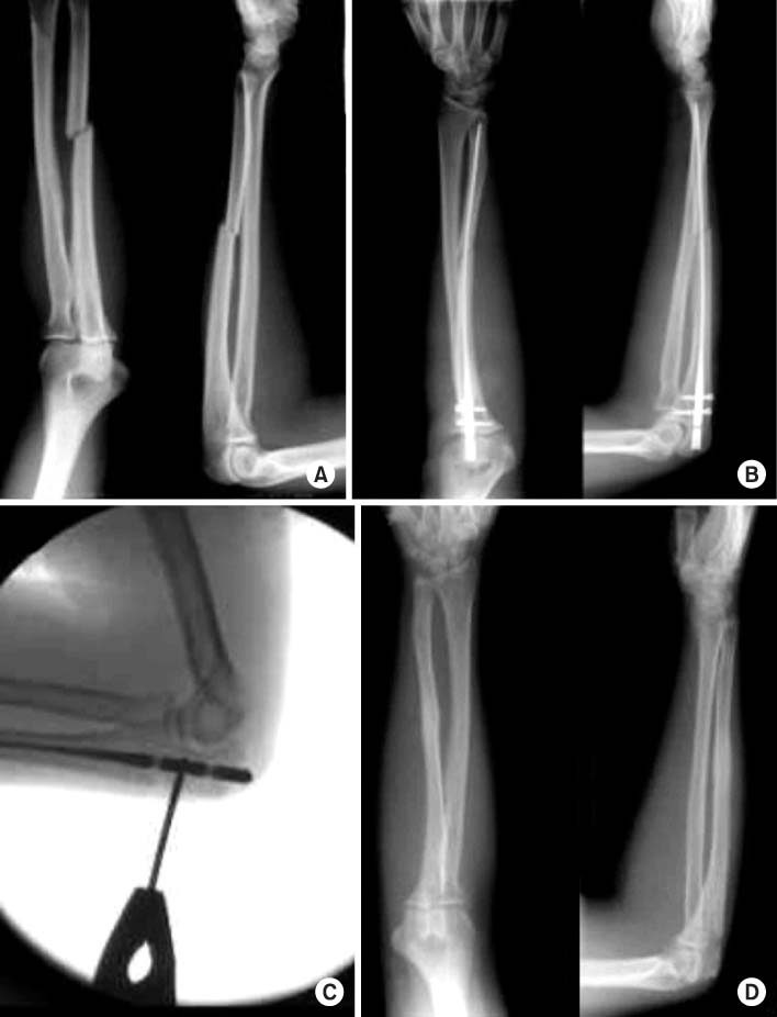
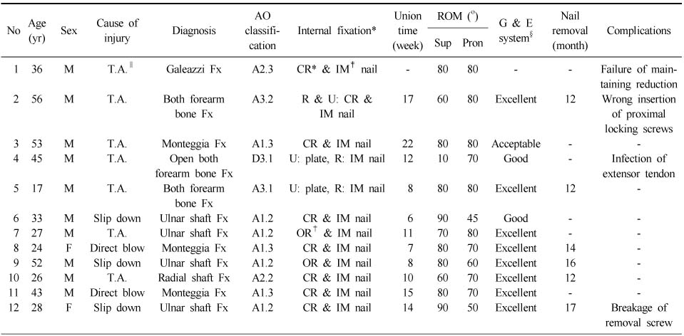
Figure & Data
REFERENCES
Citations
Citations to this article as recorded by 
- Comparison of minimally invasive plate osteosynthesis (MIPO) and open reduction and internal fixation (ORIF) for the treatment of radial shaft fractures: a retrospective study
Hyun-Tak Kang, Yang-Hoon Jo, Hong-Je Kang
BMC Musculoskeletal Disorders.2025;[Epub] CrossRef - Distal blocking screw augmentation in ulnar intramedullary nail fixation of adult forearm diaphyseal fractures
Yong Woo Kim, Sang Ki Lee, Young Sun An
Journal of Orthopaedic Surgery.2024;[Epub] CrossRef - Comparison of Bending Strength among Plate, Steinmann Pin, and Headless Compression Screw Fixations for Proximal Ulnar Shaft Fracture in Sawbones
Jinyoung Han, Jin Rok Oh, Jaewoong Um
Archives of Hand and Microsurgery.2020; 25(4): 267. CrossRef
 Cite
CiteInterlocking Intramedullary Nailing of Forearm Shaft Fractures in Adults


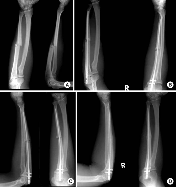

Fig. 1
(A) Photograph shows the left radial rod (anterior view), right radial rod (anterior view), right radial rod (lateral view), ulnar rod (anterior view), and ulnar rod (lateral view) from left to right.
(B) The radial rod is assembled with the targeting guide. The drill guide for an interlocking screw is inserted through the cannula.
(C) The ulnar rod is inserted with the targeting guide.
(D) The probe is tapped on the skin to indicate the incision site for the screw.
Fig. 2
(A) Preoperative radiographs of the forearm of 17-year-old male patient showed ulnar oblique and radial transverse shaft fracture after slip-down from the stairs.
(B) Ulna was fixated with plate and screws and radius was reduced closely and stabilized with the intramedullary nail.
(C) Fracture gap was not observed on the radiograph and the patient had no tenderness on the fracture site at 8 weeks postoperatively.
(D) At postoperative one year, metal was removed. No complication had been observed till one year after removal with 'excellent' functional result in the Grace and Eversmann rating system.
Fig. 3
(A) Preoperative X-ray showed ulnar shaft comminuted fracture.
(B) Immediate postoperative X-ray showed that ulna was fixated with intramedullary nail.
(C) Radiographs showed increased gap in the fracture site at four weeks postoperatively and patient reported pain during the motion of wrist and elbow. Then, they were immobilized with long arm splint until the pain was subsided for about two weeks.
(D) Radiographs at postoperative one year showed solid union without additional bone grafting.
Fig. 4
(A) Preoperative radiographs of the forearm of 28-year-old female patient showed ulnar transverse shaft fracture.
(B) Immediate postoperative X-ray showed that ulna was fixated with intramedullary nail.
(C) Intraoperative C-arm film showed removal of IM nail.
(D) At postoperative 17 months, metal was removed.
Fig. 1
Fig. 2
Fig. 3
Fig. 4
Interlocking Intramedullary Nailing of Forearm Shaft Fractures in Adults
Patient data
*CR: Closed reduction, †OR: Open reduction, ‡IM nail: Intramedullary nail, §G & E system: Grace and Eversmann rating system, ∥T.A.: Traffic accident.
Table 1
Patient data
*CR: Closed reduction, †OR: Open reduction, ‡IM nail: Intramedullary nail, §G & E system: Grace and Eversmann rating system, ∥T.A.: Traffic accident.

