 E-submission
 TOTA
TOTA TOTS
TOTS
Articles
- Page Path
- HOME > J Musculoskelet Trauma > Volume 28(3); 2015 > Article
-
Technical Trick
- Extraction of Misplaced Endcap during Tibia Intramedullary Nailing by 'Fish-Hook' Technique: Technical Note
- Se-Hyeok Yun, M.D., Jae-Hyuk Yang, M.D., Ph.D.
-
Journal of the Korean Fracture Society 2015;28(3):194-197.
DOI: https://doi.org/10.12671/jkfs.2015.28.3.194
Published online: July 22, 2015
Department of Orthopedic Surgery, Veterans Health Service Medical Center, Seoul, Korea.
- Address reprint requests to: Jae-Hyuk Yang, M.D., Ph.D. Department of Orthopedic Surgery, Veterans Health Service Medical Center, 53 Jinhwangdo-ro 61-gil, Gangdong-gu, Seoul 134-791, Korea. Tel: 82-2-2225-1609, Fax: 82-2-2225-1910, jaekorea@gmail.com
• Received: January 13, 2015 • Revised: February 12, 2015 • Accepted: April 9, 2015
Copyright © 2015 The Korean Fracture Society. All rights reserved.
This is an Open Access article distributed under the terms of the Creative Commons Attribution Non-Commercial License (http://creativecommons.org/licenses/by-nc/4.0/) which permits unrestricted non-commercial use, distribution, and reproduction in any medium, provided the original work is properly cited.
- 684 Views
- 0 Download
Abstract
- Endcap placement after intramedullary nailing can be cumbersome. Misplacement of the endcap which may be difficult to extract may occur. In this report, a simple Kirschner wire device with 'fish-hook' technique may ease the procedure without further violating bony or soft tissues.
Case Reports
Discussion
- 1. Alan RK, Baig R, Voss FR. Exchange femoral nailing: a new technique for removal of a broken nail. Am J Orthop (Belle Mead NJ), 2007;36:500-502.
- 2. Karladani AH. Removal of a broken nail using a guide wire and a screw. Acta Orthop, 2006;77:986-988.Article
- 3. Marwan M, Ibrahim M. Simple method for retrieval of distal segment of the broken interlocking intramedullary nail. Injury, 1999;30:333-335.Article
- 4. Liodakis E, Krettek C, Kenawey M, Hankemeier S. A new technique for removal of an incarcerated expandable femoral nail. Clin Orthop Relat Res, 2010;468:1405-1409.Article
- 5. Acharya M, Alani A, Almedghio S. The fish hook technique of extracting broken intramedullary nails. Acta Orthop Belg, 2008;74:686-688.
REFERENCES
Fig. 1
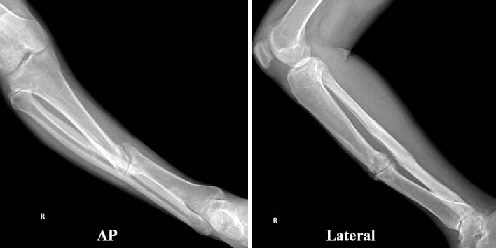
Initial presentation radiograph at our clinic. Nonunion and varus angulation of the previous fracture site is demonstrated. Notice the hypertrophied fibular. AP: Anteroposterior.
Fig. 2

(A) Endcap misplaced at the metaphyseal area of the proximal tibia posterior to the intramedullary nail. (B) Curved long Kelly forceps appear to displace the endcap further distally and anteriorly. (C) Without violating the anterior cruciate ligament footprint area, the fish hook K-wire was successful in holding the misplaced endcap. (D, E) Finally, the endcap was extracted.
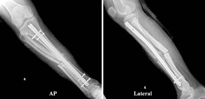
Figure & Data
REFERENCES
Citations
Citations to this article as recorded by 
 Cite
CiteExtraction of Misplaced Endcap during Tibia Intramedullary Nailing by 'Fish-Hook' Technique: Technical Note

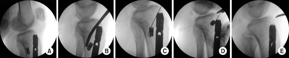

Fig. 1
Initial presentation radiograph at our clinic. Nonunion and varus angulation of the previous fracture site is demonstrated. Notice the hypertrophied fibular. AP: Anteroposterior.
Fig. 2
(A) Endcap misplaced at the metaphyseal area of the proximal tibia posterior to the intramedullary nail. (B) Curved long Kelly forceps appear to displace the endcap further distally and anteriorly. (C) Without violating the anterior cruciate ligament footprint area, the fish hook K-wire was successful in holding the misplaced endcap. (D, E) Finally, the endcap was extracted.
Fig. 3
Immediate postoperative radiographs. AP: Anteroposterior.
Fig. 1
Fig. 2
Fig. 3
Extraction of Misplaced Endcap during Tibia Intramedullary Nailing by 'Fish-Hook' Technique: Technical Note

