 E-submission
 TOTA
TOTA TOTS
TOTS
Articles
- Page Path
- HOME > J Musculoskelet Trauma > Volume 23(1); 2010 > Article
-
Surgical Technique
- The 'beta-wire Technique' for the Fixation of Ulnar Styloid Process Fracture: Surgical Technique
- Jee Hyoung Kim, M.D., Jin Hak Kim, M.D., Song Lee, M.D., Seung Jin Yang, M.D., Chang Wook Yoo, M.D., Tae Hwan Chun, M.D.
-
Journal of the Korean Fracture Society 2010;23(1):104-108.
DOI: https://doi.org/10.12671/jkfs.2010.23.1.104
Published online: January 31, 2010
Department of Orthopedic Surgery, Seoul Sacred Heart General Hospital, Seoul, Korea.
- Address reprint requests to: Jin Hak Kim, M.D. Department of Orthopedic Surgery, Seoul Sacred Heart General Hospital, 40-12, Chungryangri-dong, Dongdaemoon-gu, Seoul 130-011, Korea. Tel: 82-2-966-1616, Fax: 82-2-968-2394, benikim@paran.com
• Received: May 29, 2009 • Revised: August 9, 2009 • Accepted: October 30, 2009
Copyright © 2010 The Korean Fracture Society
- 1,139 Views
- 5 Download
- 1 Crossref
Abstract
- For the fixation of ulnar styloid process fracture, we want to introduce the 'β-wire technique', which is easy to learn and practice and thought to give a compressive force to the fracture site.
- 1. Bowers WH. DPG. The distal radioulnar joint. Operative hand surgery, 1993;1st ed. New York, Churchill Lingvingstone. 973-1019.
- 2. Hauck RM, Skahen J 3rd, Palmer AK. Classification and treatment of ulnar styloid nonunion. J Hand Surg Am, 1996;21:418-422.Article
- 3. Kim BG, Chung YG, Lee JY, Song SW, Rhee SK. Treatment of ulnar styloid fractures using miniscrew and tension-band suture augmentation. J Korean Soc Surg Hand, 2007;12:206-211.
- 4. Knirk JL, Jupiter JB. Intra-articular fractures of the distal end of the radius in young adults. J Bone Joint Surg, 1986;68:647-659.Article
- 5. May MM, Lawton JN, Blazar PE. Ulnar styloid fractures associated with distal radius fractures: incidence and implications for distal radioulnar joint instability. J Hand Surg Am, 2002;27:965-971.Article
- 6. Zenke Y, Sakai A, Oshige T, Moritani S, Nakamura T. The effect of an associated ulnar styloid frature on the outcome after fixation of a fracture of the distal radius. J Bone Joint Surg Br, 2009;91:102-107.
REFERENCES
Fig. 1
(A) The 'β-wire' in AP view, (B) lateral view and (C) oblique view. Be sure that the straight part and the hooked part nearly contact each other.

Fig. 2
(A) Holding the 'sigmoid' part of the 'β-wire', insert the 'straight' part in the distal fragment.
(B) After that using the 'joy stick method', reduce the distal part to the proximal part and insert the wire deeply.
(C) One or two taps using a small mallet would be applicable or not.

Fig. 3
(A) Tracking the hooked part proximally, be sure to watch the extent of it.
(B) Using a drill, make a hole.
(C) Tracking the hooked part proximally, insert it into the hole.
(D) To secure the insertion, one or two taps using a small mallet would be applicable.
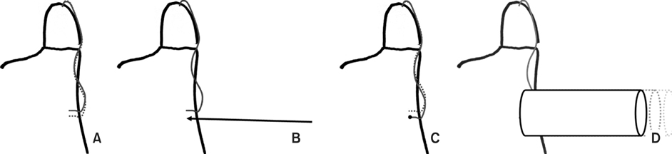
Fig. 4
(A, B) Intraoperative films show the ulnar styloid process fracture.
(C~E) Post operative films show anatomical reduction using the 'β-wire'.
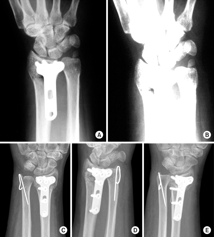
Fig. 5
(A, B) Other postoperative films show loss of elasticity of wires because the wire was too thin and the distance between the straight part and the hooked part was wide at first. Using a drill makes it difficult to feel a penetrating sense; we could find the wire was not inside of the bone during operation.

Figure & Data
REFERENCES
Citations
Citations to this article as recorded by 
- Korean Medicine Treatments for the Angular Deformity of Wrist Fracture with Disuse Osteopenia: A Case Report
Myung Jin Oh
Korean Journal of Acupuncture.2018; 35(4): 234. CrossRef
 Cite
CiteThe 'beta-wire Technique' for the Fixation of Ulnar Styloid Process Fracture: Surgical Technique
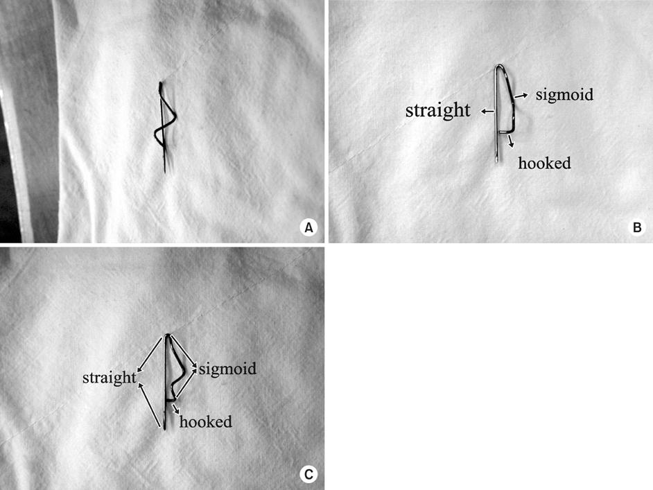
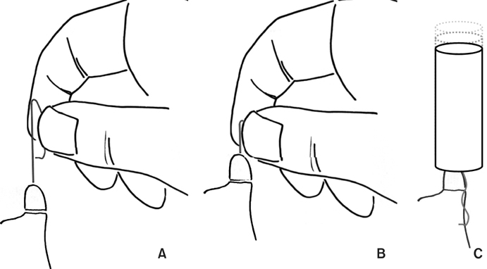


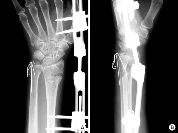
Fig. 1
(A) The 'β-wire' in AP view, (B) lateral view and (C) oblique view. Be sure that the straight part and the hooked part nearly contact each other.
Fig. 2
(A) Holding the 'sigmoid' part of the 'β-wire', insert the 'straight' part in the distal fragment.
(B) After that using the 'joy stick method', reduce the distal part to the proximal part and insert the wire deeply.
(C) One or two taps using a small mallet would be applicable or not.
Fig. 3
(A) Tracking the hooked part proximally, be sure to watch the extent of it.
(B) Using a drill, make a hole.
(C) Tracking the hooked part proximally, insert it into the hole.
(D) To secure the insertion, one or two taps using a small mallet would be applicable.
Fig. 4
(A, B) Intraoperative films show the ulnar styloid process fracture.
(C~E) Post operative films show anatomical reduction using the 'β-wire'.
Fig. 5
(A, B) Other postoperative films show loss of elasticity of wires because the wire was too thin and the distance between the straight part and the hooked part was wide at first. Using a drill makes it difficult to feel a penetrating sense; we could find the wire was not inside of the bone during operation.
Fig. 1
Fig. 2
Fig. 3
Fig. 4
Fig. 5
The 'beta-wire Technique' for the Fixation of Ulnar Styloid Process Fracture: Surgical Technique

