 E-submission
 TOTA
TOTA TOTS
TOTS
Articles
- Page Path
- HOME > J Musculoskelet Trauma > Volume 27(1); 2014 > Article
-
Case Report
- Isolated Avulsion Fracture of the Lesser Tuberosity of the Humerus: A Case Report
- Tae-Ho Kim, M.D., Ki-Do Hong, M.D., Sung-Sik Ha, M.D., Jae-Chun Sim, M.D., Min-Chul Sung, M.D.
-
Journal of the Korean Fracture Society 2014;27(1):72-76.
DOI: https://doi.org/10.12671/jkfs.2014.27.1.72
Published online: January 17, 2014
Department of Orthopedic Surgery, Sahmyook Medical Center, Seoul, Korea.
- Address reprint requests to: Ki-Do Hong, M.D. Department of Orthopedic Surgery, Sahmyook Medical Center, 82 Mangu-ro, Dongdaemun-gu, Seoul 130-711, Korea. Tel: 82-2-2210-3580, Fax: 82-2-2217-1897, oskimth@naver.com
• Received: September 13, 2013 • Revised: October 17, 2013 • Accepted: November 5, 2013
Copyright © 2014 The Korean Fracture Society. All rights reserved.
This is an Open Access article distributed under the terms of the Creative Commons Attribution Non-Commercial License (http://creativecommons.org/licenses/by-nc/3.0/) which permits unrestricted non-commercial use, distribution, and reproduction in any medium, provided the original work is properly cited.
- 1,303 Views
- 5 Download
- 1 Crossref
Abstract
- Isolated avulsion of the lesser tuberosity of the humerus is a rare injury. The mechanism of injury is the avulsion of the lesser tuberosity from forced contractions of the subscapularis muscle when the arm is forced into an externally rotated position on shoulder abduction. Authors report a case for isolated avulsion of the lesser tuberosity of the humerus which was treated with open reduction and transosseous suture fixation, as well as a view of the literature.
- 1. Coates MH, Breidahl W. Humeral avulsion of the anterior band of the inferior glenohumeral ligament with associated subscapularis bony avulsion in skeletally immature patients. Skeletal Radiol, 2001;30:661-666.ArticlePDF
- 2. Kim YH, Kim KW, Min HJ, Yoon ES, Kim HO, Lee SH. Isolated avulsion fracture of the lesser tuberosity of the humerus: a case report. J Korean Fract Soc, 2002;15:181-184.Article
- 3. Levine B, Pereira D, Rosen J. Avulsion fractures of the lesser tuberosity of the humerus in adolescents: review of the literature and case report. J Orthop Trauma, 2005;19:349-352.
- 4. Ogawa K, Takahashi M. Long-term outcome of isolated lesser tuberosity fractures of the humerus. J Trauma, 1997;42:955-959.Article
- 5. Robinson CM, Teoh KH, Baker A, Bell L. Fractures of the lesser tuberosity of the humerus. J Bone Joint Surg Am, 2009;91:512-520.Article
- 6. Scheibel M, Martinek V, Imhoff AB. Arthroscopic reconstruction of an isolated avulsion fracture of the lesser tuberosity. Arthroscopy, 2005;21:487-494.Article
- 7. Sohn HS, Chung DM, Shin SJ. Arthroscopic treatment of lesser tuberosity malunion: a case report. J Korean Arthrosc Soc, 2008;12:217-221.
- 8. Sugalski MT, Hyman JE, Ahmad CS. Avulsion fracture of the lesser tuberosity in an adolescent baseball pitcher: a case report. Am J Sports Med, 2004;32:793-796.ArticlePDF
- 9. van Laarhoven HA, te Slaa RL, van Laarhoven EW. Isolated avulsion fracture of the lesser tuberosity of the humerus. J Trauma, 1995;39:997-999.Article
- 10. Yum JK, Chung HJ, Lee SL, Choi EO. Isolated avulsion of the lesser tuberosity of the humerus in an adolescent judo player: a case report. J Korean Shoulder Elbow Soc, 2006;9:119-123.Article
REFERENCES
Fig. 1Preoperative radiographs. (A) Anteroposterior view shows blunting of the inferior glenoid in right shoulder with an area of increased opacity. (B) Axillary lateral view shows a bony avulsion of the lesser tuberosity in right shoulder.

Fig. 2Preoperative computed tomography shows an avulsion of the lesser tuberosity in right shoulder. (A) Axial view. (B) Coronal view. (C) Three-dimensional view.

Fig. 3Arthroscopic view showing avulsion of the lesser tuberosity in right shoulder with intact biceps tendon. Arrow: lesser tuberosity bony fragments. HH: Humeral head, B: Biceps long head.
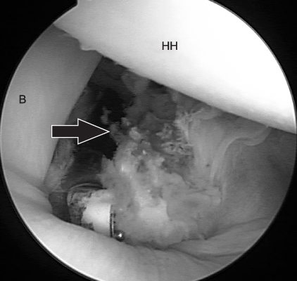


Figure & Data
REFERENCES
Citations
Citations to this article as recorded by 
- What are the patient-reported outcomes, functional limitations, and complications after lesser tuberosity fractures? a systematic review of 172 patients
Reinier W.A. Spek, Bram J.A. Schoolmeesters, Chantal den Haan, Ruurd L. Jaarsma, Job N. Doornberg, Michel P.J. van den Bekerom
JSES International.2021; 5(4): 754. CrossRef
 Cite
CiteIsolated Avulsion Fracture of the Lesser Tuberosity of the Humerus: A Case Report
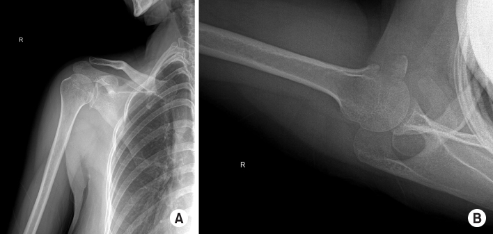
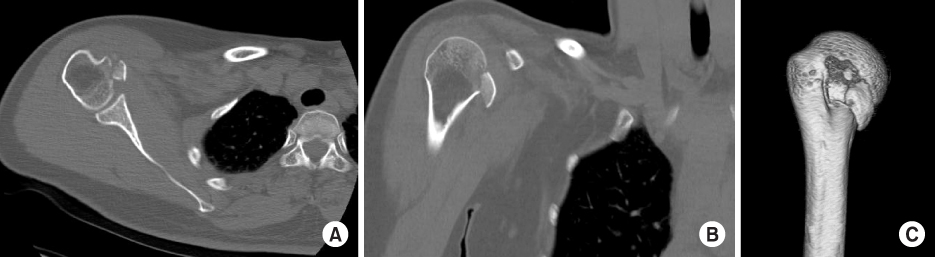

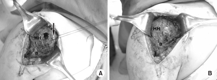
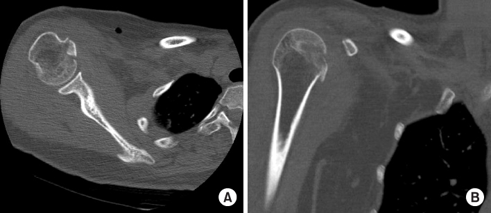
Fig. 1
Preoperative radiographs. (A) Anteroposterior view shows blunting of the inferior glenoid in right shoulder with an area of increased opacity. (B) Axillary lateral view shows a bony avulsion of the lesser tuberosity in right shoulder.
Fig. 2
Preoperative computed tomography shows an avulsion of the lesser tuberosity in right shoulder. (A) Axial view. (B) Coronal view. (C) Three-dimensional view.
Fig. 3
Arthroscopic view showing avulsion of the lesser tuberosity in right shoulder with intact biceps tendon. Arrow: lesser tuberosity bony fragments. HH: Humeral head, B: Biceps long head.
Fig. 4
Photograph of the operation field. (A) Avulsion of the lesser tuberosity at exploration. (B) Reattachment of the lesser tuberosity with transosseous sutures. Arrow: lesser tuberosity bony fragment. HH: Humeral head, B: Biceps brachii.
Fig. 5
Computed tomography 4 days postoperatively shows the reduced bony fragments. (A) Axial view. (B) Coronal view.
Fig. 1
Fig. 2
Fig. 3
Fig. 4
Fig. 5
Isolated Avulsion Fracture of the Lesser Tuberosity of the Humerus: A Case Report

