 E-submission
 TOTA
TOTA TOTS
TOTS
Articles
- Page Path
- HOME > J Musculoskelet Trauma > Volume 29(1); 2016 > Article
-
Case Report
- Functional Recovery of the Shoulder after Correcting Malrotation of the Distal Humerus: A Case Report
- Hyunseong Yoo, M.D., Jaehui Han, M.D., Youngsoo Byun, M.D., Ph.D., Daegeun Jeong, M.D., Dongju Shin, M.D.
-
Journal of the Korean Fracture Society 2016;29(1):73-78.
DOI: https://doi.org/10.12671/jkfs.2016.29.1.73
Published online: January 19, 2016
Department of Orthopaedic Surgery, Daegu Fatima Hospital, Daegu, Korea.
- Address reprint requests to: Dongju Shin, M.D. Department of Orthopaedic Surgery, Daegu Fatima Hospital, 99 Ayang-ro, Dong-gu, Daegu 41199, Korea. Tel: 82-53-940-7324, Fax: 82-53-954-7417, osaabga@gmail.com
• Received: May 26, 2015 • Revised: July 13, 2015 • Accepted: December 26, 2015
Copyright © 2016 The Korean Fracture Society. All rights reserved.
This is an Open Access article distributed under the terms of the Creative Commons Attribution Non-Commercial License (http://creativecommons.org/licenses/by-nc/4.0) which permits unrestricted non-commercial use, distribution, and reproduction in any medium, provided the original work is properly cited.
- 730 Views
- 3 Download
Abstract
- Although studies on malrotation of the humerus possibly leading to dysfunction of the shoulder have been reported, studies on its causes are inadequate. The authors encountered a patient complaining of malrotation accompanied by dysfunction of the shoulder which occurred during treatment of a distal humeral fracture. The patient recovered the shoulder function by only correcting malrotation of the humerus without direct treatment on the shoulder, and we report it herein with a review of the literature.
- 1. Puloski S, Romano C, Buckley R, Powell J. Rotational malalignment of the tibia following reamed intramedullary nail fixation. J Orthop Trauma, 2004;18:397-402.Article
- 2. Buckley R, Mohanty K, Malish D. Lower limb malrotation following MIPO technique of distal femoral and proximal tibial fractures. Injury, 2011;42:194-199.Article
- 3. Wallny T, Sagebiel C, Westerman K, Wagner UA, Reimer M. Comparative results of bracing and interlocking nailing in the treatment of humeral shaft fractures. Int Orthop, 1997;21:374-379.ArticlePDF
- 4. Flury MP, Goldhahn J, Holzmann P, Simmen BR. Does Weber's rotation osteotomy induce degenerative joint disease at the shoulder in the long term? J Shoulder Elbow Surg, 2007;16:735-741.Article
- 5. Constant CR, Murley AH. A clinical method of functional assessment of the shoulder. Clin Orthop Relat Res, 1987;(214):160-164.Article
- 6. Boileau P, Bicknell RT, Mazzoleni N, Walch G, Urien JP. CT scan method accurately assesses humeral head retroversion. Clin Orthop Relat Res, 2008;466:661-669.Article
- 7. Li Y, Wang C, Wang M, Huang L, Huang Q. Postoperative malrotation of humeral shaft fracture after plating compared with intramedullary nailing. J Shoulder Elbow Surg, 2011;20:947-954.Article
- 8. Robinson CM, Bell KM, Court-Brown CM, McQueen MM. Locked nailing of humeral shaft fractures Experience in Edinburgh over a two-year period. J Bone Joint Surg Br, 1992;74:558-562.ArticlePDF
REFERENCES
Fig. 1

(A) Initial radiographs show a comminuted open fracture of the distal humerus. (B) Radiographs after the emergency operation show open reduction and internal fixation with a long anatomical plate on the anterior aspect of the medial column and a short reconstruction plate on the lateral column.
Fig. 2

Radiograph 7 months after the initial operation shows non-union of the distal humeral fracture with loss of reduction.
Fig. 3

Photographs 7 months after the initial operation show the limitation of the right shoulder motion.
Fig. 4

Bilateral anteroposterial radiographs (A) and bilateral (B) computed tomography scans show 45° of malrotation of the right humerus.

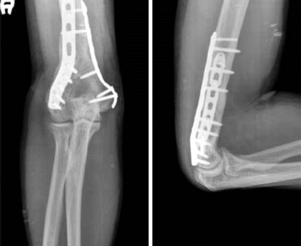

Figure & Data
REFERENCES
Citations
Citations to this article as recorded by 
 Cite
CiteFunctional Recovery of the Shoulder after Correcting Malrotation of the Distal Humerus: A Case Report
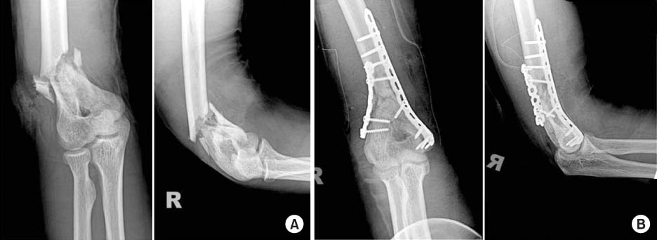
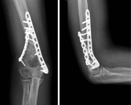
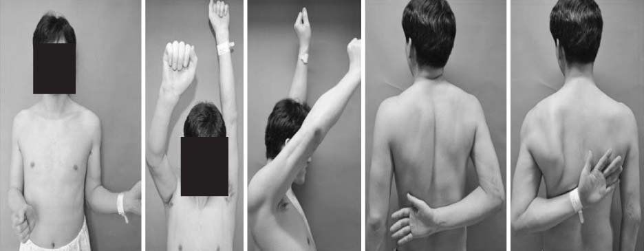
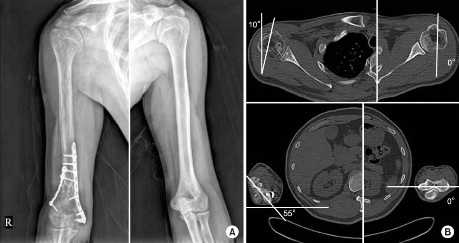
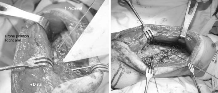

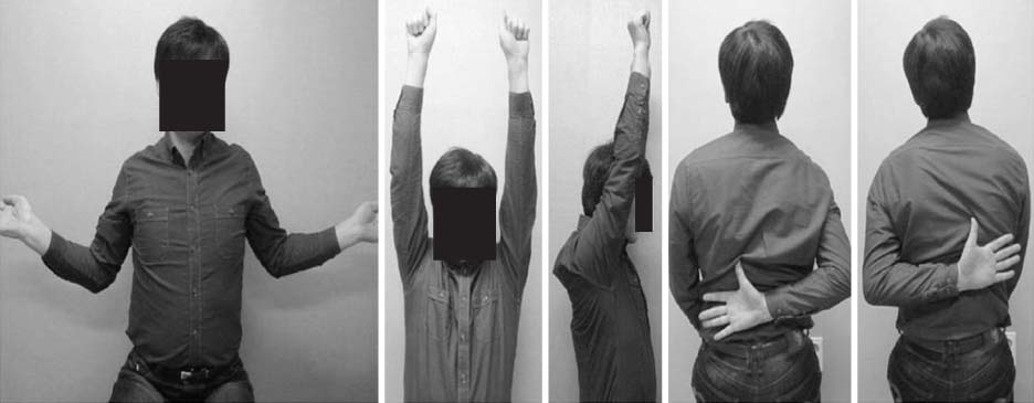
Fig. 1
(A) Initial radiographs show a comminuted open fracture of the distal humerus. (B) Radiographs after the emergency operation show open reduction and internal fixation with a long anatomical plate on the anterior aspect of the medial column and a short reconstruction plate on the lateral column.
Fig. 2
Radiograph 7 months after the initial operation shows non-union of the distal humeral fracture with loss of reduction.
Fig. 3
Photographs 7 months after the initial operation show the limitation of the right shoulder motion.
Fig. 4
Bilateral anteroposterial radiographs (A) and bilateral (B) computed tomography scans show 45° of malrotation of the right humerus.
Fig. 5
Intraoperative photographs show 45° of derotation with 2 K-wires.
Fig. 6
Final radiographs 17 months after the operation show solid union without implant loosening and loss of reduction.
Fig. 7
Photographs 2 years after the operation show a good functional result of the right shoulder.
Fig. 1
Fig. 2
Fig. 3
Fig. 4
Fig. 5
Fig. 6
Fig. 7
Functional Recovery of the Shoulder after Correcting Malrotation of the Distal Humerus: A Case Report

