 E-submission
 TOTA
TOTA TOTS
TOTS
Articles
- Page Path
- HOME > J Musculoskelet Trauma > Volume 29(1); 2016 > Article
-
Case Report
- Iatrogenic Humeral Fracture during Reduction of Shoulder Dislocation: Two Cases Report
- Hyung Lae Cho, M.D., Hyoung Min Kim, M.D., Ki Bong Park, M.D., Tae Hyun Wang, M.D., Dong Hyun Lee, M.D.
-
Journal of the Korean Fracture Society 2016;29(1):50-54.
DOI: https://doi.org/10.12671/jkfs.2016.29.1.50
Published online: January 19, 2016
Department of Orthopaedic Surgery, Good Samsun Hospital, Busan, Korea.
- Address reprint requests to: Tae Hyun Wang, M.D. Department of Orthopaedic Surgery, Good Samsun Hospital, 326 Gaya-daero, Sasang-gu, Busan 47007, Korea. Tel: 82-51-310-9289, Fax: 82-51-310-9348, socoolyo28@gmail.com
• Received: July 24, 2015 • Revised: August 26, 2015 • Accepted: November 17, 2015
Copyright © 2016 The Korean Fracture Society. All rights reserved.
This is an Open Access article distributed under the terms of the Creative Commons Attribution Non-Commercial License (http://creativecommons.org/licenses/by-nc/4.0) which permits unrestricted non-commercial use, distribution, and reproduction in any medium, provided the original work is properly cited.
- 727 Views
- 9 Download
Abstract
- Shoulder dislocation is the most common dislocation presenting to the emergency department. In old age, the attempt of closed reduction is made with caution in order to prevent iatrogenic fracture around the shoulder. We report two cases of iatrogenic fractures of humeral shaft and anatomical neck in female patients older than 70 years old, which occurred during the manual closed reduction. One patient was proved as first-time and the other was recurrent. In addition, the second case had a massive irreparable rotator cuff tear. Those patients were treated successfully with humeral nailing and reverse total shoulder arthroplasty, respectively.
- 1. Court-Brown CM, Heckman JD, McQueen MM, Tornetta P. Rockwood and Green's fractures in adults. 8th ed. Philadelphia: Lippincott Williams & Wilkins Co; 2015. p. 1531.
- 2. Atoun E, Narvani A, Even T, et al. Management of first-time dislocations of the shoulder in patients older than 40 years: the prevalence of iatrogenic fracture. J Orthop Trauma, 2013;27:190-193.
- 3. Court-Brown CM, Caesar B. Epidemiology of adult fractures: a review. Injury, 2006;37:691-697.Article
- 4. Sayegh FE, Kenanidis EI, Papavasiliou KA, Potoupnis ME, Kirkos JM, Kapetanos GA. Reduction of acute anterior dislocations: a prospective randomized study comparing a new technique with the Hippocratic and Kocher methods. J Bone Joint Surg Am, 2009;91:2775-2782.Article
- 5. Hersche O, Gerber C. Iatrogenic displacement of fracture-dislocations of the shoulder. A report of seven cases. J Bone Joint Surg Br, 1994;76:30-33.ArticlePDF
- 6. McLaughlin HL, MacLellan DI. Recurrent anterior dislocation of the shoulder. II. A comparative study. J Trauma, 1967;7:191-201.Article
- 7. Gumina S, Postacchini F. Anterior dislocation of the shoulder in elderly patients. J Bone Joint Surg Br, 1997;79:540-543.ArticlePDF
- 8. Shin DI, Shin DJ, Choi S, Park JH. Reduction of anterior shoulder dislocations by acromion upholding method (Shin's method): technical note. J Korean Orthop Assoc, 2013;48:471-474.Article
- 9. Beattie TF, Steedman DJ, McGowan A, Robertson CE. A comparison of the Milch and Kocher techniques for acute anterior dislocation of the shoulder. Injury, 1986;17:349-352.Article
- 10. Saitoh S, Nakatsuchi Y, Latta L, Milne E. Distribution of bone mineral density and bone strength of the proximal humerus. J Shoulder Elbow Surg, 1994;3:234-242.Article
REFERENCES
Fig. 1

(A, B) Shoulder radiographs show a subglenoid type of anterior glenohumeral dislocation. (C) The humeral shaft fracture occurred when attempting closed reduction detected in a post reduction radiograph. (D) A radiograph taken five months later shows fixation with humeral nailing. Postoperatively, dislocation was reduced and there were no specific complications. Bone union was observed at 5 months of final follow-up.
Fig. 2

(A, B) Shoulder radiographs show a subacromial type of anterior glenohumeral dislocation. (C) Anterior dislocation with humeral anatomical neck fracture was observed on 3-dimensional computed tomography images. (D) Intraoperative photograph shows a humeral anatomical neck fracture. (E) Coronal oblique, T2-weighted magnetic resonance imaging (MRI) scan of a full thickness tear and medial retraction of the supraspinatus tendon (white arrow). (F) And supraspinatus muscle (SSP) with Goutallier classification grade III was seen on a sagittal, T1-weighted MRI scan. (G, H) Postoperative with a reverse total shoulder arthroplasty (Zimmer, Cowpens, SC, USA) 4 months later, radiograph and clinical photograph, there were no specific complications and favorable range of motion of the right shoulder was gained at 4 months of follow-up.
Figure & Data
REFERENCES
Citations
Citations to this article as recorded by 
 Cite
CiteIatrogenic Humeral Fracture during Reduction of Shoulder Dislocation: Two Cases Report
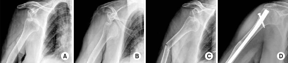
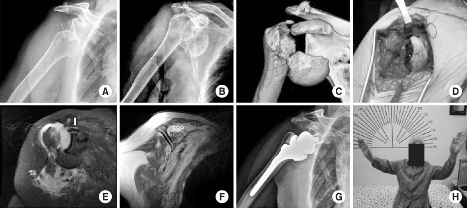
Fig. 1
(A, B) Shoulder radiographs show a subglenoid type of anterior glenohumeral dislocation. (C) The humeral shaft fracture occurred when attempting closed reduction detected in a post reduction radiograph. (D) A radiograph taken five months later shows fixation with humeral nailing. Postoperatively, dislocation was reduced and there were no specific complications. Bone union was observed at 5 months of final follow-up.
Fig. 2
(A, B) Shoulder radiographs show a subacromial type of anterior glenohumeral dislocation. (C) Anterior dislocation with humeral anatomical neck fracture was observed on 3-dimensional computed tomography images. (D) Intraoperative photograph shows a humeral anatomical neck fracture. (E) Coronal oblique, T2-weighted magnetic resonance imaging (MRI) scan of a full thickness tear and medial retraction of the supraspinatus tendon (white arrow). (F) And supraspinatus muscle (SSP) with Goutallier classification grade III was seen on a sagittal, T1-weighted MRI scan. (G, H) Postoperative with a reverse total shoulder arthroplasty (Zimmer, Cowpens, SC, USA) 4 months later, radiograph and clinical photograph, there were no specific complications and favorable range of motion of the right shoulder was gained at 4 months of follow-up.
Fig. 1
Fig. 2
Iatrogenic Humeral Fracture during Reduction of Shoulder Dislocation: Two Cases Report

