 E-submission
 TOTA
TOTA TOTS
TOTS
Articles
- Page Path
- HOME > J Musculoskelet Trauma > Volume 29(1); 2016 > Article
-
Case Report
- Periprosthetic Fracture after Hook Plate Fixation in Neer Type II Distal Clavicle Fracture: A Report of 3 Cases
- Kyung Yong Kim, M.D., Ph.D., Joon Yub Kim, M.D., Ph.D., Won Bok Lee, M.D., Ph.D., Myong Gon Jung, M.D., Jeong Hyun Yoo, M.D., Ph.D., Joo Hak Kim, M.D., Ph.D.
-
Journal of the Korean Fracture Society 2016;29(1):55-60.
DOI: https://doi.org/10.12671/jkfs.2016.29.1.55
Published online: January 19, 2016
Department of Anatomy, Chung-Ang University College of Medicine, Seoul, Korea.
*Department of Orthopaedic Surgery, Myongji Hospital, Seonam University School of Medicine, Goyang, Korea.
- Address reprint requests to: Joon Yub Kim, M.D., Ph.D. Department of Orthopaedic Surgery, Myongji Hospital, Seonam University School of Medicine, 55 Hwasu-ro 14beon-gil, Deokyanggu, Goyang 10475, Korea. Tel: 82-31-810-6530, Fax: 82-31-969-0500, doctoryub@naver.com
• Received: September 7, 2015 • Revised: October 20, 2015 • Accepted: November 17, 2015
Copyright © 2016 The Korean Fracture Society. All rights reserved.
This is an Open Access article distributed under the terms of the Creative Commons Attribution Non-Commercial License (http://creativecommons.org/licenses/by-nc/4.0) which permits unrestricted non-commercial use, distribution, and reproduction in any medium, provided the original work is properly cited.
- 897 Views
- 1 Download
- 3 Crossref
Abstract
- Hook plate fixation is a treatment method for the displaced distal clavicle fracture with favorable results regarding bone union and shoulder function, however possible complications include impingement syndromes, subacrormial erosions, acromial fractures, and periprosthetic fractures. In this report, we observed 3 cases of periprosthetic fracture after hook plate fixation. All cases of periprosthetic fractures were initiated at the medial end screw holes. The causes of these periprosthetic fractures appeared to be the off centered fixation of medial end screws near the anterior or posterior cortex which were specific during operations with hook plates with more than 6 holes and the increased stress on the medial end screw by over-reduced or inferiorly reduced position of the distal end of the clavicle by the hook plate.
- 1. Neer CS 2nd. Fracture of the distal clavicle with detachment of the coracoclavicular ligaments in adults. J Trauma, 1963;3:99-110.Article
- 2. Oh JH, Kim SH, Lee JH, Shin SH, Gong HS. Treatment of distal clavicle fracture: a systematic review of treatment modalities in 425 fractures. Arch Orthop Trauma Surg, 2011;131:525-533.ArticlePDF
- 3. Tambe AD, Motkur P, Qamar A, Drew S, Turner SM. Fractures of the distal third of the clavicle treated by hook plating. Int Orthop, 2006;30:7-10.ArticlePDF
- 4. Fann CY, Chiu FY, Chuang TY, Chen CM, Chen TH. Transacromial Knowles pin in the treatment of Neer type 2 distal clavicle fractures. A prospective evaluation of 32 cases. J Trauma, 2004;56:1102-1105.Article
- 5. Good DW, Lui DF, Leonard M, Morris S, McElwain JP. Clavicle hook plate fixation for displaced lateral-third clavicle fractures (Neer type II): a functional outcome study. J Shoulder Elbow Surg, 2012;21:1045-1048.Article
- 6. Meda PV, Machani B, Sinopidis C, Braithwaite I, Brownson P, Frostick SP. Clavicular hook plate for lateral end fractures:- a prospective study. Injury, 2006;37:277-283.Article
- 7. Flinkkilä T, Ristiniemi J, Hyvönen P, Hämäläinen M. Surgical treatment of unstable fractures of the distal clavicle: a comparative study of Kirschner wire and clavicular hook plate fixation. Acta Orthop Scand, 2002;73:50-53.Article
- 8. Nadarajah R, Mahaluxmivala J, Amin A, Goodier DW. Clavicular hook-plate: complications of retaining the implant. Injury, 2005;36:681-683.Article
- 9. Charity RM, Haidar SG, Ghosh S, Tillu AB. Fixation failure of the clavicular hook plate: a report of three cases. J Orthop Surg (Hong Kong), 2006;14:333-335.ArticlePDF
- 10. Ding M, Ni J, Hu J, Song D. Rare complication of clavicular hook plate: clavicle fracture at the medial end of the plate. J Shoulder Elbow Surg, 2011;20:e18-e20.Article
REFERENCES
Fig. 1

(A) Neer type IIa distal clavicle fracture. (B) Open reduction and internal fixation with clavicular hook plate. (C) Medial end of the clavicular hook plate had migrated superiorly and medial end screw lost the fixation to inferior cortex (white arrow). (D) Axial cut of 2-dimensional computed tomography. The medial end fracture at the medial screw of the hook plate was observed (white arrow). (E) Open reduction and internal fixation with clavicular anatomical plate for newly onset fracture after removal of the hook plate.
Fig. 2
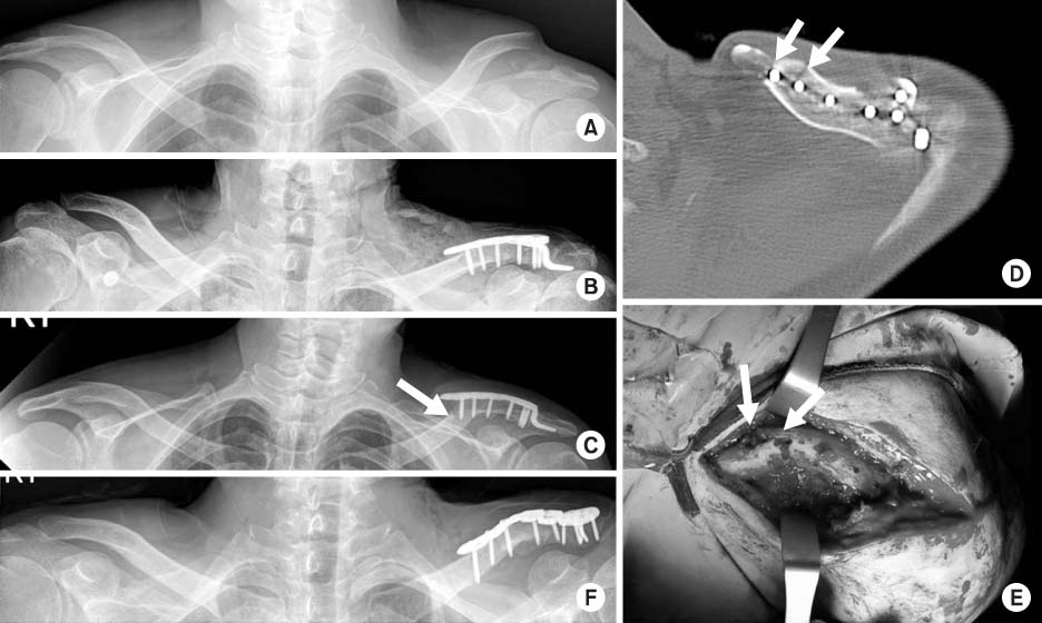
(A) Neer type IIa distal clavicle fracture. (B) Open reduction and internal fixation with clavicular hook plate. (C) Fracture at the medial screw of the hook plate was observed and medial end screw lost the fixation to inferior cortex (white arrow). (D) Axial cut of 2-dimensional computed tomography (CT) showed the fracture line communicating the medial two holes of the hook plate (2 white arrows). (E) Clinical photo corresponded to the CT findings (2 white arrows). (F) Open reduction and internal fixation with clavicular plate with cerclage wiring with No. 2 fiber wire between coracoid process and clavicular shaft.
Fig. 3

(A) Neer type IIb distal clavicle fracture. (B) Open reduction and internal fixation with clavicular hook plate. (C) Medial end of the plate had migrated superiorly (white arrow). (D) Axial cut of 2-dimensional computed tomography of the medial end fracture. The fracture started from inferior portion of medial end screw to the unused drill hole which was located anterolaterally to the medial end screw (2 white arrows). (E) Open reduction and internal fixation with clavicular anatomical plate and reconstruction plate as well as K-wires was performed for the revision surgery.
Fig. 4
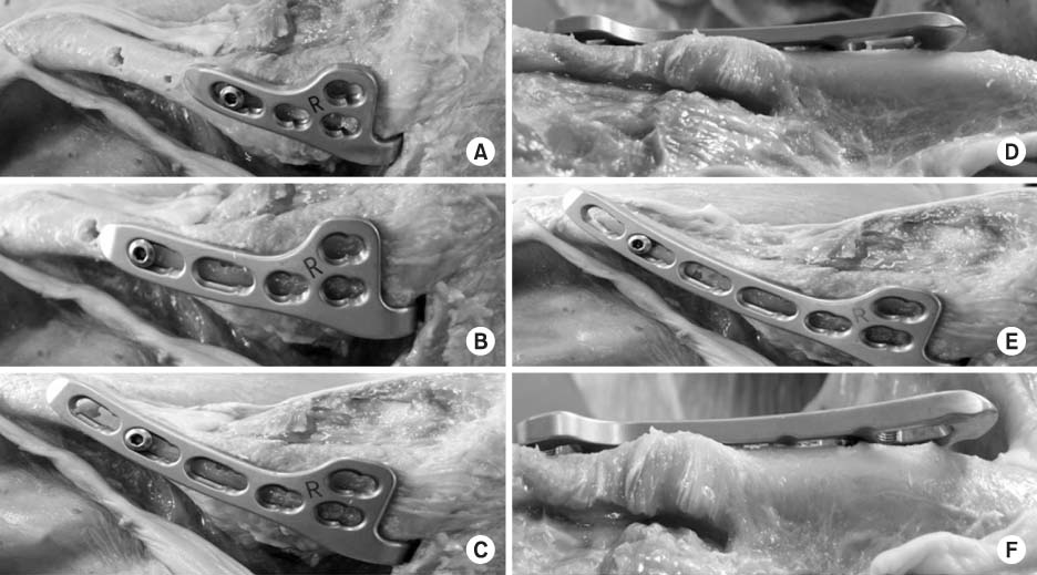
(A) A 4-hole clavicular hook plate was applied and all holes were in appropriate position on the clavicle. (B) A 5-hole clavicular hook plate was applied and all holes were in appropriate position on the clavicle. (C, D) A 6-hole clavicular hook plate was applied and the most medial hole was slightly off centered and anteriorly positioned to the clavicle (C: from cranial to caudal view, D: from caudal to cranial view). (E, F) A 7-hole clavicular hook plate was applied and the most medial hole was definitely off centered and anteriorly positioned to the clavicle (E: from cranial to caudal view, F: from caudal to cranial view).
Figure & Data
REFERENCES
Citations
Citations to this article as recorded by 
- Comparison of a novel hybrid hook locking plate fixation method with the conventional AO hook plate fixation method for Neer type V distal clavicle fractures
Joongbae Seo, Kang Heo, Seong-Jun Kim, Jun-Kyom Kim, Hee-Jung Ham, Jaesung Yoo
Orthopaedics & Traumatology: Surgery & Research.2020; 106(1): 67. CrossRef - Comparative analysis of a locking plate with an all-suture anchor versus hook plate fixation of Neer IIb distal clavicle fractures
Joong-Bae Seo, Kwon-young Kwak, Jae-Sung Yoo
Journal of Orthopaedic Surgery.2020;[Epub] CrossRef - Comparison of Locking Compression Plate Superior Anterior Clavicle Plate with Suture Augmentation and Hook Plate for Treatment of Distal Clavicle Fractures
Jun-Cheol Choi, Woo-Suk Song, Woo-Sung Kim, Jeong-Muk Kim, Chan-Woong Byun
Archives of Hand and Microsurgery.2017; 22(4): 247. CrossRef
 Cite
CitePeriprosthetic Fracture after Hook Plate Fixation in Neer Type II Distal Clavicle Fracture: A Report of 3 Cases


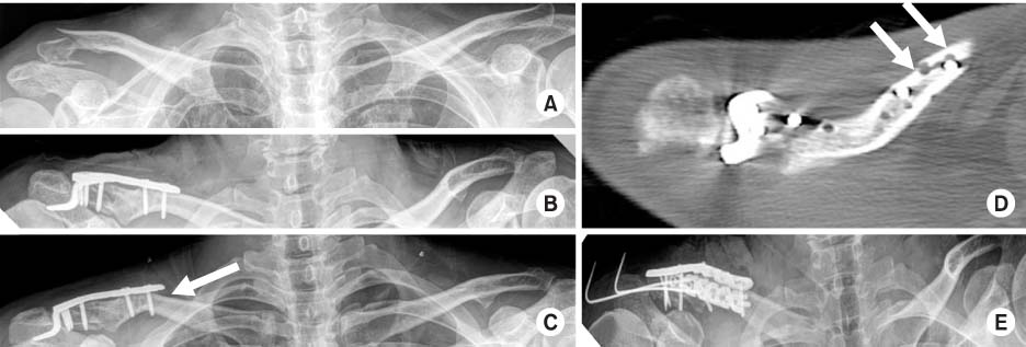

Fig. 1
(A) Neer type IIa distal clavicle fracture. (B) Open reduction and internal fixation with clavicular hook plate. (C) Medial end of the clavicular hook plate had migrated superiorly and medial end screw lost the fixation to inferior cortex (white arrow). (D) Axial cut of 2-dimensional computed tomography. The medial end fracture at the medial screw of the hook plate was observed (white arrow). (E) Open reduction and internal fixation with clavicular anatomical plate for newly onset fracture after removal of the hook plate.
Fig. 2
(A) Neer type IIa distal clavicle fracture. (B) Open reduction and internal fixation with clavicular hook plate. (C) Fracture at the medial screw of the hook plate was observed and medial end screw lost the fixation to inferior cortex (white arrow). (D) Axial cut of 2-dimensional computed tomography (CT) showed the fracture line communicating the medial two holes of the hook plate (2 white arrows). (E) Clinical photo corresponded to the CT findings (2 white arrows). (F) Open reduction and internal fixation with clavicular plate with cerclage wiring with No. 2 fiber wire between coracoid process and clavicular shaft.
Fig. 3
(A) Neer type IIb distal clavicle fracture. (B) Open reduction and internal fixation with clavicular hook plate. (C) Medial end of the plate had migrated superiorly (white arrow). (D) Axial cut of 2-dimensional computed tomography of the medial end fracture. The fracture started from inferior portion of medial end screw to the unused drill hole which was located anterolaterally to the medial end screw (2 white arrows). (E) Open reduction and internal fixation with clavicular anatomical plate and reconstruction plate as well as K-wires was performed for the revision surgery.
Fig. 4
(A) A 4-hole clavicular hook plate was applied and all holes were in appropriate position on the clavicle. (B) A 5-hole clavicular hook plate was applied and all holes were in appropriate position on the clavicle. (C, D) A 6-hole clavicular hook plate was applied and the most medial hole was slightly off centered and anteriorly positioned to the clavicle (C: from cranial to caudal view, D: from caudal to cranial view). (E, F) A 7-hole clavicular hook plate was applied and the most medial hole was definitely off centered and anteriorly positioned to the clavicle (E: from cranial to caudal view, F: from caudal to cranial view).
Fig. 1
Fig. 2
Fig. 3
Fig. 4
Periprosthetic Fracture after Hook Plate Fixation in Neer Type II Distal Clavicle Fracture: A Report of 3 Cases

