 E-submission
 TOTA
TOTA TOTS
TOTS
Articles
- Page Path
- HOME > J Musculoskelet Trauma > Volume 30(3); 2017 > Article
-
Case Report
- Extensive Multiple Morel-Lavallée Lesions: A Case Report
-
Kyu-Dong Shim, M.D.
 , Won Rak Choi, M.D., Ye-Soo Park, M.D.
 -
Journal of the Korean Fracture Society 2017;30(3):142-145.
DOI: https://doi.org/10.12671/jkfs.2017.30.3.142
Published online: July 21, 2017
Department of Orthopaedic Surgery, Guri Hospital, Hanyang University College of Medicine, Guri, Korea.
- Correspondence to: Ye-Soo Park, M.D. Department of Orthopaedic Surgery, Guri Hospital, Hanyang University College of Medicine, 153 Gyeongchun-ro, Guri 11923, Korea. Tel: +82-31-560-2181, Fax: +82-31-557-8781, hyparkys@hanyang.ac.kr
• Received: January 13, 2017 • Revised: April 17, 2017 • Accepted: April 27, 2017
Copyright © 2017 The Korean Fracture Society. All rights reserved.
This is an Open Access article distributed under the terms of the Creative Commons Attribution Non-Commercial License (http://creativecommons.org/licenses/by-nc/4.0) which permits unrestricted non-commercial use, distribution, and reproduction in any medium, provided the original work is properly cited.
- 688 Views
- 1 Download
Abstract
- Morel-Lavallée is a rare lesion caused by post-traumatic soft tissue injury. It usually occurs around the greater trochanter, and it occurs very rarely in the lumbar region. It is often difficult to be diagnosed in the emergency room. Delayed diagnosis may result in the need for open surgery. The authors report a patient with extensive multiple Morel-Lavallée lesions in the thoracolumbar, buttock, and thigh after trauma and provide a literature review.
- 1. Letournel E, Judet R. Fractures of the acetabulum. 2nd ed. Berlin: Springer; 1993.
- 2. Parra JA, Fernandez MA, Encinas B, Rico M. Morel-Lavallée effusions in the thigh. Skeletal Radiol, 1997;26:239-241.ArticlePDF
- 3. Kalaci A, Karazincir S, Yanat AN. Long-standing Morel-Lavallée lesion of the thigh simulating a neoplasm. Clin Imaging, 2007;31:287-291.Article
- 4. El Kininy W, Davy S, Sayana M. Unusual Morel-Lavallee lesion of the knee region in an elderly patient. BMJ Case Rep, 2017;2017:bcr2016218577. Article
- 5. Luria S, Applbaum Y, Weil Y, Liebergall M, Peyser A. Talc sclerodhesis of persistent Morel-Lavallée lesions (posttraumatic pseudocysts): case report of 4 patients. J Orthop Trauma, 2006;20:435-438.
- 6. Tseng S, Tornetta P 3rd. Percutaneous management of Morel-Lavallee lesions. J Bone Joint Surg Am, 2006;88:92-96.Article
- 7. Hudson DA, Knottenbelt JD, Krige JE. Closed degloving injuries: results following conservative surgery. Plast Reconstr Surg, 1992;89:853-855.
- 8. Zairi F, Wang Z, Shedid D, Boubez G, Sunna T. Lumbar Morel-Lavallée lesion: case report and review of the literature. Orthop Traumatol Surg Res, 2016;102:525-527.Article
- 9. Tran W, Foran J, Wang M, Schwartz A. Postsurgical bleeding following treatment of a chronic Morel-Lavallée lesion. Orthopedics, 2008;31:814. Article
- 10. Neal C, Jacobson JA, Brandon C, Klaume-Brigido M, Morag Y, Girish G. Sonography of Morel-Lavallée lesions. J Ultrasound Med, 2008;27:1077-1081.ArticlePDF
REFERENCES
Fig. 1

Sagittal T2-weighted fat-suppressed image of the thoracolumbar spine (A) shows a large fluid collection within the subcutaneous tissues, from the T10 level to the S1 level. Coronal T2-weighted fat-suppressed image of the left thigh (B) and axial image of the right hip (C) show a fluid collection between the fascia and the subcutaneous layer.


Figure & Data
REFERENCES
Citations
Citations to this article as recorded by 
 Cite
CiteExtensive Multiple Morel-Lavallée Lesions: A Case Report
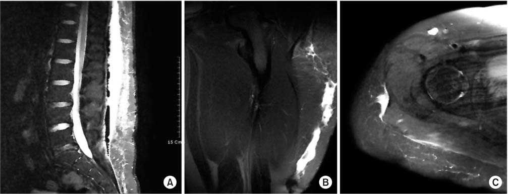
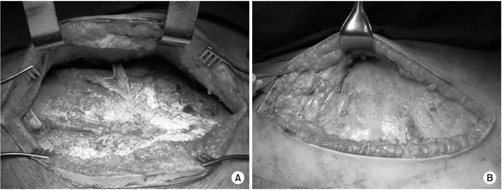
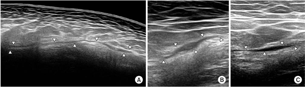
Fig. 1
Sagittal T2-weighted fat-suppressed image of the thoracolumbar spine (A) shows a large fluid collection within the subcutaneous tissues, from the T10 level to the S1 level. Coronal T2-weighted fat-suppressed image of the left thigh (B) and axial image of the right hip (C) show a fluid collection between the fascia and the subcutaneous layer.
Fig. 2
Intraoperative appearance of Morel-Lavallée lesions at the thoracolumbar spine (A) and left thigh (B).
Fig. 3
Longitudinal ultrasound image of the thoracolumbar spine (A), and transverse ultrasound image of the left thigh (B) and right hip (C).
Fig. 1
Fig. 2
Fig. 3
Extensive Multiple Morel-Lavallée Lesions: A Case Report

