 E-submission
 TOTA
TOTA TOTS
TOTS
Articles
- Page Path
- HOME > J Musculoskelet Trauma > Volume 24(1); 2011 > Article
-
Case Report
- Bilateral Open Transcalcaneal Fracture with Talonavicular Dislocation: A Case Report
- Hun Park, M.D., Sung Jin Shin, M.D., Sang Rim Kim, M.D., Kwang Woo Nam, M.D., Sung Wook Choi, M.D., Kyu Bum Seo, M.D., Jun Young Seo, M.D.
-
Journal of the Korean Fracture Society 2011;24(1):87-91.
DOI: https://doi.org/10.12671/jkfs.2011.24.1.87
Published online: January 21, 2011
Department of Orthopedic Surgery, Chonbuk National University School of Medicine, Research of Clinical Medicine, Jeonju, Korea.
*Department of Orthopedic Surgery, Jeju National University College of Medicine, Jeju, Korea.
- Address reprint requests to: Sung Jin Shin, M.D. Department of Orthopedic Surgery, Jeju National University College of Medicine, 1753-3, Ara 1-dong, Jeju 690-767, Korea. Tel: 82-64-717-1690·Fax: 82-64-757-8276, ggeep@naver.com
• Received: January 17, 2010 • Revised: March 5, 2010 • Accepted: October 20, 2010
Copyright © 2011 The Korean Fracture Society
- 902 Views
- 2 Download
- 1 Crossref
Abstract
- Although calcaneal fracture is relatively common in ankle injury, open intraarticular calcaneal fracture with dorsal dislocation of the navicular from talus is extremely rare and severe injury. There are few data which are available concerning the injury mechanism and treatment options. The purpose of this report is to describe a case with bilateral open transcalcaneal fracture with talonavicular dislocation and to discuss the prevalence, mechanism of this injury, and treatment options.
- 1. Byun YS, Cho YH, Park JW, Lee JS, Kim JH. Early postoperative complications of calcaneal fractures following operative treatment by a lateral extensile approach. J Korean Fract Soc, 2004;17:323-327.Article
- 2. Coltart WD. Aviator's astragalus. J Bone Joint Surg Br, 1952;34:545-566.ArticlePDF
- 3. Ebraheim NA, Savolaine ER, Paley K, Jackson WT. Comminuted fracture of the calcaneus associated with subluxation of the talus. Foot Ankle, 1993;14:380-384.ArticlePDF
- 4. Folk JW, Starr AJ, Early JS. Early wound complications of operative treatment of calcaneus fractures: analysis of 190 fractures. J Orthop Trauma, 1999;13:369-372.Article
- 5. Jung HG, Kim YJ, Jeon SH. Primary subtalar arthrodesis for the treatment of intra-articular calcaneal comminuted fractures. J Korean Fract Soc, 2006;19:418-423.Article
- 6. Kleiger B. Injuries of the talus and its joints. Clin Orthop Relat Res, 1976;121:243-262.Article
- 7. Marsh JL, Saltzman CL, Iverson M, Shapiro DS. Major open injuries of the talus. J Orthop Trauma, 1995;9:371-376.Article
- 8. Ricci WM, Bellabarba C, Sanders R. Transcalcaneal talonavicular dislocation. J Bone Joint Surg Am, 2002;84:557-561.Article
- 9. Sanders R, Fortin P, DiPasquale T, Walling A. Operative treatment in 120 displaced intraarticular calcaneal fractures. Results using a prognostic computed tomography scan classification. Clin Orthop Relat Res, 1993;290:87-95.
- 10. Sanders R, Clare M. Coughlin MJ, Mann RA, Saltzman CL. Fractures of the calcaneus. In: Surgery of the foot and ankle, 2007;7th ed. St. Louis, Mosby. 2017-2073.
REFERENCES
Fig. 1
26-year-old woman sustained an open fractures of the both calcaneus and fracture of the lumbar spine by a fall from a height. Initial (A) right and (B) left lateral radiographs show severe comminuted intra-articular calcaneus fracture with talonavicular dislocation.
(C) Lateral radiograph of the lumbar spine shows compression fracture of 2nd, 3rd and 4th lumbar vertebral body.
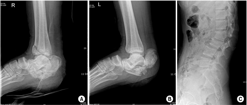
Fig. 2
(A) Right and (B) left CT semi-coronal view shows the Sanders type IV with severe comminution.
(C) CT axial view of the 2nd lumbar vertebra shows compromising the spinal canal by bursting fracture.
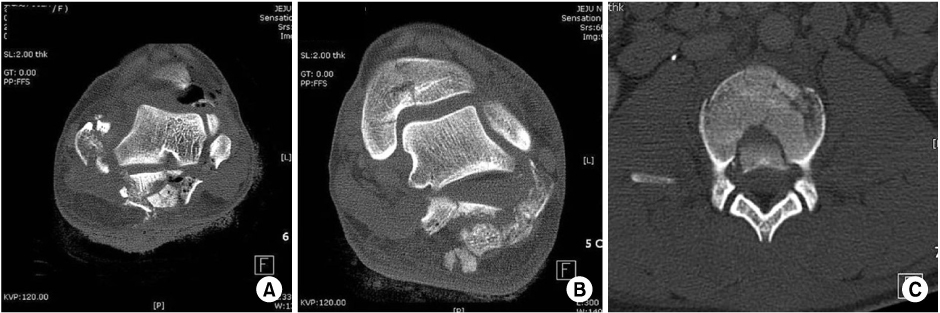
Fig. 3After irrigation and debridement, closed reduction and percutaneous K-wires and Steinmann pins fixation were performed due to contaminated open fracture.
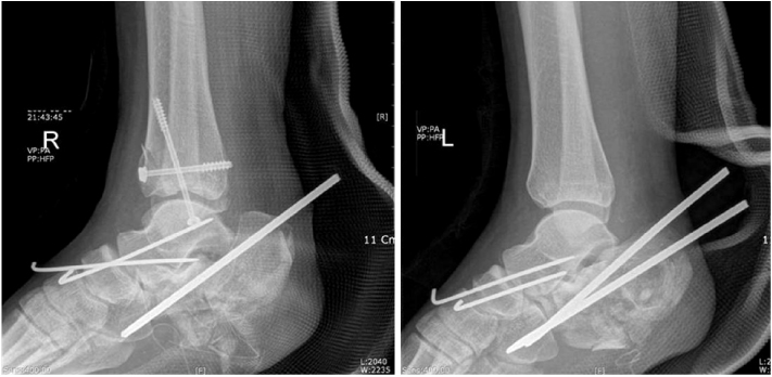


Figure & Data
REFERENCES
Citations
Citations to this article as recorded by 
- Results in Operative Treatment of Open Calcaneal Fracture
Ba Rom Kim, Jun Young Lee, Donghyuk Cha
Journal of Korean Foot and Ankle Society.2021; 25(3): 133. CrossRef
 Cite
CiteBilateral Open Transcalcaneal Fracture with Talonavicular Dislocation: A Case Report


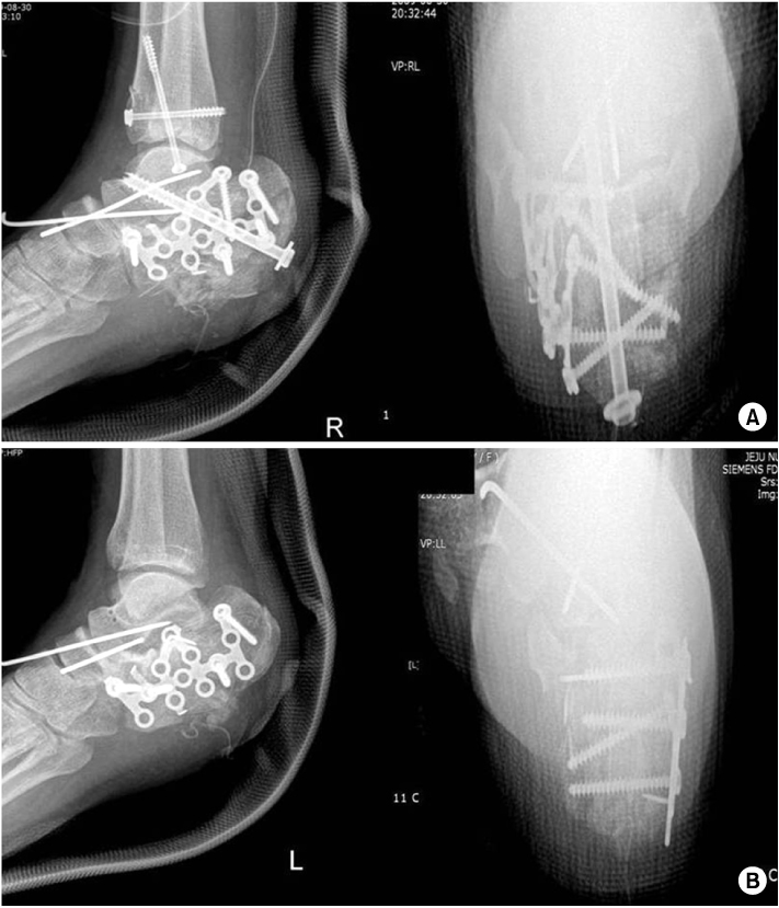
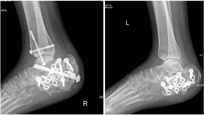
Fig. 1
26-year-old woman sustained an open fractures of the both calcaneus and fracture of the lumbar spine by a fall from a height. Initial (A) right and (B) left lateral radiographs show severe comminuted intra-articular calcaneus fracture with talonavicular dislocation.
(C) Lateral radiograph of the lumbar spine shows compression fracture of 2nd, 3rd and 4th lumbar vertebral body.
Fig. 2
(A) Right and (B) left CT semi-coronal view shows the Sanders type IV with severe comminution.
(C) CT axial view of the 2nd lumbar vertebra shows compromising the spinal canal by bursting fracture.
Fig. 3
After irrigation and debridement, closed reduction and percutaneous K-wires and Steinmann pins fixation were performed due to contaminated open fracture.
Fig. 4
(A) Open reduction and internal fixation with sub-talar arthrodesis by plate and cannulated screw were performed due to severe comminuted intra-articular fracture.
(B) Open reduction and internal fixation were performed by extended lateral approach.
Fig. 5
Lateral radiographs of both calcaneus show maintenance of the reduction of talonavicular joint and good bony union of calcaneus at 14 weeks after operation.
Fig. 1
Fig. 2
Fig. 3
Fig. 4
Fig. 5
Bilateral Open Transcalcaneal Fracture with Talonavicular Dislocation: A Case Report

