 E-submission
 TOTA
TOTA TOTS
TOTS
Articles
- Page Path
- HOME > J Musculoskelet Trauma > Volume 26(2); 2013 > Article
-
Case Report
- Excessive Sliding of the Helical Blade and the Femoral Neck Fracture after Insertion of Proximal Femoral Nail Anti-Rotation for Type A2 Intertrochanteric Fractures - A Case Report -
- Bong-Ju Park, M.D., Hong-Man Cho, M.D., Ju-Han Kim, M.D., Woo-Jin Sin, M.D.
-
Journal of the Korean Fracture Society 2013;26(2):151-155.
DOI: https://doi.org/10.12671/jkfs.2013.26.2.151
Published online: April 22, 2013
Department of Orthopedic Surgery, Gwangju Veterans Hospital, Gwangju, Korea.
- Address reprint requests to: Hong-Man Cho, M.D. Department of Orthopedic Surgery, Gwangju Veterans Hospital, 91 Sanwol-ro, Gwangsan-gu, Gwangju 506-705, Korea. Tel: 82-62-602-6162, Fax: 82-62-602-6164, chm1228@naver.com
• Received: December 6, 2012 • Revised: February 5, 2013 • Accepted: February 5, 2013
Copyright © 2013 The Korean Fracture Society
- 1,345 Views
- 5 Download
- 4 Crossref
Abstract
- Proximal femoral nail anti-rotation (PFNA) with a lag screw that is shaped like a spiral blade shape is an orthopedic implant to fix trochanteric fractures of the proximal femur. In addition the reason of the biomechanical advantages, PFNA widely been used recently. We report an 83-year-old man with excessive sliding of the helical blade and a femoral neck fracture after AO/OTA type A2 intertrochanteric fracture, which was fixed with a PFNA.
- 1. Brunner A, Jöckel JA, Babst R. The PFNA proximal femur nail in treatment of unstable proximal femur fractures--3 cases of postoperative perforation of the helical blade into the hip joint. J Orthop Trauma, 2008;22:731-736.Article
- 2. Byun YS, Yoo CH, Nam JM, Cho YH, Shin DJ. Unstable trochanteric fractures of the femur treated with a condylar blade plate. J Korean Soc Fract, 2002;15:320-327.Article
- 3. Jang SA, Cho YH, Byun YS, Cho HS, Choi S, Yoo HS. A case report of unique complications of PFNA penetration of the blade into the hip joint. J Korean Hip Soc, 2011;23:318-322.Article
- 4. Mereddy P, Kamath S, Ramakrishnan M, Malik H, Donnachie N. The AO/ASIF proximal femoral nail antirotation (PFNA): a new design for the treatment of unstable proximal femoral fractures. Injury, 2009;40:428-432.ArticlePubMed
- 5. Niikura T, Lee SY, Matsumoto T, et al. Backout of the helical blade of proximal femoral nail antirotation and accompanying fracture nonunion. Orthopedics, 2012;35:e1264-e1266.Article
- 6. Park JH, Lee YS, Park JW, Wang JH, Kim JG. A comparative study of screw and helical proximal femoral nails for the treatment of intertrochanteric fractures. Orthopedics, 2010;33:81-85.ArticlePubMed
- 7. Simmermacher RK, Bosch AM, Van der Werken C. The AO/ASIF-proximal femoral nail (PFN): a new device for the treatment of unstable proximal femoral fractures. Injury, 1999;30:327-332.Article
- 8. Simmermacher RK, Ljungqvist J, Bail H, et al. AO - PFNA studygroup. The new proximal femoral nail antirotation (PFNA) in daily practice: results of a multicentre clinical study. Injury, 2008;39:932-939.ArticlePubMed
- 9. Sommers MB, Roth C, Hall H, et al. A laboratory model to evaluate cutout resistance of implants for pertrochanteric fracture fixation. J Orthop Trauma, 2004;18:361-368.Article
- 10. Takigami I, Ohnishi K, Ito Y, et al. Acetabular perforation after medial migration of the helical blade through the femoral head after treatment of an unstable trochanteric fracture with proximal femoral nail antirotation (PFNA): a case report. J Orthop Trauma, 2011;25:e86-e89.Article
REFERENCES
Fig. 1
An 83-year-old male with right hip pain.
(A) Preoperative radiology showed AO/OTA type A2.2 intertrochanteric fracture.
(B) Preoperative 3-dimensional computerized tomography showed AO/OTA type A2.2 intertrochanteric fracture.
(C) Immediate postoperative radiology showed fixation with proximal femoral nail anti-rotation.
(D) Radiologic finding on his postoperative 16 weeks showed slight sliding of the helical blade.
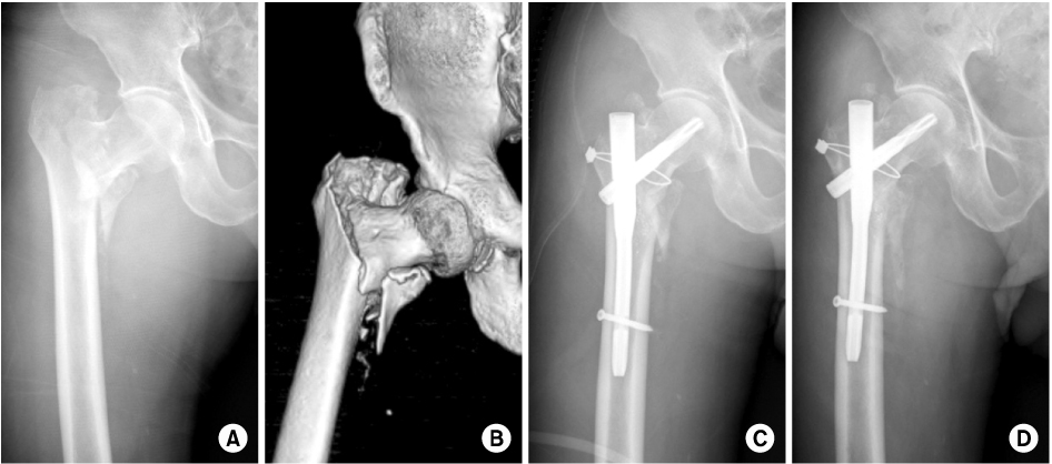


Figure & Data
REFERENCES
Citations
Citations to this article as recorded by 
- Helical Blade Locking Sleeve Disassembly Following Failed Femur Intertrochanter Fracture - A Case Report -
Soon Ho Huh, Hong-Man Cho, Ji-Yeon Park
Journal of the Korean Fracture Society.2021; 34(3): 112. CrossRef - Retrospective Comparative Study of the Intraoperative Fracture Gap Compression in the Treatment of Intertrochanteric Fracture Using Proximal Femoral Nail Antirotation
Se Jin Kim, Hong Man Cho, Jiyeon Park, Ki Yong An, Young Woo Chung, Woojin Shin
Journal of the Korean Fracture Society.2020; 33(4): 179. CrossRef - Failure of a Rotation Control Gamma 3 Lag Screw Used to Treat a Trochanteric Fracture
Kyungho Choi, Yongtae Kim, Shicheng Zhou, Jihyo Hwang
Hip & Pelvis.2018; 30(2): 129. CrossRef - Femoral neck fractures after internal fixation of trochanteric fractures with implants in situ in adults: A systematic review
Antonio Barquet, Peter V. Giannoudis, Andrés Gelink
Injury.2018; 49(12): 2121. CrossRef
 Cite
CiteExcessive Sliding of the Helical Blade and the Femoral Neck Fracture after Insertion of Proximal Femoral Nail Anti-Rotation for Type A2 Intertrochanteric Fractures - A Case Report -

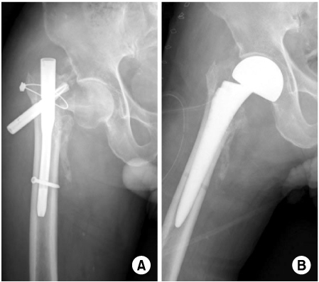
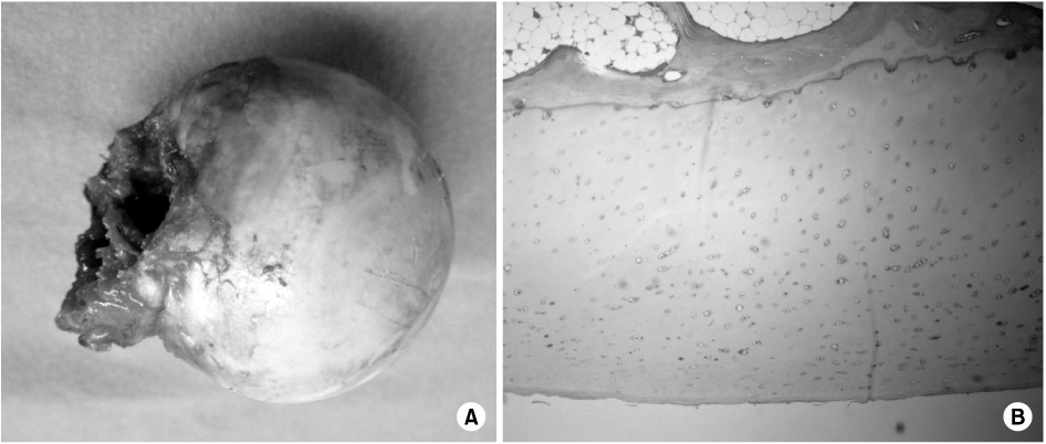
Fig. 1
An 83-year-old male with right hip pain.
(A) Preoperative radiology showed AO/OTA type A2.2 intertrochanteric fracture.
(B) Preoperative 3-dimensional computerized tomography showed AO/OTA type A2.2 intertrochanteric fracture.
(C) Immediate postoperative radiology showed fixation with proximal femoral nail anti-rotation.
(D) Radiologic finding on his postoperative 16 weeks showed slight sliding of the helical blade.
Fig. 2
(A) Anteroposterior radiograph taken 20 weeks after the operation showing back-out of the helical blade and femur neck fracture.
(B) Bipolar hemiarthroplasty was done.
Fig. 3
(A) The retrieved femoral head showing subcapital femur neck fracture.
(B) Biopsy finding was normal mature trabecular bone and marrow fat tissue (H&E, ×40).
Fig. 1
Fig. 2
Fig. 3
Excessive Sliding of the Helical Blade and the Femoral Neck Fracture after Insertion of Proximal Femoral Nail Anti-Rotation for Type A2 Intertrochanteric Fractures - A Case Report -

